. 2021 Apr 14;29(4):579-593.e5.
doi: 10.1016/j.chom.2021.02.004.

Il4ra-independent vaginal eosinophil accumulation following helminth infection exacerbates epithelial ulcerative pathology of HSV-2 infection

Affiliations

Il4ra-independent vaginal eosinophil accumulation following helminth infection exacerbates epithelial ulcerative pathology of HSV-2 infection

Alisha Chetty et al. Cell Host Microbe. .

Abstract

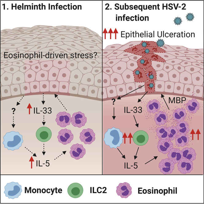
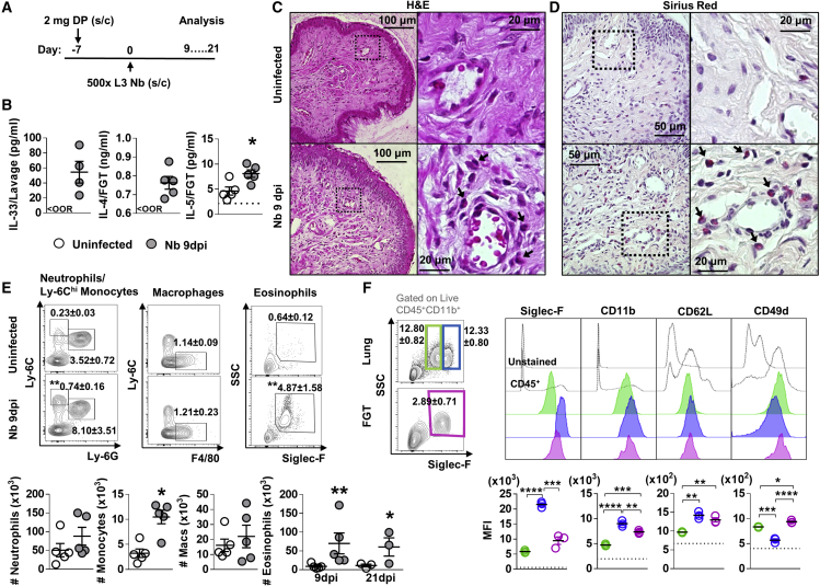
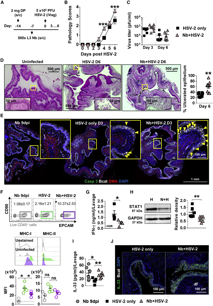
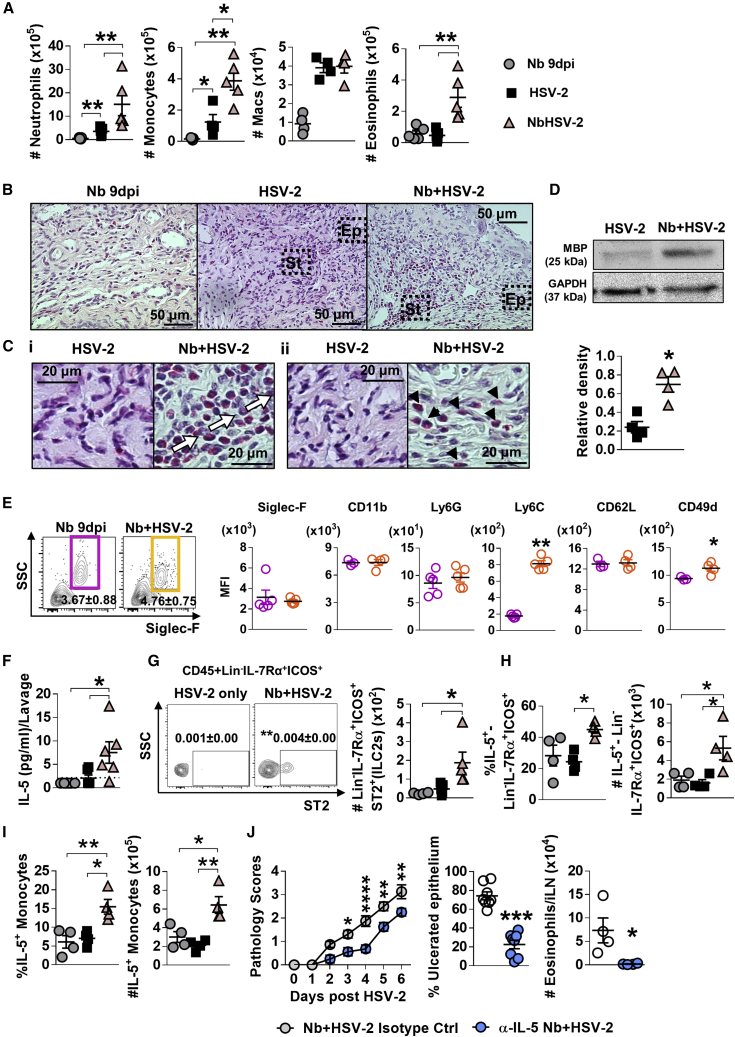
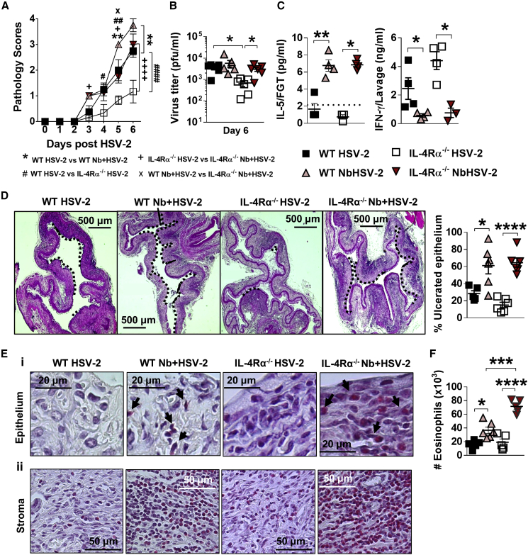
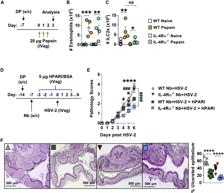
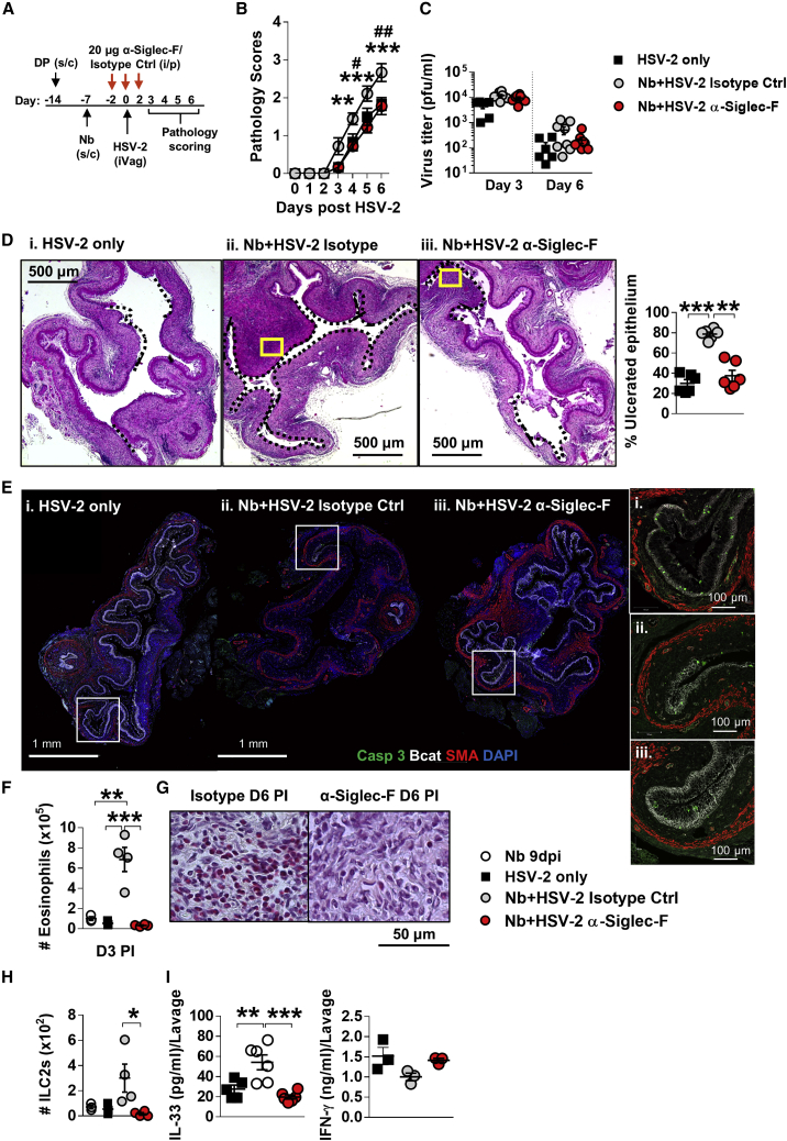
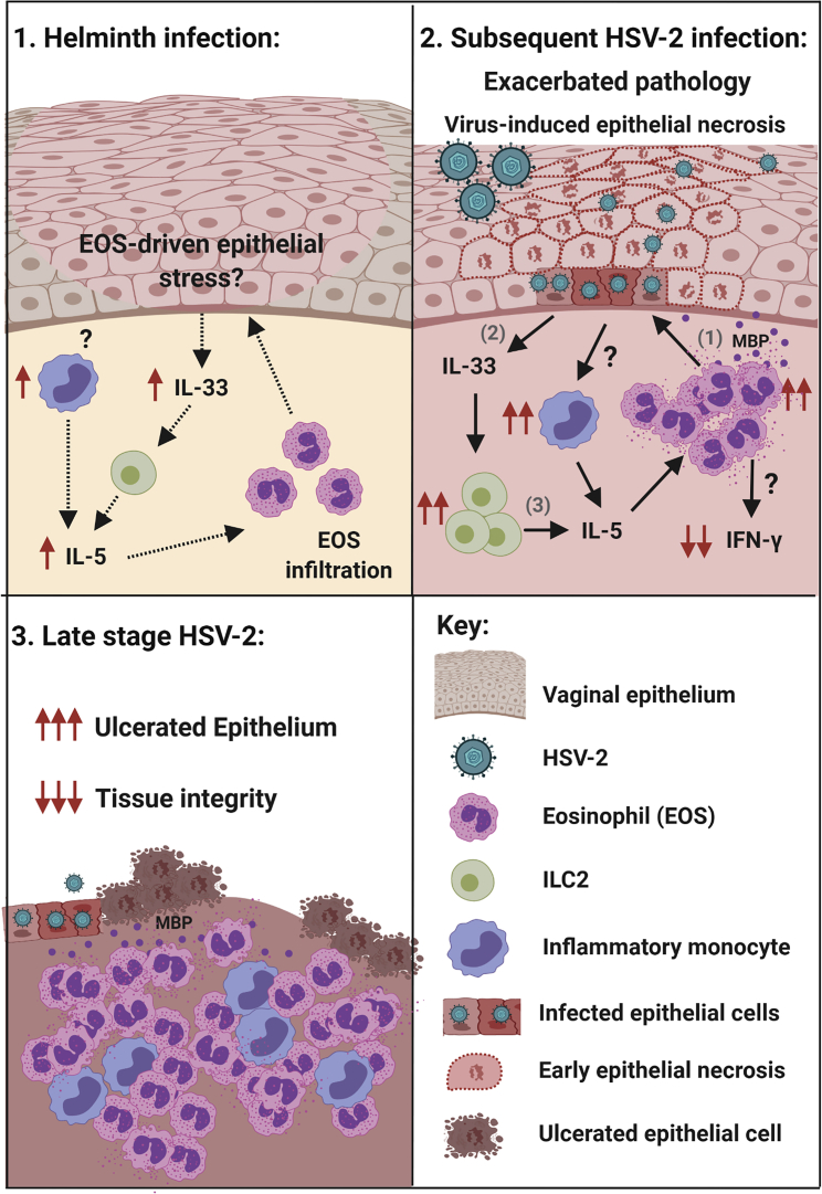
How helminths influence the pathogenesis of sexually transmitted viral infections is not comprehensively understood. Here, we show that an acute helminth infection (Nippostrongylus brasiliensis [Nb]) induced a type 2 immune profile in the female genital tract (FGT). This leads to heightened epithelial ulceration and pathology in subsequent herpes simplex virus (HSV)-2 infection. This was IL-5-dependent but IL-4 receptor alpha (Il4ra) independent, associated with increased FGT eosinophils, raised vaginal IL-33, and enhanced epithelial necrosis. Vaginal eosinophil accumulation was promoted by IL-33 induction following targeted vaginal epithelium damage from a papain challenge. Inhibition of IL-33 protected against Nb-exacerbated HSV-2 pathology. Eosinophil depletion reduced IL-33 release and HSV-2 ulceration in Nb-infected mice. These findings demonstrate that Nb-initiated FGT eosinophil recruitment promotes an eosinophil, IL-33, and IL-5 inflammatory circuit that enhances vaginal epithelial necrosis and pathology following HSV-2 infection. These findings identify a mechanistic framework as to how helminth infections can exacerbate viral-induced vaginal pathology.

Keywords: HSV-2; IL-33; IL-5; Nippostrongylus brasiliensis; eosinophils; epithelial ulceration; helminths; systemic immunity; vagina.


Conflict of interest statement

Declaration of interests The authors declare no conflict of interest.

Figures

None
Graphical abstract
Figure 1
Figure 1
Influence of N. brasiliensis exposure on uncolonized FGT, increase in FGT eosinophils following Nb exposure (A) Female mice were hormone-synchronized 7 days prior to Nb infection. (B) At day 9 post-Nb infection (Nb 9dpi), levels of IL-33, IL-4, and IL-5 in FGT homogenates or lavages were assessed by ELISA or Luminex. 3) of neutrophils (CD11b+Ly-6G+), Ly-6Chi monocytes (CD11b+Ly-6Chi), macrophages (CD11b+F480+), and eosinophils (CD11b+Siglec-F+SSChi) in the FGT of naive and Nb-infected mice. (F) Mean fluorescence intensity (MFI) of Siglec-F, CD11b, Gr-1, CD62L, and CD49d on lung (green, blue) and FGT (pink) eosinophils at Nb 9 dpi. Dotted line represents the MFI of CD45+ FGT cells. Data are representative of two independent experiments with 4–5 mice per group (mean ± SEM). Statistical significance was calculated by Mann-Whitney t test. p ≤ 0.05, ∗∗p ≤ 0.01, ∗∗∗p ≤ 0.001, ∗∗∗∗p ≤ 0.0001.
Figure 2
Figure 2
Prior Nb exposure results in earlier and exacerbated HSV-2 pathology (A) 7 days post-Nb infection, mice were infected intravaginally with 5 × 105 plaque-forming units (PFUs) HSV-2. (B) Viral progression was determined by daily pathology scoring. (C) Viral shedding (PFU/mL) was measured by plaque assay of day 3 and 6 vaginal washes. (D) At day 6 post-HSV-2 infection, vaginal tissue was analyzed by H&E staining. Representative images (n = 4) were taken at ×50 and ×400 magnification. Magnified areas are indicated by yellow boxes. HSV-2 ulcerated epithelium is indicated by black dotted lines and qualified as percentage (%) of ulcerated epithelium. (E) At day 3 post-HSV-2 infection, vaginal tissue (n = 4) was analyzed by immunofluorescent (IF) staining for β-catenin (Bcat; white), α-smooth muscle actin (SMA; red), hoechst 33342 (blue), and c-Casp-3 (green). Yellow boxes identify magnified areas. Yellow arrowheads identify “necrotic” cells i.e., large Bcat-filled nuclei that are c-Casp-3 negative. (F) At day 3 post-viral infection, vaginal epithelial cells were isolated and analyzed by flow cytometry (CD45CD90EPCAM+): MFI of MHCI and MHCII on vaginal epithelial cells from virus-only and co-infected mice. Dotted line represents the MFI of uninfected epithelial cells. (G) Levels of IFN-γ in vaginal lavages at day 2 post-HSV-2 infection, determined by ELISA. (H) At day 2 post-viral infection, levels of STAT1 (87 kDa) and GAPDH (37 kDa) were determined in HSV-2-only (H) and co-infected (N + H) FGT homogenates, by western blot. Density of STAT1 was measured relative to GAPDH. (I and J) (I) Vaginal IL-33 measured by ELISA (day 2) and (J) IF staining of vaginal tissue (day 3; n = 4). Data are representative of two independent experiments with 4–6 mice per group (mean ± SEM). Statistical significance was calculated by two-way analysis of variance (ANOVA) with Bonferroni correction for multiple comparisons and Mann-Whitney t test. p ≤ 0.05, ∗∗p ≤ 0.01, ∗∗p ≤ 0.01, ∗∗∗p ≤ 0.001, ns, not significant.
Figure 3
Figure 3
Prior Nb exposure results in elevated inflammation, eosinophil infiltration, and ILC2 presence in genital tissue, following HSV-2 vaginal infection (A) At day 9 post-Nb infection (Nb only) and day 3 post-HSV-2 infection (HSV-2 only and Nb + HSV-2), numbers of FGT myeloid cells were analyzed by flow cytometry. (B) Vaginal tissue was analyzed by Sirius red staining. Representative images (n = 4) of virus-induced ulcers were taken at ×400 magnification. (C) Black boxes indicate magnified sections of (Ci) vaginal stroma (St) and (Cii) epithelium (Ep), taken at ×1,000 magnification. White arrows indicate eosinophilic cell infiltration and migration in ulcerated vaginal tissue. Black arrowheads indicate eosinophil presence in vaginal epithelial layer. (D) At day 2 post-HSV-2 infection, levels of MBP (25 kDa) and GAPDH (37 kDa) were measured in HSV-2-only and Nb + HSV-2 FGT homogenates. Density of MBP was measured relative to that of GAPDH. (E) MFI of Siglec-F, CD11b, Ly6G, Ly6C, CD62L, and CD49d on FGT eosinophils in Nb 9 dpi and Nb + HSV-2 mice. (F) Levels of IL-5 in vaginal lavages at day 2 post-virus infection, determined by Luminex. Dotted line represents LLOQ. (G) Frequencies (mean ± SEM) and numbers of LinIL-7Rα+ICOS+ST2+ cells (ILC2s) in the FGT of Nb 9 dpi, HSV-2-only and Nb + HSV-2 mice. (H and I) Frequency and number of (H) LinIL-7Rα+ICOS+ cells and (I) Ly6Chi monocytes that are IL-5+ in the FGT, determined by flow cytometry. Data are representative of two independent experiments with 5–6 mice per group (mean ± SEM). (J) Nb-infected mice were treated with 20 μg α-IL-5 or isotype control, on day −2, 0, and 2 post-HSV-2 infection. Viral progression was determined by daily pathology scoring and HSV-2 ulcerated epithelium was qualified as percentage (%) of ulcerated epithelium. At day 6 post-HSV-2 infection, numbers of eosinophils in the iLN were determined by flow cytometry. Data are representative of two independent experiments with 4 mice per group (mean ± SEM). Statistical significance was calculated by two-way ANOVA with Bonferroni correction for multiple comparisons and Mann-Whitney t test. p ≤ 0.05, ∗∗p ≤ 0.01, ∗∗∗p ≤ 0.001, ∗∗∗∗p ≤ 0.0001.
Figure 4
Figure 4
Nb-exacerbated HSV-2 pathology and FGT eosinophil infiltration is Il4ra independent WT and Il4ra−/− mice were infected with HSV-2 following Nb exposure as previously described. (A) HSV-2 progression was determined by daily pathology scoring. (B) Viral shedding was measured by plaque assay of day 6 vaginal washes. (C) Genital levels of IL-5 and IFN-γ at day 2 post-HSV-2 infection, determined by Luminex and ELISA, respectively. Dotted line represents LLOQ of Luminex analysis. At 6 dpi, vaginal tissue was analyzed by H&E staining. (D) Representative sections (n = 3–4), displaying ulceration and inflammation of vaginal tissue. Images were taken at ×50 magnification. HSV-2-ulcerated vaginal epithelium is indicated by black dotted lines and qualified as percentage (%) of ulcerated epithelium. (E) Representative Sirius-red-stained sections (n = 3–4) of virus-induced (Ei) epithelial ulcers and (Eii) stromal inflammation. Black arrows indicate eosinophil presence in the vaginal epithelial layer. Images were taken at ×400 and ×1,000 magnification. (F) Numbers (×103) of FGT eosinophils in WT and Il4ra−/− co-infected mice compared with HSV-2-only controls. Data are representative of two independent experiments with 3–6 mice per group (mean ± SEM). Statistical significance was calculated by two-way ANOVA with Bonferroni correction for multiple comparisons. p ≤ 0.05, ∗∗p ≤ 0.01, ∗∗∗p ≤ 0.001, ∗∗∗∗p ≤ 0.0001.
Figure 5
Figure 5
Epithelial IL-33-induced FGT eosinophil inflammation in the absence of Il4ra signaling (A) WT and Il4ra−/− mice were treated intravaginally with 20 μg papain for 3 days. The next day, FGT cells were analyzed by flow cytometry. (B and C) Numbers of FGT (B) eosinophils (x103) and (C) ILC2s (×102) in papain-treated and untreated WT and Il4ra−/− mice. Data are representative of two experiments with 3–4 mice per group (mean ± SEM). (D) To inhibit vaginal IL-33, co-infected mice were treated intravaginally with helminth-derived HpARI (day −3 to 3 post-HSV-2). (E) HSV-2 progression in WT and Il4ra−/− co-infected HpARI-treated mice and BSA-treated controls was determined by daily pathology scoring (WT Nb + HSV-2 versus WT Nb + HSV-2 + HpARI, #Il4ra−/− Nb + HSV-2 versus Il4ra−/− Nb + HSV-2 + HpARI). (F) Representative H&E-stained sections (n = 4) of ulcerated vaginal tissue. Images were taken at ×50 magnification. HSV-2-ulcerated vaginal epithelium is indicated by black dotted lines and qualified as percentage (%) of ulcerated epithelium. Data are representative of two independent experiments with 4–6 mice per group. Statistical significance was calculated by two-way ANOVA with Bonferroni correction for multiple comparisons and Mann-Whitney t test. p ≤ 0.05, ∗∗p ≤ 0.01, ∗∗∗p ≤ 0.001, ∗∗∗∗p ≤ 0.0001, ns, not significant.
Figure 6
Figure 6
Depletion of eosinophils rescues HSV-2 pathology in co-infected mice (A) Co-infected mice were treated with 20 μg α-Siglec-F or isotype control antibody at days 5, 7, and 9 post-Nb infection. (B) Viral progression in HSV-2-only, α-Siglec-F-, and isotype-treated mice was determined by daily pathology scoring (Nb + HSV-2 isotype control versus Nb + HSV-2 α-Siglec-F; #HSV-2 only versus Nb + HSV-2 isotype control). (C) Viral shedding was measured by plaque assay of days 3 and 6 vaginal washes. (D) Representative H&E-stained sections (n = 3) of vaginal tissue at day 6 post-HSV-2 infection. Images were taken at ×50 magnification. Ulcerated vaginal epithelium is indicated by black dotted lines and qualified as percentage (%) of ulcerated epithelium. Yellow boxes indicate magnified sections in (G). (E) IF analysis of day 6 post-HSV-2 vaginal tissue. White boxes indicate magnified sections. (F) Numbers of FGT eosinophils at day 3 post-virus infection in isotype control and α-Siglec-F-treated Nb + HSV-2 mice compared with Nb 9dpi and HSV-2 only controls. (G) Representative magnified sections (n = 3) of Sirius-red-stained vaginal tissue at day 6 post-HSV-2. Images were taken at ×1,000 magnification. (H) Numbers of FGT ILC2s at day 3 post-HSV-2 infection. (I) At day 2 post-HSV-2 infection, lavage or FGT levels of IL-33 and IFN-γ were measured by ELISA. Data are representative of two independent experiments with 3 mice per group (mean ± SEM). Statistical significance was calculated by two-way ANOVA with Bonferroni correction for multiple comparisons and Mann-Whitney t test. p ≤ 0.05, ∗∗p ≤ 0.01, ∗∗∗p ≤ 0.001.
Figure 7
Figure 7
Helminth-induced FGT eosinophils mediate exacerbated vaginal pathology during subsequent lytic viral infection (A) We hypothesize that Nb-induced infiltration of eosinophils and inflammatory monocytes in uncolonized FGT, results in epithelial stress and release of epithelial “alarmin” IL-33, which supports the local activation of ILC2 and release of IL-5, essential for eosinophil survival. (B) The consequence of this during a subsequent virus infection was exacerbated pathology caused by virus-induced epithelial necrosis. Type 2 immunity and inflammation is amplified following HSV-2 infection: (1) eosinophil release of granule proteins promotes epithelial necrosis and (2) further release of IL-33, which (3) expands ILC2s that are a source of IL-5, along with infiltrating monocytes. Impaired anti-viral IFN-γ responses associated with eosinophil accumulation in the FGT. (C) During late-stage disease, epithelial ulceration is increased, and tissue integrity is lost. Created with BioRender.com.

Comment in

Similar articles

Cited by

References

    1. Akuthota P., Weller P.F. Eosinophilic pneumonias. Clin. Microbiol. Rev. 2012;25:649–660. - PMC - PubMed
    1. Blackwell A.D., Tamayo M.A., Beheim B., Trumble B.C., Stieglitz J., Hooper P.L., Martin M., Kaplan H., Gurven M. Helminth infection, fecundity, and age of first pregnancy in women. Science. 2015;350:970–972. - PMC - PubMed
    1. Blaho J.A., Morton E.R., Yedowitz J.C. Herpes simplex virus: propagation, quantification, and storage. Curr. Protoc. Microbiol. 2005:14E.1.1–14E.1.23. - PubMed
    1. Bobat S., Darby M., Mrdjen D., Cook C., Logan E., Auret J., Jones E., Schnoeller C., Flores-Langarica A., Ross E.A. Natural and vaccine-mediated immunity to Salmonella Typhimurium is impaired by the helminth Nippostrongylus brasiliensis. PLoS Negl. Trop. Dis. 2014;8:e3341. - PMC - PubMed
    1. Bonilla W.V., Fröhlich A., Senn K., Kallert S., Fernandez M., Johnson S., Kreutzfeldt M., Hegazy A.N., Schrick C., Fallon P.G. The alarmin interleukin-33 drives protective antiviral CD8⁺ T cell responses. Science. 2012;335:984–989. - PubMed

Publication types

MeSH terms