. 2021 Jun 1:190:108564.
doi: 10.1016/j.neuropharm.2021.108564. Epub 2021 Apr 20.

Altered excitatory transmission in striatal neurons after chronic ethanol consumption in selectively bred crossed high alcohol-preferring mice

Affiliations

Altered excitatory transmission in striatal neurons after chronic ethanol consumption in selectively bred crossed high alcohol-preferring mice

Claudia Rangel-Barajas et al. Neuropharmacology. .

Abstract

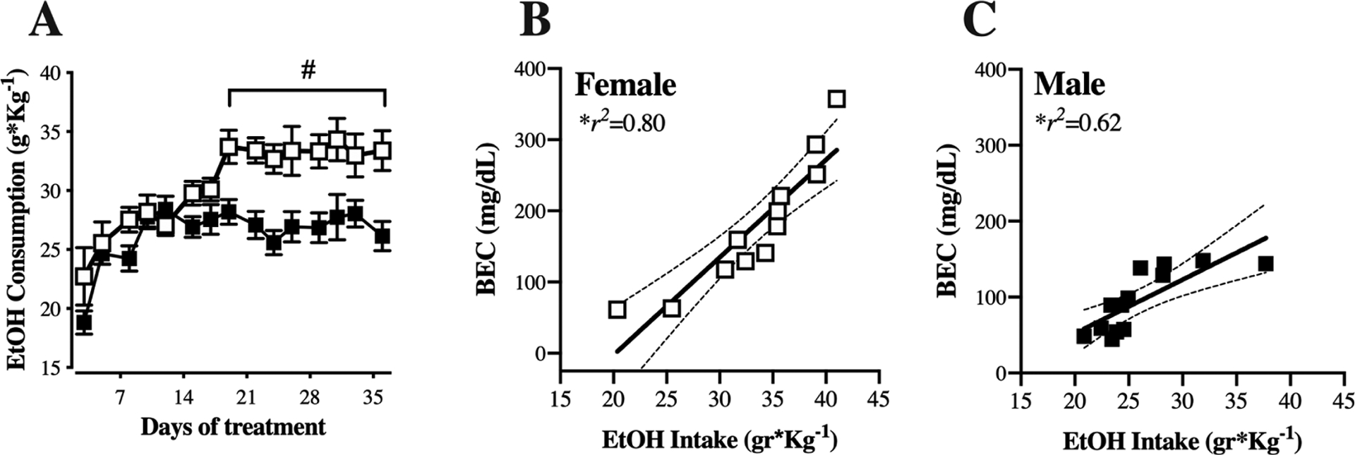
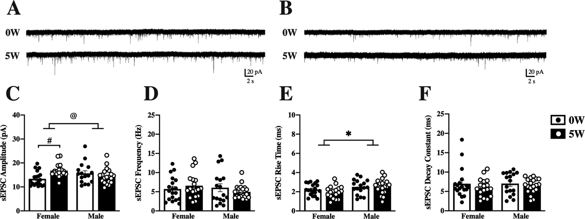
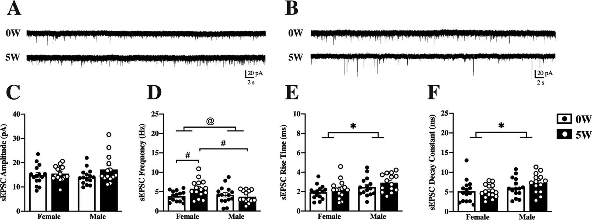
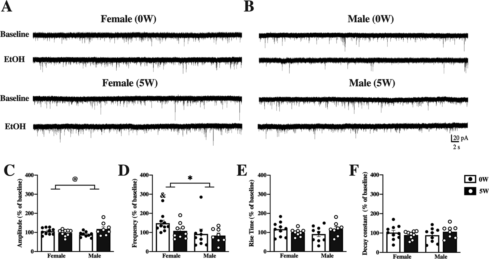
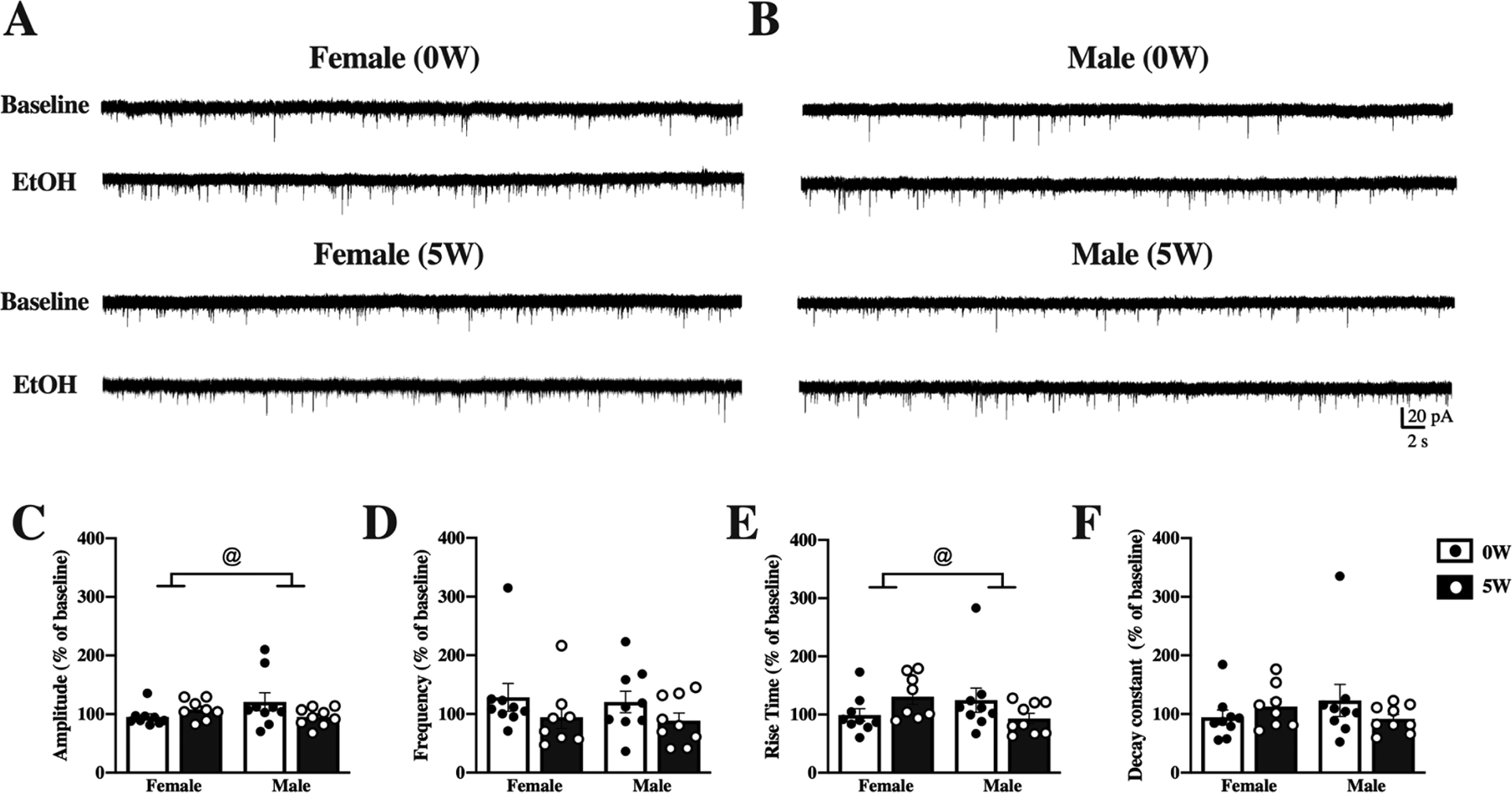
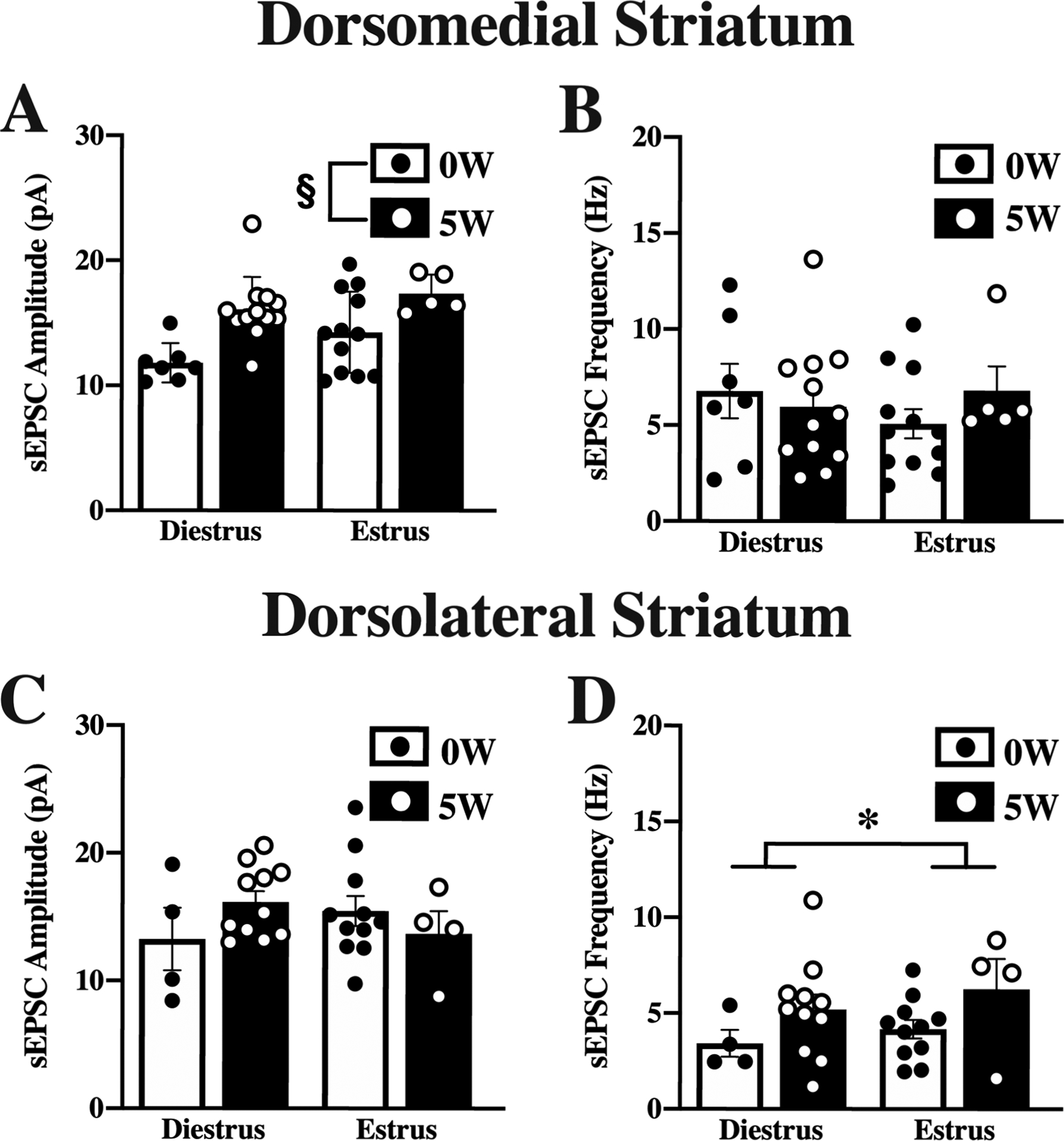
Genetic predisposition to heavy drinking is a risk factor for alcohol misuse. We used selectively bred crossed high alcohol-preferring (cHAP) mice to study sex differences in alcohol drinking and its effect on glutamatergic activity in dorsolateral (DLS) and dorsomedial (DMS) striatum. We performed whole-cell patch-clamp recording in neurons from male and female cHAP mice with 5-week alcohol drinking history and alcohol-naïve controls. In DMS, alcohol-naïve males' neurons displayed lower cell capacitance and higher membrane resistance than females' neurons, both effects reversed by drinking. Conversely, in DLS neurons, drinking history increased capacitance only in males and changed membrane resistance only in females. Altered biophysical membrane properties were accompanied by disrupted glutamatergic transmission. Drinking history increased spontaneous excitatory postsynaptic current (sEPSC) amplitude in DMS and frequency in DLS female neurons, compared to alcohol-naïve females, without effect in males. Acute ethanol differentially impacted DMS and DLS neurons by sex and drinking history. In DMS, acute alcohol significantly increased sEPSC frequency only in neurons from alcohol-naïve females, an effect that disappeared after drinking history. In DLS, acute alcohol had opposing effects in males and females based on drinking history. Estrous cycle also impacted DMS and DLS neurons differently: sEPSC amplitudes were higher in DMS cells from drinking history than alcohol-naïve females, whereas estrous cycle, not drinking history, modified DLS firing rate. Our data show sex differences in cHAP ethanol consumption and neurophysiology, suggesting differential dysregulation of glutamatergic drive onto DMS and DLS after chronic ethanol consumption.

Keywords: Alcohol misuse; Dorsal striatum; Estrous cycle; Genetically selected lines; Glutamate.


Figures

Figure 1.
Figure 1.. Females consume more alcohol and display greater blood ethanol levels than males.
(A) Male (black symbols) and female (white symbols) cHAP mice were provided two-bottle choice access to 10% (v/v) alcohol and water in the home cage for 5 weeks. Intake was measured and bottle position alternated 3 times per week. Data are presented as mean ± SEM weight-normalized alcohol intake, calculated as (g alcohol consumed)/(body weight in kg), averaged across 2 or 3 days. (B-C) Correlation between weight-normalized alcohol intake on the day prior to euthanasia and the blood ethanol content (BEC) obtained at the time of euthanasia for females (B) and males (C). # p<0.01, female vs. male. n=12 female, n=13 male.
Figure 2.
Figure 2.. Sex and alcohol history affect baseline properties of DMS neurons.
Mice were given access to 10% (v/v) alcohol and water in the home cage for 5 weeks (5W, filled bars & white dots) or water alone (0W, open bars & black dots) prior to euthanasia. Whole-cell spontaneous excitatory postsynaptic currents (sEPSCs) were recorded from dorsomedial striatal (DMS) neurons and properties of the currents measured and averaged across a 2-minute time window. Representative traces are shown for female (A) and male (B) neurons following 0W (top) and 5W (bottom) drinking history. (C) Amplitude of sEPSCs increased after 5W alcohol intake, compared to 0W, for females only. (D) Frequency of sEPSCs did not differ by sex or drinking history. (E) Rise time was higher for males than females, regardless of drinking history. (F) Sex and drinking history did not significantly impact decay time. Histograms represent mean ± SEM. @ p<0.05, sex by drinking history interaction, # p<0.05, 0W vs. 5W, * p<0.05 main effect of sex, as indicated. Number of cells: n=19, 0W female, n=18, 5W female, n=17, 0W male, n=20, 5W male; from 6–8 mice per group.
Figure 3.
Figure 3.. Sex and alcohol history affect baseline properties of DLS neurons.
Mice were given access to 10% (v/v) alcohol and water in the home cage for 5 weeks (5W, filled bars & white dots) or water alone (0W, open bars & black dots) prior to euthanasia. Whole-cell spontaneous excitatory postsynaptic currents (sEPSCs) were recorded from dorsolateral striatal (DLS) neurons and properties of the currents measured and averaged across a 2-minute time window. Representative traces are shown for female (A) and male (B) neurons following 0W (top) and 5W (bottom) drinking history. (C) Amplitude of sEPSCs did not differ by sex or drinking history. (D) Frequency of sEPSCs was increased after drinking in females, and frequencies were higher in 5W female than 5W male neurons. (E) Rise time was higher for males than females, regardless of drinking history. (F) Decay time was higher in male than in female neurons, regardless of drinking history. Histograms represent mean ± SEM. @ p<0.05, sex by drinking history interaction, # p<0.05 between groups, as indicated; * p<0.05 main effect of sex, as indicated. Number of cells: n=15, 0W female, n=15, 5W female, n=15, 0W male, n=14, 5W male; from 6 to 8 mice per group.
Figure 4.
Figure 4.. Acute effects of alcohol on DMS neurons differ by sex and drinking history.
Mice were given access to 10% (v/v) alcohol and water in the home cage for 5 weeks (5W, filled bars & white dots) or water alone (0W, open bars & black dots) prior to euthanasia. Whole-cell spontaneous excitatory postsynaptic currents (sEPSCs) were recorded from dorsomedial striatal (DMS) neurons and properties of the currents measured and averaged across a 2-minute time window before (baseline) and after application of 50 mM ethanol (EtOH) to the slices. Representative traces at baseline and after EtOH treatment are shown for neurons from female (A) and male (B) mice with 0W (top) and 5W (bottom) drinking history. (C) Acute alcohol treatment slightly increased sEPSC amplitude in 5W males, compared to 0W, but slightly decreased amplitude in 5W females, compared to 0W. (D) Acute alcohol changed sEPSC frequency more in females than in males, significantly increasing frequency only in 0W females. Acute alcohol application did not alter rise time (E) or decay time (F). Histograms represent mean ± SEM baseline-normalized values, calculated as (sEPSC parameter after 50 mM EtOH application)/(sEPSC parameter at baseline). @ p<0.05, sex by drinking history interaction, * p<0.05, main effect of sex; & p<0.05 vs. 100% (one-sample t-test). Number of cells: n=10, 0W female, n=9, 5W female, n=9, 0W male n=8, 5W male; from 4 to 6 mice per group.
Figure 5.
Figure 5.. Acute effects of alcohol on DLS neurons differ by sex and drinking history.
Mice were given access to 10% (v/v) alcohol and water in the home cage for 5 weeks (5W, filled bars & white dots) or water alone (0W, open bars & black dots) prior to euthanasia. Whole-cell spontaneous excitatory postsynaptic currents (sEPSCs) were recorded from dorsolateral striatal (DLS) neurons and properties of the currents measured and averaged across a 2-minute time window before (baseline) and after application of 50 mM ethanol (EtOH) to the slices. Representative traces at baseline and after EtOH treatment are shown for neurons from female (A) and male (B) mice with 0W (top) and 5W (bottom) drinking history. (C) Acute EtOH changed sEPSC amplitude differently by sex and drinking history. (D) Frequency of sEPSCs did not differ by sex or drinking history. (E) Acute EtOH increased rise times in 5W but not 0W females and 0W but not 5W males. (F) Decay constants did not differ by sex or drinking history. Histograms represent mean ± SEM baseline-normalized values, calculated as (sEPSC parameter after 50 mM EtOH application)/(sEPSC parameter at baseline). @ p<0.05, sex by drinking history interaction. Number of cells: n=9, 0W female, n=8, 5W female, n=8, 0W male, n=9 D, 5W male; from 4 to 6 mice per group.
Figure 6.
Figure 6.. Estrous cycle effects on spontaneous excitatory postsynaptic potentials.
Mice were given access to 10% (v/v) alcohol and water in the home cage for 5 weeks (5W, filled bars & white dots) or water alone (0W, open bars & black dots) prior to euthanasia. Estrous cycle was determined at euthanasia. Whole-cell spontaneous excitatory postsynaptic currents (sEPSCs) were recorded from dorsomedial striatal (DMS; A,B) and dorsolateral striatal (DLS; C,D) neurons and properties of the currents measured and averaged across a 2-minute time window. In DMS, amplitude (A) but not frequency (B) differed by drinking history, with higher amplitude currents after 5W EtOH, independent of cycle. In DLS, amplitude (C) did not differ by drinking history or estrous cycle, but frequency (D) was higher in estrus than in diestrus. Histograms represent mean ± SEM. § p<0.05, main effect of drinking history, * p<0.05, main effect of cycle, as indicated. Number of cells from DMS: 0W; diestrus, n=7; estrus, n=12, 5W; diestrus, n=12; estrus, n=5. Number of cells from DLS: 0W; diestrus, n=4; estrus, n=11, 5W; diestrus, n=11, estrus, n=4. Number of animals, 6 to 8 mice per group.

References

    1. Abburi C, Wolfman SL, Metz RA, Kamber R, McGehee DS, McDaid J, 2016. Tolerance to ethanol or nicotine results in increased ethanol self-administration and long-term depression in the dorsolateral striatum. eNeuro 3, ENEURO.0112–15.2016. - PMC - PubMed
    1. Abrahao KP, Salinas AG, Lovinger DM, 2017. Alcohol and the brain: Neuronal molecular targets, synapses, and circuits. Neuron 96, 1223–1238. - PMC - PubMed
    1. Agrawal A, Lynskey MT, 2008. Are there genetic influences on addiction: Evidence from family, adoption and twin studies. Addiction 103, 1069–1081. - PubMed
    1. Balleine BW, O’Doherty JP, 2010. Human and rodent homologies in action control: Corticostriatal determinants of goal-directed and habitual action. Neuropsychopharmacology 35, 48–69. - PMC - PubMed
    1. Bell RL, Hauser SR, Liang T, Sari Y, Maldonado-Devincci A, Rodd ZA, 2017. Rat animal models for screening medications to treat alcohol use disorders. Neuropharmacology 122, 201–243. - PMC - PubMed

Publication types