

Cerebral venous sinus thrombosis and thrombocytopenia after COVID-19 vaccination - A report of two UK cases
- PMID: 33857630
- PMCID: PMC8056834
- DOI: 10.1016/j.bbi.2021.04.006
Cerebral venous sinus thrombosis and thrombocytopenia after COVID-19 vaccination - A report of two UK cases
Abstract
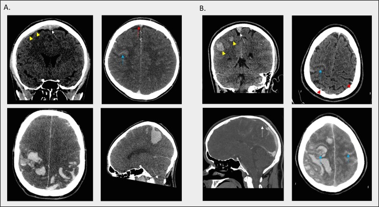
Recent reports have highlighted rare, and sometimes fatal, cases of cerebral venous sinus thrombosis (CVST) and thrombocytopenia following the Vaxzevria vaccine. An underlying immunological mechanism similar to that of spontaneous heparin-induced thrombocytopenia (HIT) is suspected, with the identification of antibodies to platelet factor-4 (PF4), but without previous heparin exposure. This unusual mechanism has significant implications for the management approach used, which differs from usual treatment of CVST. We describe the cases of two young males, who developed severe thrombocytopenia and fatal CVST following the first dose of Vaxzevria. Both presented with a headache, with subsequent rapid neurological deterioration. One patient underwent PF4 antibody testing, which was positive. A rapid vaccination programme is essential in helping to control the COVID-19 pandemic. Hence, it is vital that such COVID-19 vaccine-associated events, which at this stage appear to be very rare, are viewed through this lens. However, some cases have proved fatal. It is critical that clinicians are alerted to the emergence of such events to facilitate appropriate management. Patients presenting with CVST features and thrombocytopenia post-vaccination should undergo PF4 antibody testing and be managed in a similar fashion to HIT, in particular avoiding heparin and platelet transfusions.
Keywords: COVID-19; Cerebral venous sinus thrombosis; Cerebrovascular disease; Immunology; Neurology; Stroke; Thrombocytopenia; Vaccine.
Copyright © 2021 Elsevier Inc. All rights reserved.
Conflict of interest statement
The authors declare that they have no known competing financial interests or personal relationships that could have appeared to influence the work reported in this paper.
Figures
References
-
- British Society for Haematology: A message from BSH President, Professor Adele Fielding - March 2021. https://b-s-h.org.uk/about-us/news/a-message-from-bsh-president-professo... (accessed 29 March 2021).
-
- Coutinho J.M., Zuurbier S.M., Aramideh M., Stam J. The incidence of cerebral venous thrombosis: A cross-sectional study. Stroke. 2012;43(12):3375–3377. - PubMed
-
- Devasagayam S., Wyatt B., Leyden J., Kleinig T. Cerebral Venous sinus thrombosis incidence is higher than previously thought. A retrospective population-based study. Stroke. 2016;47(9):2180–2182. - PubMed
MeSH terms
Substances
LinkOut - more resources
Full Text Sources
Other Literature Sources
Medical
Miscellaneous
