. 2021 May:221:153419.
doi: 10.1016/j.prp.2021.153419. Epub 2021 Mar 26.

Endothelial-Mesenchymal Transition in COVID-19 lung lesions

Affiliations

Endothelial-Mesenchymal Transition in COVID-19 lung lesions

Monica Falleni et al. Pathol Res Pract. 2021 May.

Abstract

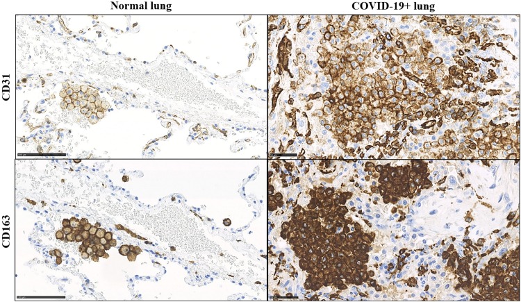
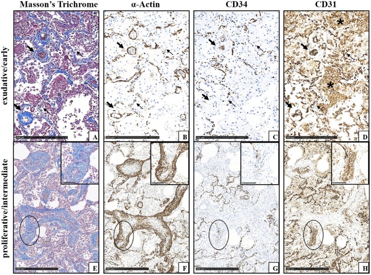
Sars-Cov-2 infection is still a healthcare emergency and acute respiratory distress failure with Diffuse Alveolar Damage (DAD) features is the main causes of patients' death. Pathogenic mechanisms of the disease are not clear yet, but new insights are necessary to improve therapeutic management, to prevent fatal irreversible multi-organ damage and to adequately follow up those patients who survive. Here we investigated, by histochemistry and immunohistochemistry, a wide number of mapped lung specimens taken from whole body autopsies of 7 patients dead of COVID-19 disease. Our data confirm morphological data of other authors, and enlarge recent reports of the literature suggesting that Endothelial-Mesenchymal Transition might be central to COVID-19 lung fibrosing lesions. Furthermore, based upon recent acquisition of new roles in immunity and vascular pathology of the CD31 molecule, we hypothesize that this molecule might be important in the development and treatment of COVID-19 pulmonary lesions. These preliminary findings need further investigations to shed light on the complexity of Sars-Cov-2 disease.

Keywords: CD31; COVID-19; Diffuse alveolar damage; Endothelial-Mesenchymal Transition; Fibrosis.


Conflict of interest statement

The authors report no declarations of interest.

Figures

Fig. 1
Fig. 1
Aberrant CD31 and CD163 expression in alveolar macrophage accumulated in COVID-19+ patients. In normal lung parenchyma, small groups of macrophages are detectable in the alveolar spaces; membrane cytoplasmic immunostaining for CD31 and CD163 is appreciable in these cells. In COVID-19+ patient macrophages are increased in number and show CD31 and CD163 overexpression at both membrane and cytoplasmic levels.
Fig. 2
Fig. 2
COVID-19+ lung lesion features in different phases (same patient, consecutive tissue sections from paraffin-embedded specimens blocks). A-D) Early lung damage with exudative DAD features in the alveolar septa (thin arrows) and around medium-sized vessels (thick arrows). A) Initial blue collagen deposition in the alveolar septa and around vessels highlighted by Masson’s Trichrome staining. B) α-Actin immunoreactivity is easily appreciated in the walls of vessels, and is fragmented in the capillaries of the alveolar septa. C) CD34 immunoreactivity is decreased in vessels and in septal capillaries, where it is also fragmented. D) CD31 immunostaining in vascular compartment at all levels is strong, but appears fragmented in alveolar capillaries; CD31+ macrophages accumulated in alveolar spaces are appreciable (asterisks). E-H) Advanced lung damage with myofibroblastic foci with proliferative DAD features (circles). E) Myofibroblastic foci are here organized as bundles of spindle cells; blue collagen production is detectable both in the cytoplasm of myofibroblastic cells and, to a lesser extent, in the extracellular matrix (inset of the circled area). Masson’s Trichrome staining. F) α-Actin immunoreactivity is easily appreciable in the outer myofibroblastic cells of fibroblastic foci (inset of the circled area). G) CD34 immunostaining is lost in the outer areas of organizing fibrosis (inset of the circled area). H) In myofibroblastic foci with disappeared/minimal CD34 immunoreactivity, CD31 staining is still present in myofibroblastic cells and in filamentous elongated structures in the centre of the proliferation (inset of the circled area).

References

    1. Bussani R., Schneider E., Zentilin L., et al. Persistence of viral RNA, pneumocyte syncytia and thrombosis are hallmarks of advanced COVID-19 pathology. EBioMedicine. 2020;61 - PMC - PubMed
    1. Calabrese F., Pezzuto F., Fortarezza F., et al. Pulmonary pathology and COVID-19: lessons from autopsy. The experience of European Pulmonary Pathologists. Virchows Archiv. 2020;477:359–372. - PMC - PubMed
    1. Eapen M.S., Lu W., Gaikwad A.V., et al. Endothelial to mesenchymal transition: a precursor to post-COVID-19 interstitial pulmonary fibrosis and vascular obliteration? Eur. Respir. J. 2020;56(4) - PMC - PubMed
    1. Bulfamante G.P., Perrucci G.L., Falleni M., et al. Evidence of SARS-CoV-2 transcriptional activity in Cardiomyocytes of COVID-19 patients without clinical signs of cardiac involvement. Biomedicines. 2020;8:626–639. - PMC - PubMed
    1. Dorward D.A., Russell C.D., Um I.H., et al. Tissue-specific immunopathology in fatal COVID-19. Am. J. Respir. Crit. Care Med. 2021;203:192–201. - PMC - PubMed