. 2021;13(4):225-241.
doi: 10.1159/000514885. Epub 2021 Apr 15.

Interleukin 8 Elicits Rapid Physiological Changes in Neutrophils That Are Altered by Inflammatory Conditions

Affiliations

Interleukin 8 Elicits Rapid Physiological Changes in Neutrophils That Are Altered by Inflammatory Conditions

Stefan Bernhard et al. J Innate Immun. 2021.

Abstract

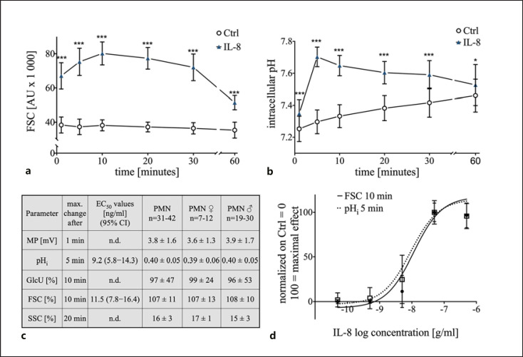
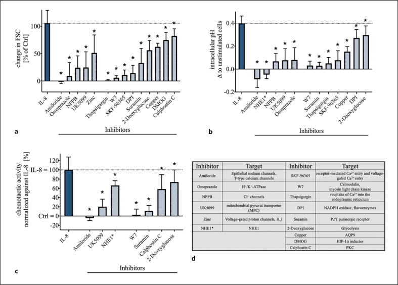
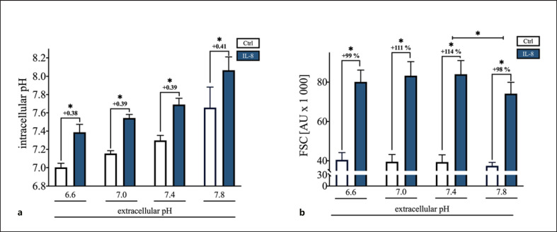
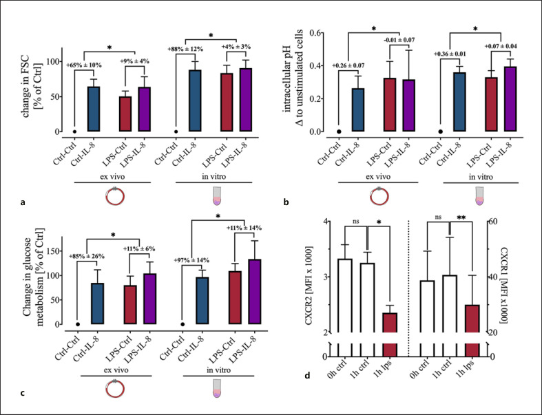
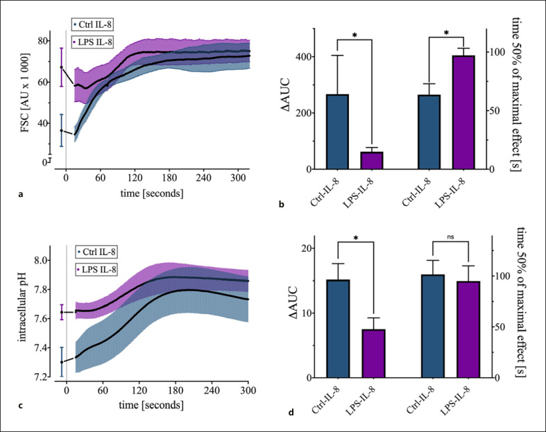
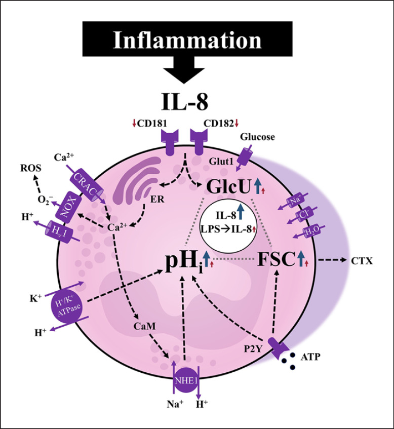
A sufficient response of neutrophil granulocytes stimulated by interleukin (IL)-8 is vital during systemic inflammation, for example, in sepsis or severe trauma. Moreover, IL-8 is clinically used as biomarker of inflammatory processes. However, the effects of IL-8 on cellular key regulators of neutrophil properties such as the intracellular pH (pHi) in dependence of ion transport proteins and during inflammation remain to be elucidated. Therefore, we investigated in detail the fundamental changes in pHi, cellular shape, and chemotactic activity elicited by IL-8. Using flow cytometric methods, we determined that the IL-8-induced cellular activity was largely dependent on specific ion channels and transporters, such as the sodium-proton exchanger 1 (NHE1) and non-NHE1-dependent sodium flux. Exposing neutrophils in vitro to a proinflammatory micromilieu with N-formyl-Met-Leu-Phe, LPS, or IL-8 resulted in a diminished response regarding the increase in cellular size and pH. The detailed kinetics of the reduced reactivity of the neutrophil granulocytes could be illustrated in a near-real-time flow cytometric measurement. Last, the LPS-mediated impairment of the IL-8-induced response in neutrophils was confirmed in a translational, animal-free human whole blood model. Overall, we provide novel mechanistic insights for the interaction of IL-8 with neutrophil granulocytes and report in detail about its alteration during systemic inflammation.

Keywords: Flow cytometry; Interleukin 8; Intracellular pH; Lipopolysaccharide; Neutrophil granulocytes; Sodium-proton exchanger 1.


Conflict of interest statement

The authors declare that the research was conducted in the absence of any commercial or financial relationship that could be construed as a potential conflict of interest.

Figures

Fig. 1
Fig. 1
Concentration dependency and maximal response during the first 60 min of IL-8-induced changes of human neutrophils: a Time-dependent effect of the IL-8-induced (50 ng/mL) increase in neutrophil cell shape assessed by FSC; n = 31–42; ***, p < 0.001 compared to the corresponding Ctrl (Mann-Whitney test). b Time course of the IL-8-induced (50 ng/mL) change in pHi in neutrophils; n = 31–42; *, p < 0.05; ***, p < 0.001 compared to the corresponding Ctrl (Mann-Whitney test). c Maximal effect for each measured parameter displayed for all test persons and differentiated by sex and calculated values for EC50. d Dose response of the change in cellular shape (FSC) after 10 min and pHi after 5 min incubation with 0.05–500 ng/mL IL-8; n = 6–42. IL, interleukin; pHi, intracellular pH; FSC, forward scatter area; Ctrl, control; EC50, half-maximal effective concentration; SSC, side scatter area; GlcU, glucose uptake; MP, membrane potential; Ctrl, control; n.d., not determined.
Fig. 2
Fig. 2
Comparison of multiple stimuli on the neutrophil cell shape (FSC), pHi, MP, and granularity (SSC). a Influence of various stimuli on the neutrophil cell size after 10 min of incubation with the indicated molecule; n = 6–12; *p < 0.05 versus Ctrl (Wilcoxon signed-rank test). b Change in pHi after 5 min of stimulation with the indicated molecule; n = 6–12; *p < 0.05 versus Ctrl (Wilcoxon signed-rank test). c Effects of numerous stimuli on the resting MP after 1 min of stimulation; n = 6–12; *p < 0.05 versus Ctrl (Wilcoxon signed-rank test). d Change in neutrophil granularity approximated by the SSC after 20 min of stimulation n = 6–12; *p < 0.05 versus Ctrl (Wilcoxon signed-rank test). FSC, forward scatter area; pHi, intracellular pH; MP, membrane potential; SSC, side scatter area; Ctrl, control; fMLF, N-formyl-Met-Leu-Phe; C5a, complement activation product 5a.
Fig. 3
Fig. 3
Effect of inhibition of various ion channels and cell signaling molecules on the IL-8-induced change in neutrophil cell shape, intracellular alkalization, and chemotactic activity. a Change in IL-8-induced (50 ng/mL) increase in neutrophil cell shape after 5 min modulated by various inhibitors; n = 5–6; *p < 0.05 versus IL-8 (Wilcoxon signed-rank test). b Influence of the indicated inhibitors on the intracellular alkalization initiated by IL-8 (50 ng/mL) after 5 min; n = 5–6; *p < 0.05 versus IL-8 (Wilcoxon signed-rank test). c Influence of key inhibitors of IL-8-induced cell swelling and intracellular alkalization on the chemotactic activity, normalized to the activity mediated by IL-8; n = 6–15; *p < 0.05 versus IL-8 (Wilcoxon signed-rank test). d Summary of the used inhibitors and their suggested target proteins (NHE1* = [4-cyanobenzo[b]thiophene-2-carbonyl]guanidine, methanesulfonate). IL, interleukin; NHE1, sodium proton exchanger 1; FSC, forward scatter area; MPC, mitochondrial pyruvate carrier; Hv1, voltage-gated H+ channel; PKC, protein kinase C; Ctrl, control.
Fig. 4
Fig. 4
Influence of a 10-min incubation with various pHe. a Dependency of the pHi and independency of the IL-8-induced (50 ng/mL) intracellular alkalization from the pHe; n = 6; *p < 0.05 versus Ctrl of the related external pH (Mann-Whitney test). b Effect of the variation in pHe on the change in neutrophil cell shape elicited by IL-8 (50 ng/mL); n = 6; *p < 0.05 FSC of pHe 7.8 versus IL-8 column with the external pH of 7.4 and FSC of IL-8-stimulated cells with the control of the related external pH (Mann-Whitney test). IL, interleukin; Ctrl, control; pHe, extracellular pH; pHi, intracellular pH; FSC, forward scatter area.
Fig. 5
Fig. 5
Impact of the preincubation for 1 h with LPS (100 ng/mL), LPS with LBP (2 ng/mL), fMLF (10 μM), and IL-8 (50 ng/mL), on I, F, and HBSS+/+ (as Ctrl) induced change in neutrophil cell shape and pHi. a In vitro effect of a preincubation with PAMPs or chemotactic agents on the neutrophil cell shape and on the ability of IL-8 and fMLF to alter the cell size. b pHi after in vitro exposure to the indicated molecules. The data are illustrated as merged violin plots with 2,000 neutrophils from independent donors to report precisely the distribution of the data. Discrepancies between individual donors did not explain the bimodal distribution of FSC and pHi on exposure to LPS. Preincubation with LPS reduced the IL-8-mediated response in FSC (c) and pHi (d). e Dose-effect response of the changes in FSC and pHi after stimulation with 0.1–1,000 ng/mL LPS and 2 ng/mL LBP for 1 h; n = 5–11. IL, interleukin; pHi, intracellular pH; HBSS+/+, Hank's Balanced Salt Solution; LBP, LPS-binding protein; PAMP, pathogen-associated molecular pattern; fMLF, N-formyl-Met-Leu-Phe; I, IL-8; F, fMLF; C, HBSS+/+; Ctrl, control; FSC, forward scatter area.
Fig. 6
Fig. 6
Comparison of the influence of a 1 h LPS (+LBP for in vitro experiments) preincubation on the IL-8 induced changes in FSC, pHi, and glucose metabolism between an ex vivo whole blood model and an in vitro model. The delta ± SD between IL-8-stimulated cells and the respective control are shown. a LPS caused a significant reduction of the IL-8-induced change in the FSC both in vitro and ex vivo, mainly by raising the control level; n = 6; *p < 0.05 (Mann-Whitney test). b LPS creates an intracellular alkalization and disrupts the pH shift elicited by IL-8; n = 6 *p < 0.05 (Mann-Whitney test). c Interaction of LPS with the glucose uptake and reduction of the IL-8-induced increase; n = 4–6 *p < 0.05 (Mann-Whitney test). d Downregulation of IL-8 receptors CXCR1 (CD181) and CXCR2 (CD182) on the cell surface by LPS treatment for 1 h; n = 6; *p < 0.05, **p< 0.01 versus Ctrl (one-way ANOVA followed by Dunn's multiple comparisons test). IL, interleukin; pHi, intracellular pH; LBP, LPS-binding protein; FSC, forward scatter area; Ctrl, control.
Fig. 7
Fig. 7
Near-real-time description of the cellular response to IL-8 with a 1-h exposure to LPS (100 ng/ml) + LBP (2 ng/ml) or PBS as Ctrl. a Kinetics of the IL-8-induced reaction in the FSC challenged by LPS; n = 6. b Effect of LPS pretreatment on the AUC and on the time at which 50% of the maximum cell swelling is attained; n = 6, *p < 0.05 (Mann-Whitney test). c Influence of LPS on the IL-8-induced alkalization; n = 6. d Impact of LPS preincubation on the IL-8-induced intracellular alkalization measured by the ΔAUC and the time at which 50% of the maximum alkalization is attained; n = 6 *p < 0.05 (Mann-Whitney test). ΔAUC was calculated as the area under the curve subtracted by the rectangular area defined by the initial measurement and zero. LBP, LPS-binding protein, IL, interleukin; FSC, forward scatter area; AUC, area under the curve; Ctrl, control.
Fig. 8
Fig. 8
Graphical summary of the IL-8-induced alterations of neutrophils during health and LPS-mediated inflammation. The IL-8-induced response comprises a significant intracellular alkalization, increased glucose uptake, and change in the cellular shape as indicated by the forward scatter. The transient intracellular alkalization as a major switch of the cellular metabolism was dependent on calcium signaling and the activity of specific ion transporters, predominantly the NHE1. On exposing the neutrophils to LPS in a clinically relevant ex vivo whole blood model system, the cells changed their baseline activity, resulting in a largely diminished additional response that could be triggered by IL-8. Blue/red arrows: IL-8-induced response of neutrophils without/with prior LPS exposure, respectively. IL-8, interleukin 8; CRAC, calcium release-activated channels; ROS, reactive oxygen species; NOX, NADPH oxidase; CaM, calmodulin; NHE1, sodium-proton exchanger 1; pHi, intracellular pH; FSC, forward scatter area used as indicator for cellular shape change (elongation); GlcU, glucose uptake; CTX, chemotaxis.

References

    1. Hotchkiss RS, Karl IE. The pathophysiology and treatment of sepsis. N Engl J Med. 2003 Jan 9;348((2)):138–50. - PubMed
    1. Kolaczkowska E, Kubes P. Neutrophil recruitment and function in health and inflammation. Nat Rev Immunol. 2013 Mar;13((3)):159–75. - PubMed
    1. Baggiolini M, Clark-Lewis I. Interleukin-8, a chemotactic and inflammatory cytokine. FEBS Lett. 1992 Jul 27;307((1)):97–101. - PubMed
    1. Alves-Filho JC, de Freitas A, Spiller F, Souto FO, Cunha FQ. The role of neutrophils in severe sepsis. Shock. 2008 Oct;30((Suppl 1)):3–9. - PubMed
    1. Shen XF, Cao K, Jiang JP, Guan WX, Du JF. Neutrophil dysregulation during sepsis: an overview and update. J Cell Mol Med. 2017 Sep;21((9)):1687–97. - PMC - PubMed

Publication types