

Acute Diffusion Tensor and Kurtosis Imaging and Outcome following Mild Traumatic Brain Injury
- PMID: 33858218
- PMCID: PMC8403189
- DOI: 10.1089/neu.2021.0074
Acute Diffusion Tensor and Kurtosis Imaging and Outcome following Mild Traumatic Brain Injury
Abstract
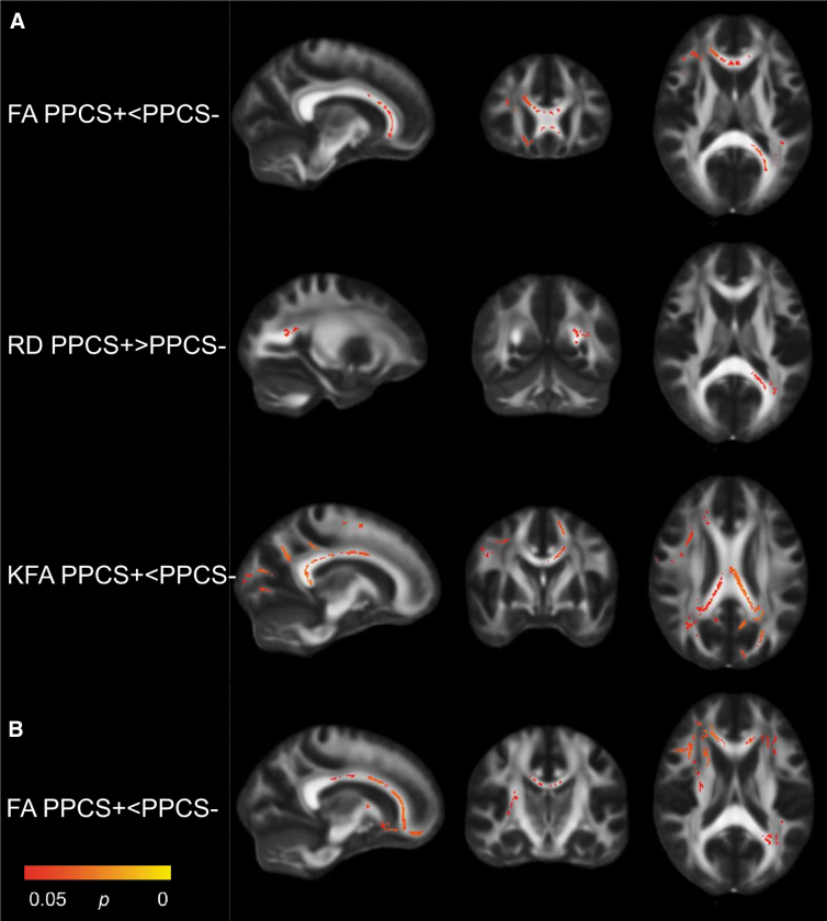
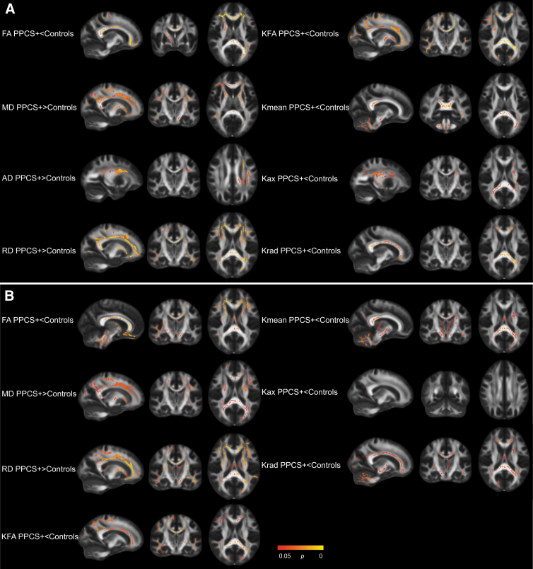
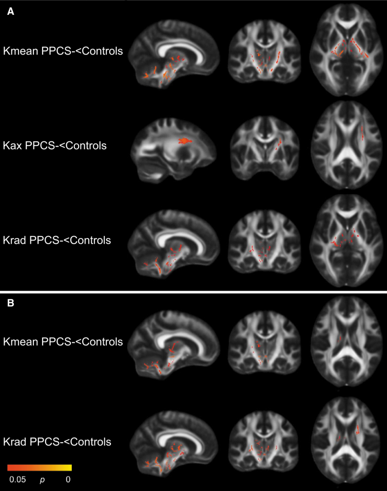
In this prospective cohort study, we investigated associations between acute diffusion tensor imaging (DTI) and diffusion kurtosis imaging (DKI) metrics and persistent post-concussion symptoms (PPCS) 3 months after mild traumatic brain injury (mTBI). Adult patients with mTBI (n = 176) and community controls (n = 78) underwent 3 Tesla magnetic resonance imaging (MRI) within 72 h post-injury, estimation of cognitive reserve at 2 weeks, and PPCS assessment at 3 months. Eight DTI and DKI metrics were examined with Tract-Based Spatial Statistics. Analyses were performed in the total sample in uncomplicated mTBI only (i.e., without lesions on clinical MRI), and with cognitive reserve both controlled for and not. Patients with PPCS (n = 35) had lower fractional anisotropy (in 2.7% of all voxels) and kurtosis fractional anisotropy (in 6.9% of all voxels), and higher radial diffusivity (in 0.3% of all voxels), than patients without PPCS (n = 141). In uncomplicated mTBI, only fractional anisotropy was significantly lower in patients with PPCS. Compared with controls, patients with PPCS had widespread deviations in all diffusion metrics. When including cognitive reserve as a covariate, no significant differences in diffusion metrics between patients with and without PPCS were present, but patients with PPCS still had significantly higher mean, radial, and axial diffusivity than controls. In conclusion, patients who developed PPCS had poorer white matter microstructural integrity acutely after the injury, compared with patients who recovered and healthy controls. Differences became less pronounced when cognitive reserve was controlled for, suggesting that pre-existing individual differences in axonal integrity accounted for some of the observed differences.
Keywords: biomarkers; brain concussion; diffusion kurtosis imaging; diffusion tensor imaging; post-concussion syndrome.
Conflict of interest statement
No competing financial interests exist.
Figures
Similar articles
-
Widespread White Matter Abnormalities in Concussed Athletes Detected by 7T Diffusion Magnetic Resonance Imaging.J Neurotrauma. 2024 Jul;41(13-14):1533-1549. doi: 10.1089/neu.2023.0099. Epub 2024 May 3. J Neurotrauma. 2024. PMID: 38481124 Free PMC article.
-
Diffusion Tensor and Kurtosis Imaging Findings the First Year following Mild Traumatic Brain Injury.J Neurotrauma. 2023 Mar;40(5-6):457-471. doi: 10.1089/neu.2022.0206. Epub 2022 Dec 9. J Neurotrauma. 2023. PMID: 36305387 Free PMC article.
-
Longitudinal Changes in Diffusion Tensor Imaging Following Mild Traumatic Brain Injury and Correlation With Outcome.Front Neural Circuits. 2019 May 7;13:28. doi: 10.3389/fncir.2019.00028. eCollection 2019. Front Neural Circuits. 2019. PMID: 31133818 Free PMC article.
-
Diffusion tensor imaging (DTI) findings in adult civilian, military, and sport-related mild traumatic brain injury (mTBI): a systematic critical review.Brain Imaging Behav. 2018 Apr;12(2):585-612. doi: 10.1007/s11682-017-9708-9. Brain Imaging Behav. 2018. PMID: 28337734
-
Evidence for Altered White Matter Organization After Mild Traumatic Brain Injury: A Scoping Review on the Use of Diffusion Magnetic Resonance Imaging and Blood-Based Biomarkers to Investigate Acute Pathology and Relationship to Persistent Post-Concussion Symptoms.J Neurotrauma. 2025 Apr;42(7-8):640-667. doi: 10.1089/neu.2024.0039. Epub 2024 Aug 21. J Neurotrauma. 2025. PMID: 39096132
Cited by
-
The Relationship Between Cognitive Functioning and Symptoms of Depression, Anxiety, and Post-Traumatic Stress Disorder in Adults with a Traumatic Brain Injury: a Meta-Analysis.Neuropsychol Rev. 2022 Dec;32(4):758-806. doi: 10.1007/s11065-021-09524-1. Epub 2021 Oct 25. Neuropsychol Rev. 2022. PMID: 34694543 Review.
-
Predicting recovery in patients with mild traumatic brain injury and a normal CT using serum biomarkers and diffusion tensor imaging (CENTER-TBI): an observational cohort study.EClinicalMedicine. 2024 Aug 8;75:102751. doi: 10.1016/j.eclinm.2024.102751. eCollection 2024 Sep. EClinicalMedicine. 2024. PMID: 39720677 Free PMC article.
-
Transcranial Direct Current Stimulation as a Treatment Tool for Mild Traumatic Brain Injury.Brain Sci. 2021 Jun 18;11(6):806. doi: 10.3390/brainsci11060806. Brain Sci. 2021. PMID: 34207004 Free PMC article. Review.
-
A 72-h sedated porcine model of traumatic spinal cord injury.Brain Spine. 2024 Apr 16;4:102813. doi: 10.1016/j.bas.2024.102813. eCollection 2024. Brain Spine. 2024. PMID: 38681174 Free PMC article.
-
Aberrant brain gray matter and functional networks topology in end stage renal disease patients undergoing maintenance hemodialysis with cognitive impairment.Front Neurosci. 2022 Aug 10;16:967760. doi: 10.3389/fnins.2022.967760. eCollection 2022. Front Neurosci. 2022. PMID: 36033631 Free PMC article.
References
-
- Panenka, W.J., Lange, R.T., Bouix, S., Shewchuk, J.R., Heran, M.K.S., Brubacher, J.R., Eckbo, R., Shenton, M.E., Iverson, G.L., and Kassubek, J. (2015). Neuropsychological outcome and diffusion tensor imaging in complicated versus uncomplicated mild traumatic brain injury. PLoS One 10, e0122746. - PMC - PubMed
-
- Wallace, E.J., Mathias, J.L., and Ward, L. (2018). Diffusion tensor imaging changes following mild, moderate and severe adult traumatic brain injury: a meta-analysis. Brain Imaging Behav. 12, 1607–1621 - PubMed
-
- Strauss, X.S.B., Kim, X.N., Branch, X.C.A., Kahn, X.M.E., Kim, X.M., Lipton, X.R.B., Provataris, X.J.M., Scholl, X.H.F., Zimmerman, X.M.E., and Lipton, X.M.L. (2016). Bidirectional changes in anisotropy are associated with outcomes in mild traumatic brain injury. Am. J. Neuroradiol. 37, 1983–1991 - PMC - PubMed
Publication types
MeSH terms
LinkOut - more resources
Full Text Sources
Other Literature Sources
Medical