. 2021 Apr 15;21(1):71.
doi: 10.1186/s12880-021-00603-6.

The value of multimodality imaging in diagnosis and treatment of cardiac lipoma

Affiliations

The value of multimodality imaging in diagnosis and treatment of cardiac lipoma

Shenglei Shu et al. BMC Med Imaging. .

Abstract

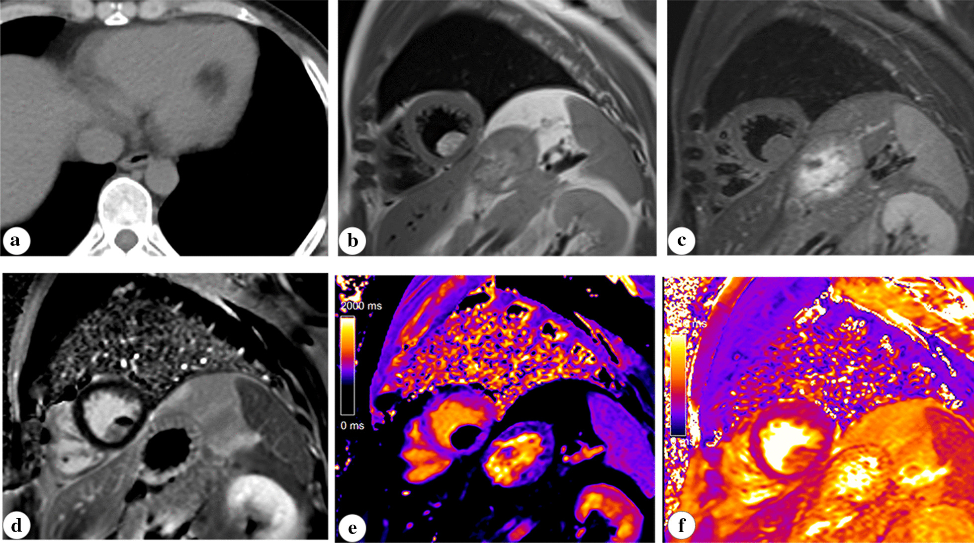
Background: Cardiac lipoma is a rare primary tumor in the heart and pericardium. Multimodality imaging methods, especially magnetic resonance imaging (MRI), are crucial in detecting and diagnosing cardiac lipomas. Besides, they are of significant importance in management of cardiac lipomas. The aim of this study was to evaluate the value of multimodality imaging methods in diagnosing and treatment of cardiac lipoma by describing a series of cases of cardiac lipoma.

Materials and methods: Data of patients with cardiac lipoma at a local institution were retrospectively collected. Their imaging findings on echocardiography, computed tomography (CT), and cardiac MRI and clinical management were described in detail.

Results: 12 patients with cardiac lipoma were retrospectively included with thirteen lipomas found within heart and pericardium. Two patients' lipoma were symptomatic, while lipomas in other 10 patients were found incidentally. Most lipomas were sensitively detected with echocardiography. Accurate diagnoses were achieved with CT and MRI in all cases. Surgical resection was performed in one symptomatic patient due to the obstruction of the left ventricular outflow tract, while the removal of pericardial lipoma in another symptomatic patient was not possible due to diffuse myocardial infiltration observed in MRI. Based on MRI findings, two patients without clinical symptoms also underwent surgery to prevent the risk of detachment of ventricular lipoma with a narrow pedicle in one patient and potential further thinning of the myocardium by pericardial lipoma growth in another patient.

Conclusions: Cardiac lipoma could be sensitively detected and accurately diagnosed with multiple noninvasive imaging tools. Comprehensive evaluation with multimodality imaging methods should also be conducted for better management planning and follow-up in all patients.

Keywords: Cardiac lipoma; Computed tomography; Magnetic resonance imaging; Noninvasive imaging.


Conflict of interest statement

The authors declare that they have no competing interests.

Figures

Fig. 1
Fig. 1
Images of a ventricular lipoma in an asymptomatic male patient (part of pictures have been previously published in the review by our griup [13]). The ventricular lipoma presented as a well-defined homogenous hyperechoic mass within left ventricle (a). The lipomas showed homogenous low density (-100 HU) on CT (b). On cardiac MRI, the lovely heart-shaped lipoma showed same signal intensity with subcutaneous fat in all sequences including cine sequence (c), T1 weighted image (d), T2 weight fat suppression image (e). Particularly, the narrow pedicle of the lipoma was depicted in short axis image attached to the endomyocardium (f, arrowhead)
Fig. 2
Fig. 2
CT and cardiac MR images of a sessile ventricular lipoma in an asymptomatic male patient. The attenuation value in CT (a, − 45 HU) and characteristic findings in MRI (b, c, d) of the mass confirmed the nature of fat tissue. Besides, the ventricular mass showed same T1 (244 ms) and T2 values (61 ms) as subcutaneous fat in tissue mapping sequences (e, f)
Fig. 3
Fig. 3
MR images of a giant pericardial lipoma of a female patient with history of pericardial lipoma resection. The T1 weighted (a, b, c, d) and T2 weighted fat suppression (e, f) images confirmed the diagnosis of lipoma. Diffuse infiltration of the lipoma to pulmonary arterial wall (a) and ventricular myocardium (b, c, d) was clearly observed (arrowhead)
Fig. 4
Fig. 4
Images of a giant silent pericardial lipoma in a female patient (part of pictures have been previously published in the review by our group [13]). The giant lipoma within pericardium presented as hypoechoic mass in echocardiogram (a). CT image showed a homogenous low-density mass (− 105 HU) within pericardium (b). The MR images demonstrated a pericardial mass with same signal intensity with fat tissue (c, d, e, f). Besides, regional thinning of the inferior and apical wall of the left ventricle was observed without enhancement in the late gadolinium scanning image (f)

References

    1. Butany J, Nair V, Naseemuddin A, Nair GM, Catton C, Yau T. Cardiac tumours: diagnosis and management. Lancet Oncol. 2005;6(4):219–228. doi: 10.1016/S1470-2045(05)70093-0. - DOI - PubMed
    1. McAllister HAJ, Fenoglio JJJ, Fine G. Tumors of the cardiovascular system. (Atlas of tumor pathology, second series, Fascicle 15.) New York: Armed Force Institute of Pathology; 1978. pp. 44–46.
    1. Puvaneswary M, Edwards JR, Bastian BC, Khatri SK. Pericardial lipoma: ultrasound, computed tomography and magnetic resonance imaging findings. Aust Radiol. 2000;44(3):321–324. doi: 10.1046/j.1440-1673.2000.00821.x. - DOI - PubMed
    1. Li J, Ho SY, Becker AE, Jones H. Multiple cardiac lipomas and sudden death: a case report and literature review. Cardiovasc Pathol. 1998;7(1):51–55. doi: 10.1016/S1054-8807(97)00058-6. - DOI - PubMed
    1. Monti L, Scardino C, Nardi B, Balzarini L. Lipoma of the interventricular septum. Eur Heart J. 2015;36(44):3073. doi: 10.1093/eurheartj/ehv238. - DOI - PubMed

LinkOut - more resources