

Differentiation of brain metastases from small and non-small lung cancers using apparent diffusion coefficient (ADC) maps
- PMID: 33858368
- PMCID: PMC8048287
- DOI: 10.1186/s12880-021-00602-7
Differentiation of brain metastases from small and non-small lung cancers using apparent diffusion coefficient (ADC) maps
Abstract
Background: Brain metastases are particularly common in patients with small cell lung cancer (SCLC) and non-small cell lung cancer (NSCLC), with NSCLC showing a less aggressive clinical course and lower chemo- and radio sensitivity compared to SCLC. Early adequate therapy is highly desirable and depends on a reliable classification of tumor type. The apparent diffusion coefficient is a noninvasive neuroimaging marker with the potential to differentiate between major histological subtypes. Here we determine the sensitivity and specificity of the apparent diffusion coefficient to distinguish between NSCLC and SCLC.
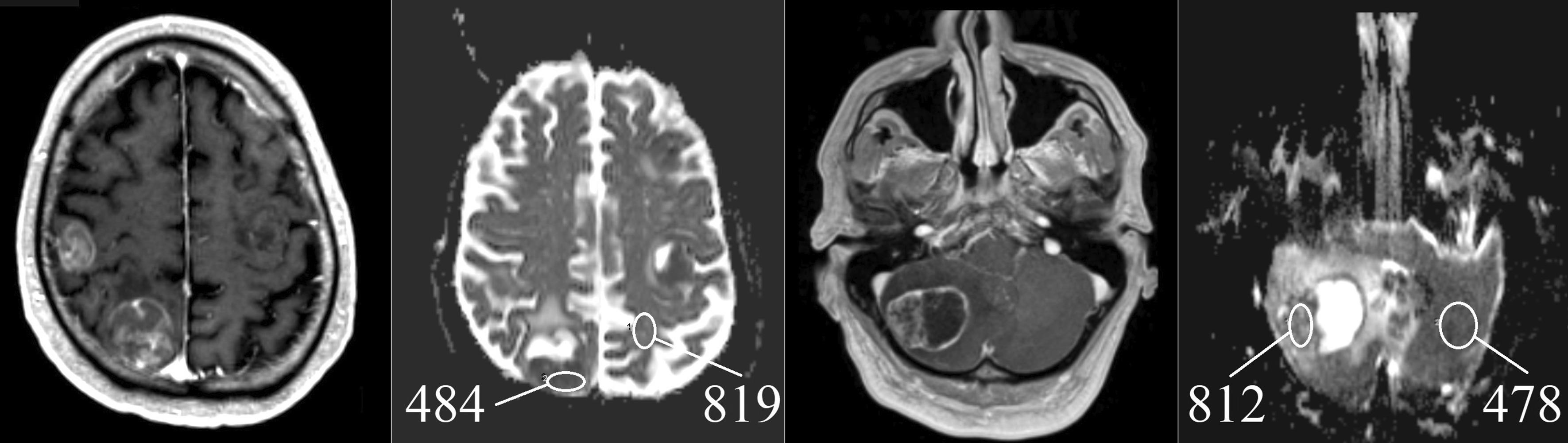
Methods: We enrolled all NSCLC and SCLC patients diagnosed between 2008 and 2019 at the University Medical Center Göttingen. Cranial MR scans were visually inspected for brain metastases and the ratio of the apparent diffusion coefficient (ADC) was calculated by dividing the ADC measured within the solid part of a metastasis by a reference ADC extracted from an equivalent region in unaffected tissue on the contralateral hemisphere.
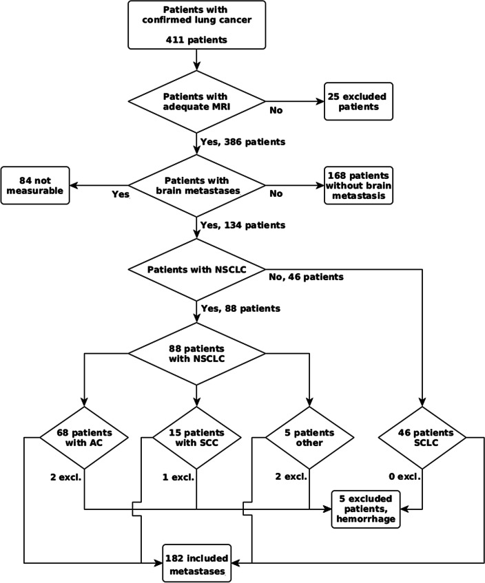
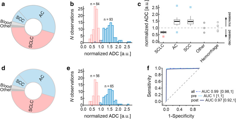
Results: Out of 411 enrolled patients, we detected 129 patients (83 NSCLC, 46 SCLC) with sufficiently large brain metastases with histologically classified lung cancer and no hemorrhage. We analyzed 185 brain metastases, 84 of SCLC and 101 of NSCLC. SCLC brain metastases showed an ADC ratio of 0.68 ± 0.12 SD, and NSCLC brain metastases showed an ADC ratio of 1.47 ± 0.31 SD. Receiver operating curve statistics differentiated brain metastases of NSCLC from SCLC with an area under the curve of 0.99 and a 95% CI of 0.98 to 1, p < 0.001. Youden's J cut-point is 0.97 at a sensitivity of 0.989 and a specificity of 0.988.
Conclusions: In patients with lung cancer and brain metastases with solid tumor parts, ADC ratio enables an ad hoc differentiation of SCLC and NSCLC, easily achieved during routine neuroradiological examination. Non-invasive MR imaging enables an early-individualized management of brain metastases from lung cancer.
Trial registration: The study was registered in the German Clinical Trials Register (DRKS00023016).
Keywords: ADC; Brain metastases; Diffusion; NSCLC; SCLC.
Conflict of interest statement
The authors declare that they have no competing interests.
Figures
Similar articles
-
Histogram Analysis of ADC Maps for Differentiating Brain Metastases From Different Histological Types of Lung Cancers.Can Assoc Radiol J. 2021 May;72(2):271-278. doi: 10.1177/0846537120933837. Epub 2020 Jun 30. Can Assoc Radiol J. 2021. PMID: 32602365
-
Differentiation of brain metastases originating from lung and breast cancers using apparent diffusion coefficient histogram analysis and the relation of histogram parameters with Ki-67.Neuroradiol J. 2022 Jun;35(3):370-377. doi: 10.1177/19714009211049082. Epub 2021 Oct 5. Neuroradiol J. 2022. PMID: 34609916 Free PMC article.
-
Diffusion Tensor Imaging Can Discriminate the Primary Cell Type of Intracranial Metastases for Patients with Lung Cancer.Magn Reson Med Sci. 2022 Jul 1;21(3):425-431. doi: 10.2463/mrms.mp.2020-0183. Epub 2021 Mar 4. Magn Reson Med Sci. 2022. PMID: 33658441 Free PMC article.
-
[Present role of prophylactic cranial irradiation].Bull Cancer. 2013 Jan 1;100(1):35-43. doi: 10.1684/bdc.2012.1678. Bull Cancer. 2013. PMID: 23306141 Review. French.
-
Gastric metastasis of small cell lung carcinoma: Three case reports and review of literature.World J Gastroenterol. 2024 Aug 21;30(31):3717-3725. doi: 10.3748/wjg.v30.i31.3717. World J Gastroenterol. 2024. PMID: 39193003 Free PMC article. Review.
Cited by
-
De novo GTP synthesis is a metabolic vulnerability for the interception of brain metastases.Cell Rep Med. 2024 Oct 15;5(10):101755. doi: 10.1016/j.xcrm.2024.101755. Epub 2024 Oct 4. Cell Rep Med. 2024. PMID: 39366383 Free PMC article.
-
Differentiation of multiple brain metastases and glioblastoma with multiple foci using MRI criteria.BMC Med Imaging. 2024 Jan 2;24(1):3. doi: 10.1186/s12880-023-01183-3. BMC Med Imaging. 2024. PMID: 38166651 Free PMC article.
-
Magnetic resonance imaging characteristics of brain metastases in small cell lung cancer.Cancer Med. 2023 Jul;12(14):15199-15206. doi: 10.1002/cam4.6206. Epub 2023 Jun 8. Cancer Med. 2023. PMID: 37288842 Free PMC article.
-
Towards updated understanding of brain metastasis.Am J Cancer Res. 2022 Sep 15;12(9):4290-4311. eCollection 2022. Am J Cancer Res. 2022. PMID: 36225632 Free PMC article. Review.
-
Differentiation between multifocal CNS lymphoma and glioblastoma based on MRI criteria.Discov Oncol. 2024 Sep 1;15(1):397. doi: 10.1007/s12672-024-01266-9. Discov Oncol. 2024. PMID: 39217585 Free PMC article.
References
MeSH terms
Associated data
LinkOut - more resources
Full Text Sources
Other Literature Sources
Medical
