

Intraoperative optical coherence tomography guided corneal sweeping for removal of remnant Interface fluid during ultra-thin Descemet stripping automated endothelial keratoplasty
- PMID: 33858385
- PMCID: PMC8048159
- DOI: 10.1186/s12886-021-01934-2
Intraoperative optical coherence tomography guided corneal sweeping for removal of remnant Interface fluid during ultra-thin Descemet stripping automated endothelial keratoplasty
Abstract
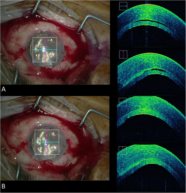
Background: Remnant interface fluid following Descemet stripping automated endothelial keratoplasty (DSAEK) is associated with postoperative detachments. The aim of this study was to assess outcomes of intraoperative optical coherence tomography (iOCT) guided meticulous peripheral corneal sweeping for removal of interface fluid during ultra-thin (UT) DSAEK.
Methods: This retrospective study included all eyes underwent iOCT guided UT-DSAEK from October 2016 to February 2018 at the Hanusch Hospital, Vienna, Austria. Peripheral meticulous corneal sweeping was performed to remove excess fluid. Central graft thickness (CGT) was measured prior to surgery, after graft bubbling and after corneal sweeping. Remnant interface fluid rates were compared between eyes that underwent rebubbling and those that did not.
Results: Overall, 28 eyes of 28 patients with a mean age of 73.9 ± 10.0 years were included. An iOCT guided meticulous peripheral sweeping was performed in 89.3% (n = 25) of the cases. Following 84% (n = 21) of the peripheral sweeping performed, remnant fluid was no longer identified. Following peripheral sweeping the interface fluid height was reduced from 17.31 ± 15.96 μm to 3.46 ± 9.52 μm (p < 0.001) and CGT was reduced by 7% (p < 0.001). Rebubbling was performed in 17.9% (n = 5) of the cases. The rebubbling group had a greater proportion of patients that had remnant fluid identified with iOCT at the end of surgery despite meticulous peripheral sweeping (60.0% versus 4.4%, p = 0.01).
Conclusion: The iOCT identified subclinical remnant fluid in nearly 90% of UT-DSAEK cases. An iOCT guided peripheral corneal sweeping led to resolution of interface fluid in a majority of cases. Eyes with persistent remnant fluid despite peripheral corneal sweeping are more likely to require subsequent rebubbling.
Keywords: Graft thickness; Interface fluid; Intraoperative optical coherence tomography; Sweeping; UT-DSAEK.
Conflict of interest statement
The authors declare that they have no competing interests.
Figures


References
-
- Hos D, Matthaei M, Bock F, Maruyama K, Notara M, Clahsen T, Hou Y, le VNH, Salabarria AC, Horstmann J, Bachmann BO, Cursiefen C. Immune reactions after modern lamellar (DALK, DSAEK, DMEK) versus conventional penetrating corneal transplantation. Prog Retin Eye Res. 2019;73:100768. doi: 10.1016/j.preteyeres.2019.07.001. - DOI - PubMed
MeSH terms
LinkOut - more resources
Full Text Sources
Other Literature Sources
Medical
