. 2021 Apr 15;12(1):2258.
doi: 10.1038/s41467-021-22565-7.

Selenoprotein W ensures physiological bone remodeling by preventing hyperactivity of osteoclasts

Affiliations

Selenoprotein W ensures physiological bone remodeling by preventing hyperactivity of osteoclasts

Hyunsoo Kim et al. Nat Commun. .

Abstract

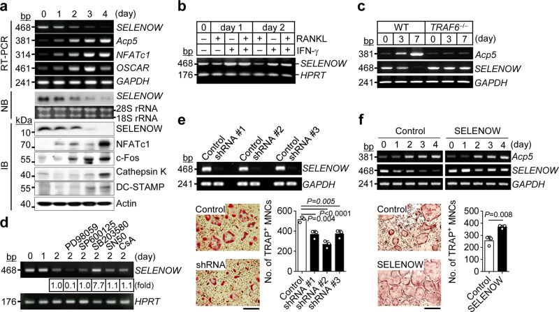
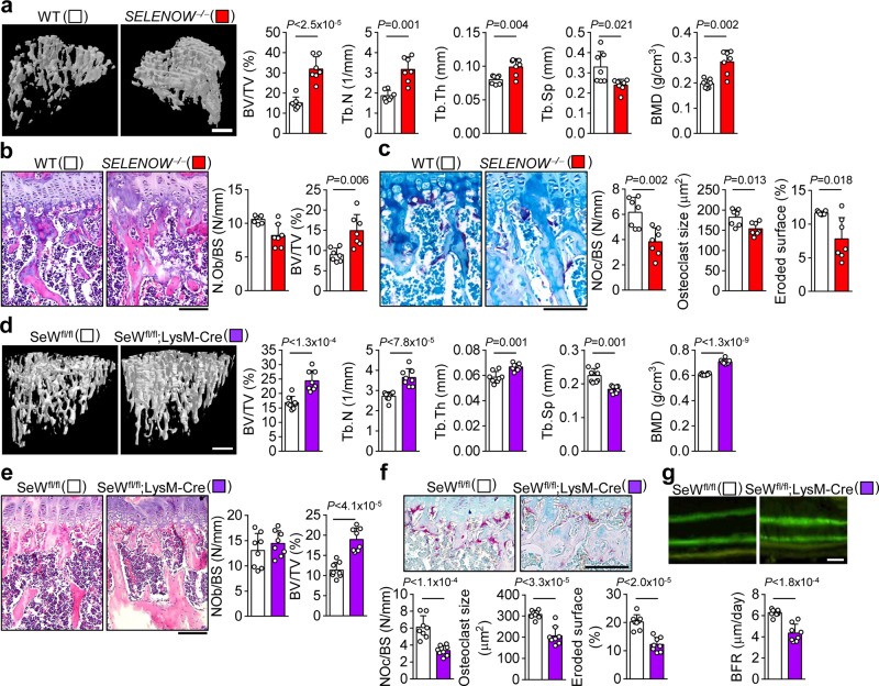
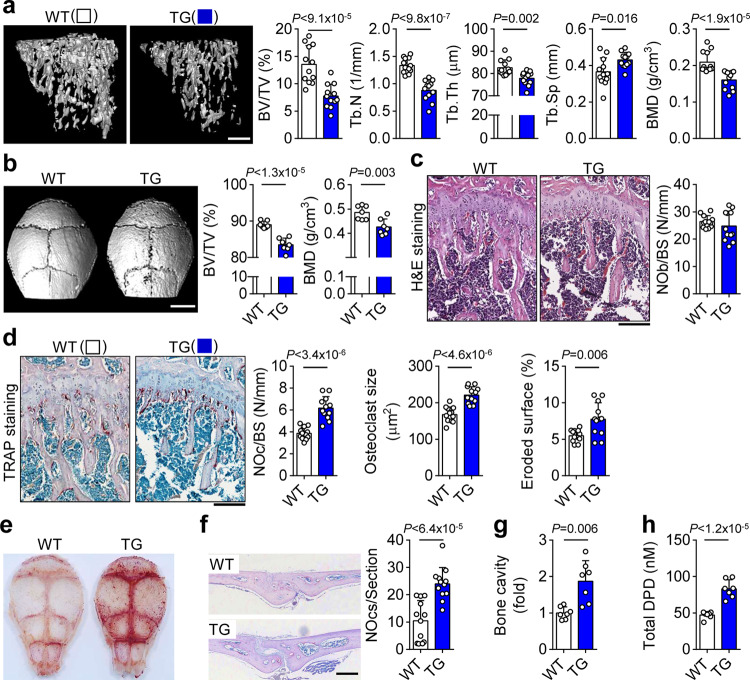
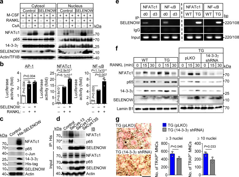
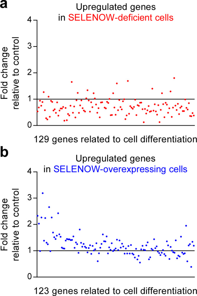
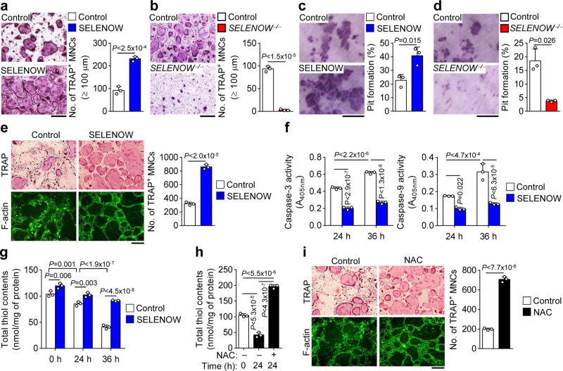
Selenoproteins containing selenium in the form of selenocysteine are critical for bone remodeling. However, their underlying mechanism of action is not fully understood. Herein, we report the identification of selenoprotein W (SELENOW) through large-scale mRNA profiling of receptor activator of nuclear factor (NF)-κΒ ligand (RANKL)-induced osteoclast differentiation, as a protein that is downregulated via RANKL/RANK/tumour necrosis factor receptor-associated factor 6/p38 signaling. RNA-sequencing analysis revealed that SELENOW regulates osteoclastogenic genes. SELENOW overexpression enhances osteoclastogenesis in vitro via nuclear translocation of NF-κB and nuclear factor of activated T-cells cytoplasmic 1 mediated by 14-3-3γ, whereas its deficiency suppresses osteoclast formation. SELENOW-deficient and SELENOW-overexpressing mice exhibit high bone mass phenotype and osteoporosis, respectively. Ectopic SELENOW expression stimulates cell-cell fusion critical for osteoclast maturation as well as bone resorption. Thus, RANKL-dependent repression of SELENOW regulates osteoclast differentiation and blocks osteoporosis caused by overactive osteoclasts. These findings demonstrate a biological link between selenium and bone metabolism.


Conflict of interest statement

The authors declare no competing interests.

Figures

Fig. 1
Fig. 1. SELENOW positively regulates osteoclastogenesis.
a Downregulation of SELENOW during osteoclastogenesis. Osteoclast precursors were cultured with RANKL and M-CSF, and SELENOW gene expression was analysed by RT-PCR, northern blotting (NB), and immunoblotting (IB). b, c RANKL/RANK/TRAF6 axis-dependent downregulation of SELENOW. Osteoclast precursors were pretreated with interferon-γ (IFN-γ; 150 U/ml), which degrades TRAF6, 30 min prior to RANKL stimulation. Osteoclast precursors treated with IFN-γ (b) and TRAF6-deficient osteoclast precursors (c) failed to induce RANKL-mediated SELENOW downregulation. d Up- and downregulation of SELENOW via ERK and p38 activation, respectively. Osteoclast precursors were pretreated with inhibitors of ERK (PD98059), JNK (SP600125), p38 (SB203580), NF-κB (SN50), and NFATc1 (cyclosporin A, CsA) for 30 min in the presence of M-CSF and then stimulated with RANKL for 2 days. The expression levels of SELENOW were analysed using RT-PCR. e, f Decreased and increased osteoclast formation following SELENOW knockdown (e) and overexpression (f), respectively. Osteoclast precursors infected with shRNA-mediated SELENOW gene-silencing lentivirus and SELENOW-overexpressing retrovirus were differentiated into osteoclasts and TRAP-positive multi-nucleated cells (TRAP + MNCs) with more than 3 nuclei were assessed (n = 3). Scale bars, 100 μm. Images are representative of three independent experiments. Data represent the mean ± SD of triplicate samples. Statistical significance was determined by Student’s two-tailed t-test (f). One-way ANOVA was performed followed by Turkey’s test (e). Source data are provided as a Source Data file.
Fig. 2
Fig. 2. Increased bone mass phenotypes in mice with SELENOW deficiency.
a μCT analysis of proximal tibiae from wild-type (WT) male littermates and age/sex-matched SELENOW−/− mice at 10 weeks. BV/TV, trabecular bone volume per tissue volume; Tb.N, trabecular bone number; Tb.Th, trabecular thickness; Tb.Sp, trabecular separation; BMD, bone mineral density. Scale bar, 0.5 mm. b, c Reduced osteoclast formation on the trabecular bone surface of SELENOW−/− mice. H&E- and TRAP-stained sections of tibiae were used to detect osteoblasts (b) and osteoclasts (c), respectively. NOb/BS, number of osteoblasts per bone surface; NOc/BS, BV/TV, trabecular bone volume per tissue volume; number of osteoclasts per bone surface. In addition, osteoclast size and eroded bone surface were analysed from the TRAP-stained sections. Scale bar, 100 μm. d Increased bone mass phenotype in μCT analysis of proximal tibiae from WT male littermates (SELENOWtm1c/tm1c; SeWfl/fl) and age/sex-matched osteoclast-specific SELENOW knockout mice (SELENOWtm/c/tm1c:LysM-Cre; SeWfl/fl;LysM-Cre) at 10 weeks. Scale bar, 0.5 mm. e Analysis of NOb/BS, number of osteoblasts per bone surface, and BV/TV in H&E-stained sections. Scale bar, 100 μm. f Analysis of NOc/BS, number of osteoclasts per bone surface, osteoclast size and eroded bone surface from TRAP-stained sections. Scale bar, 100 μm. g Histomorphometric analysis of the tibia. BFR, bone formation rate. Scale bar, 10 μm. Data represent mean ± SD (n = 7 mice per group in ac; n = 8 mice per group in dg). Statistical significance was determined by Student’s two-tailed t-test. Source data are provided as a Source Data file.
Fig. 3
Fig. 3. Osteoporotic phenotypes in mice with SELENOW overexpression.
a μCT analysis of proximal tibiae from wild-type (WT) male littermates and age/sex-matched transgenic (TG) mice at 10 weeks. Scale bar, 0.5 mm. b μCT images of calvaria and analysis of bone parameters [trabecular bone volume per tissue volume (BV/TV) and BMD]. scale bar, 3 mm. c, d Increased osteoclast formation on the trabecular bone surface of TG mice. Analysis of NOb/BS, number of osteoblasts per bone surface, from H&E-stained sections (c). Analysis of NOc/BS, number of osteoclasts per bone surface, osteoclast size and eroded bone surface from TRAP-stained sections (d). Scale bar, 100 μm. eg Whole calvaria (e) and cross-sections (f) were stained with TRAP. The number of TRAP + osteoclasts and the calvarial marrow cavity area (g), which reflects the degree of osteoporosis, were measured in whole sections. Scale bar, 1 mm. h The level of urinary DPD, a marker of osteoporosis, was measured by enzyme immunoassay. Data represent mean ± SD (n = 12 mice per group in a, c, d and f, and n = 7 mice per group in b and eh). Statistical significance was determined by Student’s two-tailed t-test. Source data are provided as a Source Data file.
Fig. 4
Fig. 4. SELENOW regulates osteoclastogenic gene expression.
a Osteoclastogenic transcription factors and SELENOW co-translocate into the nucleus. Osteoclast precursors infected with SELENOW-harbouring retrovirus were cultured with M-CSF and RANKL for 2 days. After cells were exposing to RANKL-free condition for 3 h and treated without or with an inhibitor of NFATc1 (cyclosporin A, CsA), cells were stimulated with RANKL for 20 min. Cytosolic and nuclear proteins were fractionated and NF-κB, NFATc1, and SELENOW levels were determined by immunoblotting. b Luciferase reporter assay. RAW264.7 cells were transfected with AP-1-, NF-κB-, and NFATc1-luciferase reporter or pcDNA3.1-His-tagged SELENOW (SeCys-13) vector. Cells were stimulated with RANKL for 24 h and luciferase activity was measured (n = 3). c, d SELENOW interacts with NF-κB and NFATc1. Cytosolic extracts from HEK 293 T cells expressing a His-tagged SELENOW (SeCys-13) were pulled down with an anti-His-Tag antibody (c). Also, cytosolic extracts from HEK 293T cells with a His-tagged wild-type SELENOW (SeCys-13) and His-tagged SELENOW mutants in which SeCys-13 was replaced by cysteine (SeCys13C) or serine (SeCys13S) were immunoprecipitated (IP) with anti-His-Tag antibody and then immunoblotted (IB) with the indicated antibodies (d). e ChIP assay. Osteoclast precursors were cultured with M-CSF alone (d0) or with M-CSF and RANKL for 3 days (d3; left panels). Also, osteoclast precursors from wild-type (WT) and SELENOW-overexpressing transgenic (TG) mice were cultured with M-CSF and RANKL for 3 days (right panels). Following immunoprecipitation (IP) of chromatin with anti-SELENOW antibody, ChIP assay was performed to detect the promoter for NF-κB- or NFATc1-binding sites. f, g 14-3-3γ mediates nuclear translocation of NFATc1, NF-κB, and SELENOW, and osteoclast differentiation. After osteoclast precursors from WT and TG mice were cultured with M-CSF and RANKL for 2 days to induce pre-osteoclasts, the cells were exposed to M-CSF- and RANKL-free condition for 3 h and were stimulated with RANKL for indicated times (f; left panel). In addition, this was performed in TG mice-derived pre-osteoclasts transduced with control lentivirus (pLKO) or 14-3-3γ-targeted shRNA-harbouring lentivirus (f; right panel). Nuclear proteins were fractionated and subjected to immunoblotting. Osteoclast precursors from TG mice were infected with shRNA-mediated 14-3-3γ gene-silencing lentivirus and differentiated into osteoclasts (n = 3). TRAP + MNCs with more than 3 or 10 nuclei were assessed (g). Scale bar, 100 μm. Data represent the mean ± SD of triplicate samples. Statistical significance was determined by Student’s two-tailed t-test (g). One-way ANOVA was performed followed by Turkey’s test (b). Images are representative of three independent experiments. Source data are provided as a Source Data file.
Fig. 5
Fig. 5. SELENOW upregulates osteoclast differentiation-related genes.
a, b Analysis of RNA-sequencing transcriptomic data. Osteoclast precursors from SELENOW−/− and SELENOW-transgenic mice and corresponding wild-type littermates were differentiated into osteoclasts in the presence of M-CSF and RANKL for 3 days. The relative fold change in the levels of genes in SELENOW−/− (a) and SELENOW-overexpressing osteoclasts (b) was determined. Source data are provided as a Source Data file.
Fig. 6
Fig. 6. Constitutive expression of SELENOW leads to excess pre-osteoclast fusion and osteoclastic bone resorption.
a, b Induction of pre-osteoclast fusion and osteoclastic bone resorption by SELENOW. Fusion assay in pre-osteoclasts from wild-type and SELENOW-overexpressing transgenic mice (a) or SELENOW−/− mice (b). Osteoclast precursors were treated with M-CSF and RANKL for 2 days to form pre-osteoclasts following fusion assay. Osteoclast fusion rate was determined by counting TRAP + MNCs with a diameter ≥100 μm (n = 3). c, d Pit formation. Osteoclast precursors prepared from wild-type and SELENOW-overexpressing transgenic mice (c) or SELENOW−/− mice (d) were differentiated into osteoclasts for 4 days. After mature osteoclasts were detached from the culture dish and seeded on dentine slice, cells were further cultured with M-CSF and RANKL for 2 days to allow bone resorption. Pit formation by osteoclasts is expressed as a percentage of the resorbed area on the bone slice surface (n = 3). e Anti-apoptotic effect of SELENOW. Mature osteoclasts were transduced with SELENOW-overexpressing retrovirus and cell survival was assessed 2 days later by staining with TRAP (upper panels) or FITC-labelled phalloidin (lower panels) to detect TRAP + osteoclasts with a full actin ring (n = 3). f Caspase activity was assessed at indicated times after mature osteoclasts were cultured as in (e, n = 3). g Increase in the cellular redox status by SELENOW. Osteoclasts were transduced with SELENOW-overexpressing retrovirus and total thiol content was assessed at indicated times (n = 3). h Increase in the cellular redox status by NAC. After treatment with 4 mM NAC for 24 h or no treatment, cytosolic extracts of mature osteoclasts were prepared and assayed for free thiol level (n = 3). i Increased mature osteoclast survival by NAC (n = 3). Osteoclasts were treated as described in h and then stained as in e. Scale bars, 100 μm. Data represent mean ± SD of triplicate samples. Statistical significance was determined by Student’s two-tailed t-test (ae, i). One-way ANOVA was performed followed by Turkey’s test (fh). Source data are provided as a Source Data file.

References

    1. Modell H, et al. A physiologist’s view of homeostasis. Adv. Physiol. Educ. 2015;39:259–266. doi: 10.1152/advan.00107.2015. - DOI - PMC - PubMed
    1. Mitrophanov AY, Groisman EA. Positive feedback in cellular control systems. Bioessays. 2008;30:542–555. doi: 10.1002/bies.20769. - DOI - PMC - PubMed
    1. Hancock, E. J., Ang, J., Papachristodoulou, A. & Stan, G. B. The interplay between feedback and buffering in cellular homeostasis. Cell Syst. 5, 498–508.e23 (2017). - PubMed
    1. Feng X, McDonald JM. Disorders of bone remodeling. Annu. Rev. Pathol. 2011;6:121–145. doi: 10.1146/annurev-pathol-011110-130203. - DOI - PMC - PubMed
    1. Asagiri M, et al. Autoamplification of NFATc1 expression determines its essential role in bone homeostasis. J. Exp. Med. 2005;202:1261–1269. doi: 10.1084/jem.20051150. - DOI - PMC - PubMed

Publication types

MeSH terms