. 2021 Apr 15;6(1):56.
doi: 10.1038/s41541-021-00305-8.

Meta-analysis of HIV-1 vaccine elicited mucosal antibodies in humans

Affiliations

Meta-analysis of HIV-1 vaccine elicited mucosal antibodies in humans

Kelly E Seaton et al. NPJ Vaccines. .

Abstract

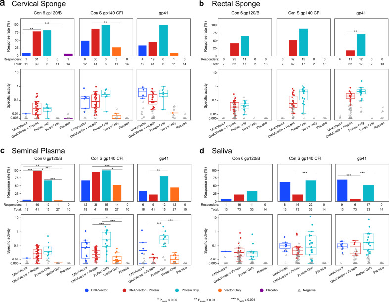
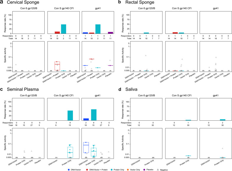
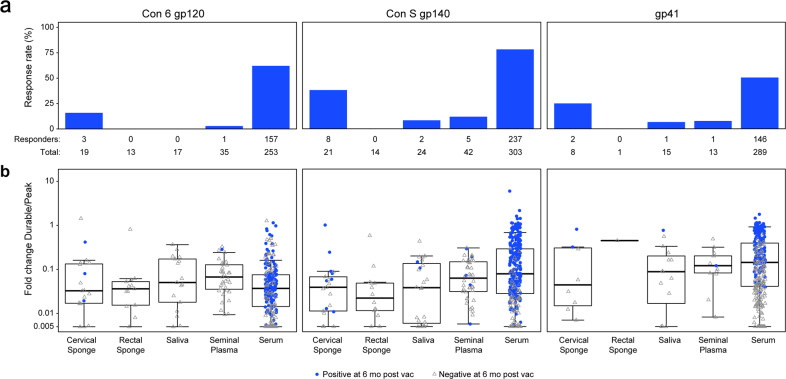
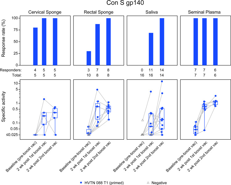
We studied mucosal immune responses in six HIV-1 vaccine trials investigating different envelope (Env)-containing immunogens. Regimens were classified into four categories: DNA/vector, DNA/vector plus protein, protein alone, and vector alone. We measured HIV-1-specific IgG and IgA in secretions from cervical (n = 111) and rectal swabs (n = 154), saliva (n = 141), and seminal plasma (n = 124) and compared to corresponding blood levels. Protein-containing regimens had up to 100% response rates and the highest Env-specific IgG response rates. DNA/vector groups elicited mucosal Env-specific IgG response rates of up to 67% that varied across specimen types. Little to no mucosal IgA responses were observed. Overall, gp41- and gp140-specific antibodies dominated gp120 mucosal responses. In one trial, prior vaccination with a protein-containing immunogen maintained durability of cervical and rectal IgG for up to 17 years. Mucosal IgG responses were boosted after revaccination. These findings highlight a role for protein immunization in eliciting HIV-1-specific mucosal antibodies and the ability of HIV-1 vaccines to elicit durable HIV-1-specific mucosal IgG.


Conflict of interest statement

The content is solely the responsibility of the authors and does not necessarily represent the official views of the National Institutes of Health. Potential competing interests: Co-authors M.A.A. and L.P. are employed by the National Institute of Allergy and Infectious Diseases (NIAID), sponsor of the clinical trials described in this manuscript. All authors are recipients of NIAID funding, and this publication is a result of activities funded by the NIAID. M.A. and L.P. are not involved with the process of funding these awards nor in their administration of scientific aspects and, in accordance with NIAID policies, are deferred from decisions regarding the funding of coauthors for a requisite period. Co-author HLR is a co-founder and employee of GeoVax Lab, Inc. and owns stocks in GeoVax Lab, Inc, and is an inventor on U.S. Patents 7,795,017, 8,623,379, 7,867,982, and 9,453,239 addressing D.N.A. and M.V.A. immunogens being developed for a Clade B HIV vaccine. Co-authors K.E.S., A.D., X.H., S.S.L., A.C., J.H., A.D., X.S., S.S., N.L.Y., P.S., G.C., P.A.G., J.M., G.G., G.P., S.G., A.K.R., Y.H., C.M., N.G., S.K., P.B.G., M.J.M., Y.H., and G.D.T. declare that there are no competing interests.

Figures

Fig. 1
Fig. 1. Protein immunization elicits a high response rate of mucosal HIV-1 gp120 envelope-specific IgG.
HIV-1 envelope-specific IgG (Con 6 gp120, Con S gp140, and gp41) were measured in secretions from a cervical sponge, b rectal sponge, c seminal plasma, and d saliva. The antibody magnitude was calculated as the HIV-specific concentration relative to total antibody concentration, noted as specific activity (SA = MFI*dilution/total IgG ng per mL). Top panels represent the response rates and bottom panels represent the response magnitudes (solid dots for positive responses and grey open triangles for negative responses). In the bottom panels, the mid-line of the boxplot denotes the median response magnitude and the ends of the boxplot denote the 25th and 75th percentiles among positive responses. Differences in the response rate between vaccine regimens were assessed using Barnard’s exact test and differences in response magnitude were assessed using the Wilcoxon rank-sum test. *PFWER < 0.05, **PFWER < 0.01, and ***PFWER < 0.001.
Fig. 2
Fig. 2. Env-specific IgA is rarely elicited in vaccinees and is predominantly to gp41.
HIV-1 envelope-specific IgA (Con 6 gp120, Con S gp140, and gp41) responses were measured in secretions from a cervical sponge, b rectal sponge, c seminal plasma, and d saliva. The antibody magnitude was calculated as the HIV-specific concentration relative to total antibody concentration noted as specific activity (SA = MFI*dilution/total IgA ng per mL). Top panels represent the response rates and bottom panels represent the response magnitudes (solid dots for positive responses and grey open triangles for negative responses). In the bottom panels, the mid-line of the boxplot denotes the median response magnitude and the ends of the boxplot denote the 25th and 75th percentiles among positive responses.
Fig. 3
Fig. 3. Rapid decline in Mucosal Env-specific IgG responses 6 months post-last vaccination from the peak.
a The response rate for detectable HIV-1 envelope-specific IgG (Con 6 gp120, Con S gp140, and gp41) in the mucosal secretions and serum at 6 months post-the last vaccination by mucosal compartment. b The fold-change in the magnitude of HIV-1 envelope-specific IgG (Con 6 gp120, Con S gp140, and gp41) from the measured peak immune responses to 6 months following the last vaccination (SA durability timepoint/SA peak time point) was determined for each mucosal compartment and serum. A fold-change of 1 indicates no decline in mucosal response magnitude at 6 months from the peak. Blue circles indicate positive vaccine responders at 6 months post-last vaccination; and open gray triangles indicate negative vaccine responders at 6 months post-last vaccination.
Fig. 4
Fig. 4. Persistent low concentrations of mucosal IgG boosted after a prolonged rest period.
Antibodies were evaluated for persistence 6–17 years (baseline) post-last vaccination in the protein-only vaccine regimen, HVTN 088, given to previously immunized participants. The response rate for detectable HIV-1 Con S gp140-specific IgG was measured in cervical and rectal secretions, saliva, and seminal plasma at baseline (pre-boost) and 2 weeks after the first and second boost vaccinations (top panels). The concentration of HIV-1 Con S gp140-specific IgG was calculated as the HIV-specific concentration relative to total antibody concentration noted as specific activity (SA = MFI*dilution/total IgG ng per mL) (bottom panels). Blue circles indicate positive vaccine responders and open gray triangles represent non-responders at each timepoint. The boxplots are for positive responses (the mid-line of the boxplot denotes the median and the ends of the boxplot denote the 25th and 75th percentiles). The grey lines connect the observations between the timepoints.
Fig. 5
Fig. 5. Circulating HIV-1 IgG in the blood exhibits a low-moderate correlation with mucosal IgG.
Spearman correlation of IgG adjusted for vaccine-type (a) between mucosal compartments and serum, and (b) between rectal and cervical sponge for women and between a rectal sponge and seminal plasma for men. *, **, and *** indicates significance at p < 0.01, p < 0.001, and p < 0.0001; respectively.

Similar articles

Cited by

References

    1. Tay MZ, et al. Antibody-mediated internalization of infectious HIV-1 virions differs among antibody isotypes and subclasses. PLoS Pathog. 2016;12:e1005817. doi: 10.1371/journal.ppat.1005817. - DOI - PMC - PubMed
    1. Mkhize NN, et al. Broadly neutralizing antibody specificities detected in the genital tract of HIV-1 infected women. Aids. 2016;30:1005–1014. doi: 10.1097/QAD.0000000000001038. - DOI - PMC - PubMed
    1. Moog C, et al. Protective effect of vaginal application of neutralizing and nonneutralizing inhibitory antibodies against vaginal SHIV challenge in macaques. Mucosal. Immunol. 2014;7:46–56. doi: 10.1038/mi.2013.23. - DOI - PubMed
    1. Julg, B. et al. Protection against a mixed SHIV challenge by a broadly neutralizing antibody cocktail. Sci. Transl. Med.9, (2017). 10.1126/scitranslmed.aao4235. - PMC - PubMed
    1. Hessell AJ, et al. Effective, low-titer antibody protection against low-dose repeated mucosal SHIV challenge in macaques. Nat. Med. 2009;15:951–954. doi: 10.1038/nm.1974. - DOI - PMC - PubMed

LinkOut - more resources