. 2021 Apr 15;11(1):8206.
doi: 10.1038/s41598-021-87592-2.

Interleukin-37 regulates innate immune signaling in human and mouse colonic organoids

Affiliations

Interleukin-37 regulates innate immune signaling in human and mouse colonic organoids

Joannie M Allaire et al. Sci Rep. .

Abstract

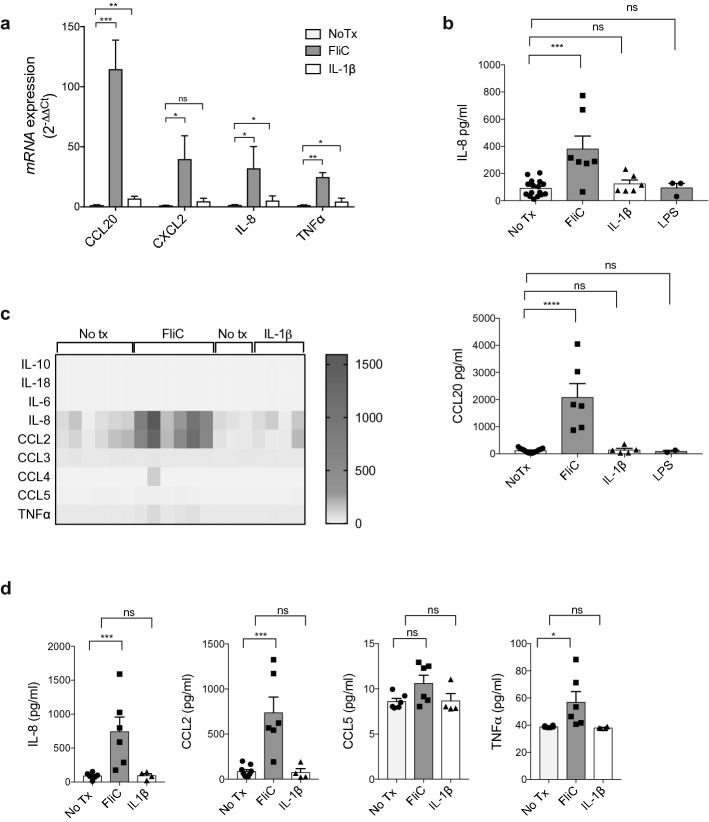
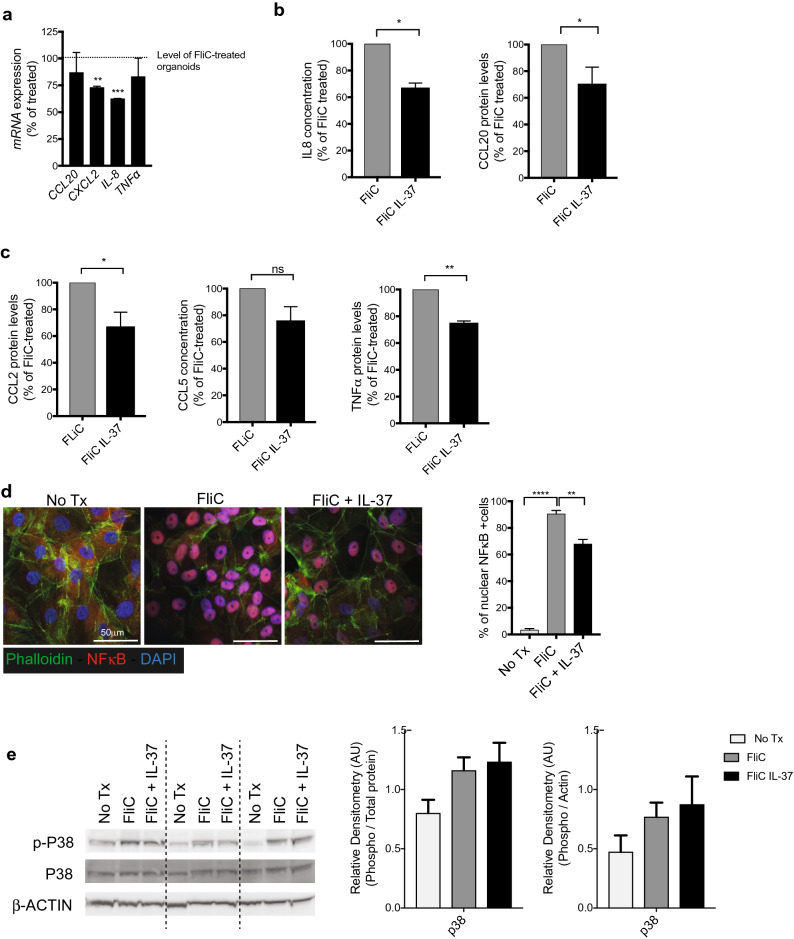
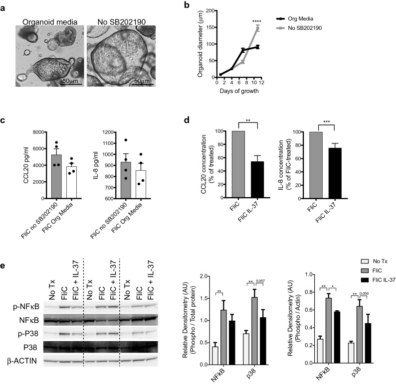
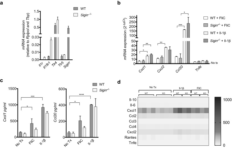
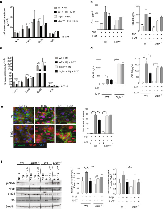
Intestinal epithelial cells (IEC) reside in close proximity to the gut microbiota and are hypo-responsive to bacterial products, likely to prevent maladaptive inflammatory responses. This is in part due to their strong expression of Single Ig IL-1 related receptor (SIGIRR), a negative regulator of interleukin (IL)-1 and toll-like receptor signaling. IL-37 is an anti-inflammatory cytokine that inhibits innate signaling in diverse cells by signaling through SIGIRR. Despite the strong expression of SIGIRR by IEC, few studies have examined whether IL-37 can suppress their innate immune signaling. We characterized innate immune responses of human and murine colonoids to bacteria (FliC, LPS) and host (IL-1β) products and the role of IL-37/SIGIRR in regulating these responses. We demonstrated that human colonoids responded only to FliC, but not to LPS or IL-1β. While colonoids derived from different donors displayed significant inter-individual variability in the magnitude of their innate responses to FliC stimulation, all colonoids released a variety of chemokines. Interestingly, IL-37 attenuated these responses through inhibition of p38 and NFκB signaling pathways. We determined that this suppression by IL-37 was SIGIRR dependent, in murine organoids. Along with species-specific differences in IEC innate responses, we show that IL-37 can promote IEC hypo-responsiveness by suppressing inflammatory signaling.


Conflict of interest statement

The authors declare no competing interests.

Figures

Figure 1
Figure 1
Human IEC responses to host (IL-1β) and bacterial stimuli (FliC, LPS). (a) qPCR analysis of inflammatory gene transcription after FliC or IL-1β stimulation for 4 h, expressed as fold change over untreated colonoids. (b) IL-8 (top) and CCL20 (bottom) protein levels secreted basolaterally by human colonoids after 4 h of stimulation with FliC or IL-1β. (c) Heat map representation of chemokine concentrations in the supernantants of colonoids after 4 h of stimulation with FliC or IL-1β. (d) Concentration of chemokines detected by Multiplex Luminex Assay in the supernatants of colonoids after 4 h of stimulation with FliC or IL-1β. Mean and SEM are indicated from n = 6–8 donors (4 adult and 2–4 pediatric). All data shown are representative of at least 2–3 independent experiments. Statistical significance calculated using unpaired Student’s t-test *, P = 0.01 to 0.05; **, P = 0.001 to 0.01; ***, P = 0.0001 to 0.001. ns = not significant.
Figure 2
Figure 2
IL-37 effect on human IEC FliC responses. (a) qPCR analysis of inflammatory genes after FliC and IL-37 exposure for 4 h expressed as percentage of change over FliC treated colonoids. (b) IL-8 and CCL20 protein levels (detected by ELISA) secreted basolaterally by human colonoids after 4 h of stimulation with FliC and IL-37. (c) CCL2, CCL5 and TNFa protein levels secreted basolaterally by colonoids after 4 h of stimulation with FliC and IL-37 (detected by Milliplex Luminex assay). (d) Immunostaining against NFkB (red), actin (Phalloidin—green) and DAPI (blue) of 2D monolayer after 30 min of stimulation with FliC with or without IL-37 (left). Counts of NFkB positive nuclei from immunostaining (right). (e) Western blot analysis of phospho and total p38, after 30 min of stimulation with FliC with or without IL-37 (left). Equal loading confirmed with β-Actin as well as total protein stain of membrane (see Fig. S4 online). Densities relative to total protein or β-Actin are shown (right). Mean and SEM are indicated from n = 4 donors (2 adult and 2 pediatric). All data shown are representative of at least 3 independent experiments. Statistical significance calculated using unpaired Student’s t-test *, P = 0.01 to 0.05; **, P = 0.001 to 0.01; ***, P = 0.0001 to 0.001; ****, P = 0.00001 to 0.0001; ns = not significant.
Figure 3
Figure 3
SB202190 effect on IL37 immunosuppressive responses. (a) Brightfield images of a representative human colonoids grown in standard organoid Media (left) or in No SB202190 media (right). b) Growth curve of colonoids grown in standard organoid media or in No SB202190 media for 12 days. (c) IL-8 and CCL20 protein levels secreted basolaterally after 4 h of stimulation with FliC of human colonoids grown in standard organoids media or in No SB202190 media. (d) IL-8 and CCL20 protein levels secreted basolaterally by human colonoids grown in No SB202190 media after 4 h of stimulation simultaneously with FliC and IL-37. (e) Western blot analysis of phospho and total NFkB, phospho and total p38 in human colonoids grown in No SB202 media after 30 min of stimulation with FliC with or without IL-37 (left). Equal loading confirmed with β-Actin as well as total protein stain of membrane (see Fig. S5 online). Densities relative to total protein or β-Actin are shown (right). Mean and SEM are indicated from n = 4 donors (2 adult and 2 pediatric). All data shown are representative of at least 3 independent experiments. Statistical significance calculated using unpaired Student’s t-test *, P = 0.01 to 0.05; **, P = 0.001 to 0.01; ***, P = 0.0001 to 0.001; ****, P = 0.00001 to 0.0001. Comparison lacking annotations are not significant.
Figure 4
Figure 4
Mouse IEC responses to host (Il-1β) and bacteria stimuli (FliC). (a) qPCR analysis of innate receptor and negative regulator gene transcription by mouse colonoids expressed as relative expression over reference genes. (b) qPCR analysis of chemokines and cytokines after 4 h of stimulation with FliC and Il-1β expressed as fold change over untreated WT (Sigirr+/+) colonoid expression. (c) Cxcl1 (left) and Ccl20 (right) protein levels secreted basolaterally by colonoids derived from WT and Sigirr−/− mice after 4 h of stimulation with FliC or Il-1β. (d) Heat map representation of various chemokine concentration in the supernatant of colonoids after 4 h of stimulation with FliC or Il-1β. Mean and SEM are indicated. Mean and SEM are indicated from n = 4 mouse organoid lines derived from each genotype. All data shown are representative of at least 2–3 independent experiments. Statistical significance calculated using one-way ANOVA *, P = 0.01 to 0.05; **, P = 0.001 to 0.01; ***, P = 0.0001 to 0.001; ***, P = 0.00001 to 0.0001. Comparison lacking annotations are not significant.
Figure 5
Figure 5
IL-37′s suppression of innate responses in mouse organoids is Sigirr dependent. (a) qPCR analysis of main inflammatory genes after FliC stimulation with IL-37 for 4 h on Sigirr+/+ (WT) and Sigirr−/− mouse colonoids expressed as fold change over untreated colonoids. (b) Cxcl1 (left) and Ccl20 (right) protein levels secreted basolaterally by WT and Sigirr−/− mouse colonoids after 4 h of stimulation with FliC and IL-37. (c) qPCR analysis of key inflammatory genes after with Il-1β stimulation, in concert with IL-37 for 4 h on Sigirr+/+ (WT) and Sigirr−/− mouse colonoids expressed as fold change over untreated colonoids. (d) Cxcl1 (left) and Ccl20 (right) protein levels secreted basolaterally by WT and Sigirr−/− mouse colonoids after 4 h of stimulation with Il-1β and IL-37. (e) Immunostaining against Nfkb (red), Actin Phalloidin (green) and DAPI (blue) of 2D monolayer after 30 min of stimulation with Il-1b with or without IL-37 (left). Counts of Nfkb positive nuclei from immunostaining (Right). (f) Western blot analysis of phospho and total NFkB, phospho and total p38 in mouse colonoids after 30 min of stimulation with Il-1β with or without IL-37 (left). Equal loading was confirmed with β-Actin as well as total protein stain of membrane (see Fig. S6 online). Densities relative to total protein or β-Actin are shown (right). Mean and SEM are indicated from n = 4 mouse organoid lines derived from each genotype. All data shown are representative of at least 3 independent experiments. Statistical significance calculated using one-way ANOVA *, P = 0.01 to 0.05; **, P = 0.001 to 0.01; ***, P = 0.0001 to 0.001; ***, P = 0.00001 to 0.0001. Comparison lacking annotations are not significant.

References

    1. Allaire JM, et al. The intestinal epithelium: central coordinator of mucosal immunity. Trends Immunol. 2018;39:677–696. doi: 10.1016/j.it.2018.04.002. - DOI - PubMed
    1. Price AE, et al. A map of toll-like receptor expression in the intestinal epithelium reveals distinct spatial, cell type-specific, and temporal patterns. Immunity. 2018;49:560–575.e6. doi: 10.1016/j.immuni.2018.07.016. - DOI - PMC - PubMed
    1. Kayisoglu O, et al. Location-specific cell identity rather than exposure to GI microbiota defines many innate immune signalling cascades in the gut epithelium. Gut. 2020 doi: 10.1136/gutjnl-2019-319919. - DOI - PMC - PubMed
    1. Abreu MT. Toll-like receptor signalling in the intestinal epithelium: How bacterial recognition shapes intestinal function. Nat. Rev. Immunol. 2010;10:131–143. doi: 10.1038/nri2707. - DOI - PubMed
    1. Frantz AL, et al. Targeted deletion of MyD88 in intestinal epithelial cells results in compromised antibacterial immunity associated with downregulation of polymeric immunoglobulin receptor, mucin-2, and antibacterial peptides. Mucosal Immunol. 2012;5:501–512. doi: 10.1038/mi.2012.23. - DOI - PMC - PubMed

Publication types

Grants and funding