. 2021 Apr 15;11(1):8214.
doi: 10.1038/s41598-021-87611-2.

Activation of creER recombinase in the mouse calvaria induces local recombination without effects on distant skeletal segments

Affiliations

Activation of creER recombinase in the mouse calvaria induces local recombination without effects on distant skeletal segments

Jue Hou et al. Sci Rep. .

Abstract

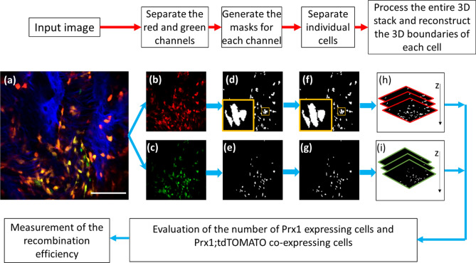
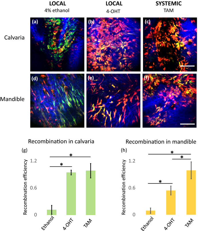
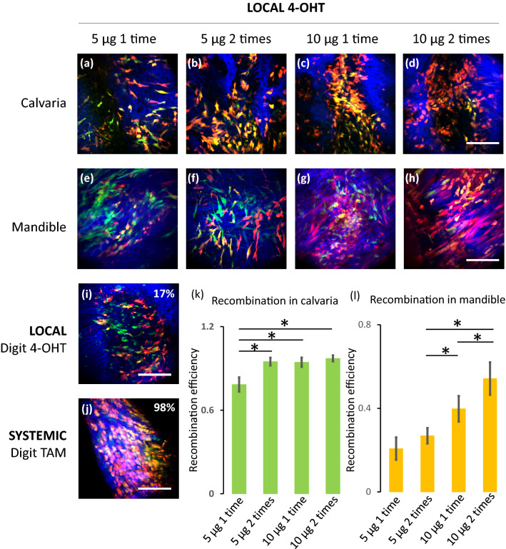
Conditional creER-mediated gene inactivation or gene induction has emerged as a robust tool for studying gene functions in mouse models of tissue development, homeostasis, and regeneration. Here, we present a method to conditionally induce cre recombination in the mouse calvarial bone while avoiding systemic recombination in distal bones. To test our method, we utilized Prx1creER-egfp;td-Tomato mice and delivered 4-hydroxytamoxifen (4-OHT) to the mouse calvaria, subperiosteally. First, we showed that two calvaria subperiosteal injections of 10 µg of 4-OHT (3.3 mg of 4-OHT/kg of body weight) can induce local recombination as efficiently as two intraperitoneal systemic injections of 200 μg of tamoxifen (70 mg of tamoxifen/kg of body weight). Then, we studied the recombination efficiency of various subperiosteal calvaria dosages and found that two subperiosteal injections of 5 µg 4-OHT (1.65 mg of 4-OHT/kg of body weight) uphold the same recombination efficiency observed with higher dosages. Importantly, the result indicated that the low dosage does not induce significant systemic recombination in remote skeletal tissues. With the proposed local low dosage protocol, the recombination efficiency at the injection site (calvarial bone) reached 94%, while the recombination efficiency at the mandible and the digits was as low as the efficiency measured in control animals.


Conflict of interest statement

The authors declare no competing interests.

Figures

Figure 1
Figure 1
Image processing and automatic cell counting workflow. (a) A representative input image uploaded into Matlab program. After acquisition, the input image is processed to separate the red channel (b) and the green channel (c), which represent the cells that express tdTomato and Prx1 (eGFP) respectively. Subsequently, a three-component Otsu thresholding is independently applied to each channel, to generate a mask for tdTomato expressing cells (d) and Prx1 (eGFP) expressing cells (e). A marker-controlled watershed algorithm is applied to further separate the connected tdTomato expressing cells (f) and Prx1 (eGFP) expressing cells (g) into individual cells. The orange insets in (d) and (f) show connected cells (in d) that are successfully separated (in f). Then, 3D reconstructed masks for the tdTomato expressing cells (h) and for the Prx1 (eGFP) expressing cells (i) are generated to calculate the final recombination efficiency.
Figure 2
Figure 2
Comparison of recombination efficiency between locally induced and systemically induced recombination. Animals were treated with 4% ethanol (subperiosteal local injection) or 10 µg of 4-OHT (subperiosteal local injection) or 200 µg of Tamoxifen (systemic intraperitoneal (IP) injection). Representative images of the animal’s calvaria and mandibular bones are displayed: (a) and (d) 4% ethanol local injection, (b) and (e) 10 µg 4-OHT local injection, and (c) and (f) IP injections of 200 µg Tamoxifen (TAM). The Prx1 (eGFP) expressing cells are shown in green, the tdTomato expressing cells are shown in red and, thus, the eGFP and tdTomato co-expressing cells are shown in yellow. Bone is visualized with second harmonic generation and is pseudo-colored in blue. The scale bar represents 100 µm. The recombination efficiency measured in the different treatment groups is shown in histograms reporting the data collected in the animals’ calvaria (g) and the data collected in the animals’ mandibles (h). The error bars indicate standard deviations and the * indicates statistically significant difference with a p value < 0.05 (n = 4).
Figure 3
Figure 3
Recombination efficiency in animals treated with different local injection protocols. Representative images are displayed: (a) and (e) 5 µg 4-OHT local injection for one time in calvaria and mandible; (b) and (f) 5 µg 4-OHT local injections for two times in calvaria and mandible; (c) and (g) 10 µg 4-OHT local injection for one time in calvaria and mandible; and (d) and (h) 10 µg 4-OHT local injections for two times in calvaria and mandible. (i) Digit bone of a mouse treated with two local injections of 5 µg 4-OHT. (j) Digit bone of a mouse treated with two systemic IP injections of 200 µg of Tamoxifen. The Prx1 (eGFP) expressing cells are shown in green, the tdTomato expressing cells are shown in red and, thus, the eGFP and tdTOMATO co-expressing cells are shown in yellow. Bone is visualized with second harmonic generation and is pseudo-colored in blue. The scale bar represents 100 µm. The recombination efficiency measured in the different treatment groups is shown in (i) and (j) for the digits (inset numbers) and in histograms for the calvaria (k) and the mandibles (l). The error bars indicate standard deviations and the * indicates statistically significant difference with a p value < 0.05 (n = 4).

Similar articles

Cited by

References

    1. Sauer B, Henderson N. Site-specific DNA recombination in mammalian cells by the Cre recombinase of bacteriophage P1. Proc. Natl. Acad. Sci. USA. 1988;85(14):5166–5170. doi: 10.1073/pnas.85.14.5166. - DOI - PMC - PubMed
    1. Metzger D, et al. Conditional site-specific recombination in mammalian cells using a ligand-dependent chimeric Cre recombinase. Proc. Natl. Acad. Sci. USA. 1995;92(15):6991–6995. doi: 10.1073/pnas.92.15.6991. - DOI - PMC - PubMed
    1. Kwan KM. Conditional alleles in mice: Practical considerations for tissue-specific knockouts. Genesis. 2002;32(2):49–62. doi: 10.1002/gene.10068. - DOI - PubMed
    1. Kühn, R. & Wurst, W. Gene knockout protocols. 2nd ed. In Methods in molecular biology, xvi, 4 p. of plates (Humana Press, 2009).
    1. Zhong ZA, et al. Optimizing tamoxifen-inducible Cre/loxp system to reduce tamoxifen effect on bone turnover in long bones of young mice. Bone. 2015;81:614–619. doi: 10.1016/j.bone.2015.07.034. - DOI - PMC - PubMed

Publication types

MeSH terms

LinkOut - more resources