. 2022 Feb;43(2):401-416.
doi: 10.1038/s41401-021-00654-z. Epub 2021 Apr 15.

TNF-α impairs EP4 signaling through the association of TRAF2-GRK2 in primary fibroblast-like synoviocytes

Affiliations

TNF-α impairs EP4 signaling through the association of TRAF2-GRK2 in primary fibroblast-like synoviocytes

Yu Tai et al. Acta Pharmacol Sin. 2022 Feb.

Abstract

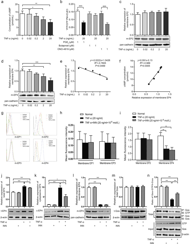
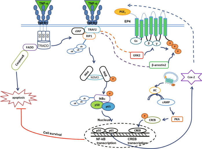
Our previous study showed that chronic treatment with tumor necrosis factor-α (TNF-α) decreased cAMP concentration in fibroblast-like synoviocytes (FLSs) of collagen-induced arthritis (CIA) rats. In this study we investigated how TNF-α impairs cAMP homeostasis, particularly clarifying the potential downstream molecules of TNF-α and prostaglandin receptor 4 (EP4) signaling that would interact with each other. Using a cAMP FRET biosensor PM-ICUE3, we demonstrated that TNF-α (20 ng/mL) blocked ONO-4819-triggered EP4 signaling, but not Butaprost-triggered EP2 signaling in normal rat FLSs. We showed that TNF-α (0.02-20 ng/mL) dose-dependently reduced EP4 membrane distribution in normal rat FLS. TNF-α significantly increased TNF receptor 2 (TNFR2) expression and stimulated proliferation in human FLS (hFLS) via ecruiting TNF receptor-associated factor 2 (TRAF2) to cell membrane. More interestingly, we revealed that TRAF2 interacted with G protein-coupled receptor kinase (GRK2) in the cytoplasm of primary hFLS and helped to bring GRK2 to cell membrane in response of TNF-α stimulation, the complex of TRAF2 and GRK2 then separated on the membrane, and translocated GRK2 induced the desensitization and internalization of EP4, leading to reduced production of intracellular cAMP. Silencing of TRAF2 by siRNA substantially diminished TRAF2-GRK2 interaction, blocked the translocation of GRK2, and resulted in upregulated expression of membrane EP4 and intracellular cAMP. In CIA rats, administration of paroxetine to inhibit GRK2 effectively improved the symptoms and clinic parameters with significantly reduced joint synovium inflammation and bone destruction. These results elucidate a novel form of cross-talk between TNFR (a cytokine receptor) and EP4 (a typical G protein-coupled receptor) signaling pathways. The interaction between TRAF2 and GRK2 may become a potential new drug target for the treatment of inflammatory diseases.

Keywords: EP4; GRK2; TNFR2; TRAF2; fibroblast-like synoviocytes; rheumatoid arthritis.


Conflict of interest statement

The authors declare no competing interests.

Figures

Fig. 1
Fig. 1. EP4 signaling is blocked in TNF-α-stimulated FLS.
Normal FLSs were obtained from the synovia of SD rats or from trauma patients who had undergone total joint replacement surgery or synovectomy. a Rat FLSs were cultured in the presence of TNF-α at final concentrations of 0, 0.02, 0.2, 2, or 20 ng/mL for 48 h, and cAMP levels were analyzed by a 125I-cAMP RIA kit. b Rat FLSs were transfected with a plasma membrane-linked cAMP biosensor, which has cyan fluorescent protein (CFP) and yellow fluorescent protein (YFP). Cyclic-AMP close to the plasma membrane binds to the EPAC1 domain of the PM-ICUE3 sensor and separates CFP and YFP, leading to a decrease in the intensity of the YFP/CFP ratio. c, d Rat FLSs were treated with different concentrations of TNF-α for 48 h, the cell membrane fraction was purified by centrifugation 30,000 × g, and EP2 and EP4 expression levels were determined by Western blotting. e The relationship between the exogenous TNF-α concentration and the relative membrane expression of EP4 was analyzed, and a negative association was observed. f A significant positive correlation between the relative membrane expression of EP4 and intracellular cAMP levels was described. gi Rat FLSs were stained with primary EP1–4 antibodies and corresponding FITC-labeled secondary antibodies after treatment with vehicle, TNF-α 20 ng/mL, or 20 ng/mL TNF-α and 1 μM INN for 48 h; cells incubated with only the fluorescently labeled secondary antibody were used as an isotype control. Flow cytometry was used to observe the membrane expression of EP1–4 on FLSs, and the mean fluorescence intensities were compared between the different treatments. jl Total, cytoplasmic and membrane levels of EP4 were measured in hFLSs treated with TNF-α (20 ng/mL) or TNF-α (20 ng/mL) and INN (1 μM) for 48 h. m Total Gαs expression was measured by Western blotting. n Coimmunoprecipitation was used to determine the level of activated GTP-bound Gαs. The ratio of Gαs-GTP to total Gαs was calculated to reflect the activation of Gαs. *P < 0.05, **P < 0.01, ***P < 0.001 (n = 6).
Fig. 2
Fig. 2. TNF-α impairs EP4 signaling in hFLSs in a TNFR2-dependent manner.
a, b TNFR1 and TNFR2 were indirectly stained with FITC-labeled secondary antibodies, and the distribution of these receptors was further observed using a laser confocal microscope. c, d The expression of membrane TNFR1 or TNFR2 on hFLSs was measured by Western blotting. e, f TNFR1 and TNFR2 gene expression was silenced by siRNA, and the protein expression of TNFR1 or TNFR2 was clearly downregulated. g, h High content screening was used to analyze hFLS proliferation (×100). i, j Laser confocal light microscopy was used to examine the expression of EP4 in hFLSs with or without TNFR1/2. *P < 0.05, **P < 0.01, ***P < 0.001 (n = 6).
Fig. 3
Fig. 3. TNF-α impairs EP4 signaling in hFLSs in a TNFR2- and TRAF2-dependent manner.
a Total expression of TRAF2 was measured in hFLSs treated with TNF-α with or without INN. b Membrane TRAF2 was examined by Western blotting. c The membrane localization of TRAF2 was confirmed using laser confocal light microscopy. d TRAF2 gene expression was silenced by siRNA, and both the protein and mRNA expression of TRAF2 were clearly downregulated. e The viability of hFLSs transfected with TRAF2 siRNA or the negative control was analyzed using MTT assays. f Intracellular cAMP levels were not decreased in TRAF2-silenced hFLSs, even after stimulation with TNF-α with or without INN. gj Total and membrane EP4 expression was increased and cytoplasmic EP4 was decreased in hFLSs cultured with TNF-α with or without INN in the absence of TRAF2. k TRAF2 silencing decreased the gene expression of EP4 in hFLSs. Gene expression was quantified relative to the internal control (GAPDH). *P < 0.05, **P < 0.01 (n = 6).
Fig. 4
Fig. 4. TNF-α triggers the translocation of the TRAF2-GRK2 complex to the cell membrane and induces complex dissociation.
ad The subcellular localization of the TRAF2-GRK2/β-Arrestin2 complex was observed in purified hFLSs after stimulation with TNF-α with or without INN. e, f GRK2 translocation in response to both treatments was examined by Western blotting. g Laser confocal light microscopy was used to demonstrate the colocalization of TRAF2 and GRK2 in hFLSs. h Changes in the interaction between TRAF2 and GRK2 in the cell membrane and cytoplasm under TNF-α stimulation were determined via coimmunoprecipitation. i Synovial tissue was collected from the rat knee joint at different time points after local injection of 20 ng/mL TNF-α. The distribution of the TRAF2-GRK2 complex was analyzed. *P < 0.05, **P < 0.01 (n = 6).
Fig. 5
Fig. 5. Molecular docking of GRK2 and TRAF2.
a Overall diagram of the GRK2 and TRAF2 docking results. Blue indicates the receptor GRK2, pink indicates the ligand TRAF2, red indicates interface ligand residues, and yellow indicates interface receptor residues. b Local plot of the TRAF2 and GRK2 docking results. The dashed line represents the existing interaction force between the ligand and receptor.
Fig. 6
Fig. 6. TRAF2 silencing reduces EP4 desensitization by decreasing membrane GRK2 translocation. Inhibition of GRK2 attenuates TNF-α-induced FLS proliferation.
ac Membrane, cytoplasmic and phospho-GRK2 levels in hFLSs treated with TNF-α with or without INN were measured by Western blotting. d High-content screening was used to analyze hFLS proliferation after treatment with TNF-α with or without paroxetine (×100). *P < 0.05, **P < 0.01, ***P < 0.001 (n = 6).
Fig. 7
Fig. 7. Paroxetine treatment attenuates the symptoms of CIA rats.
CIA was induced by immunizing rats with an emulsion of native chicken CII and incomplete Freund’s adjuvant. a, b The concentration of intracellular cAMP and cell viability were compared between normal hFLSs and RA hFLSs. c The swelling of secondary joints reached a plateau on day 28. Obvious pathological changes were detected in the ankle joint. The degree of paw swelling was evaluated by the change in volume. d, e The concentration of cAMP and cell viability were analyzed in FLSs isolated from treated CIA rats. f Body weight. g Clinical score. h Arthritis index. i Swollen joint count. j Secondary paw swelling. k H&E staining of the joints was performed, and pathogenic parameters, including inflammation, pannus formation, cartilage erosion, cellular infiltration and synoviocyte proliferation, were evaluated (scale bar: 100 μm). **P < 0.01, ***P < 0.001, vs the normal group; #P < 0.05, ##P < 0.01, ###P < 0.001, vs the RA or CIA group (n = 6).
Fig. 8
Fig. 8. Potential mechanisms of TNF-α-mediated EP4 signaling.
Signaling pathways activated by TNF-α-induced PGE2 stimulation of the human receptor EP4. EP4 is quickly phosphorylated by GRK2 and subjected to desensitization after agonist exposure. Then, β-Arrestin2 binds to phosphorylated EP4 and mediates its internalization. EP4 generates intracellular cAMP by coupling to Gs proteins, leading to the activation of PKA and CREB phosphorylation. TNF-α not only stimulates PGE2 production but also interacts with GRK2 through TRAF2 to regulate EP4 desensitization in hFLSs

Similar articles

Cited by

References

    1. Dayer JM. From supernatants to cytokines: a personal view on the early history of IL-1, IL-1Ra, TNF and its inhibitor in rheumatology. Arthritis Res Ther. 2018;20:101. - PMC - PubMed
    1. Ursini F, Leporini C, Bene F, D’Angelo S, Mauro D, Russo E, et al. Anti-TNF-alpha agents and endothelial function in rheumatoid arthritis: a systematic review and meta-analysis. Sci Rep. 2017;7:5346. - PMC - PubMed
    1. Jia XY, Chang Y, Sun XJ, Dai X, Wei W. The role of prostaglandin E2 receptor signaling of dendritic cells in rheumatoid arthritis. Int Immunopharmacol. 2014;23:163–9. - PubMed
    1. Samuels JS, Holland L, Lopez M, Meyers K, Cumbie WG, McClain A, et al. Prostaglandin E2 and IL-23 interconnects STAT3 and RoRgamma pathways to initiate Th17 CD4+ T-cell development during rheumatoid arthritis. Inflamm Res. 2018;67:589–96. - PMC - PubMed
    1. Lowin T, Apitz M, Anders S, Straub RH. Anti-inflammatory effects of N-acylethanolamines in rheumatoid arthritis synovial cells are mediated by TRPV1 and TRPA1 in a COX-2 dependent manner. Arthritis Res Ther. 2015;17:321. - PMC - PubMed

MeSH terms

Substances

LinkOut - more resources