. 2021 Apr 9:16:2715-2733.
doi: 10.2147/IJN.S296406. eCollection 2021.

Immunoassay-Amplified Responses Using a Functionalized MoS2-Based SPR Biosensor to Detect PAPP-A2 in Maternal Serum Samples to Screen for Fetal Down's Syndrome

Affiliations

Immunoassay-Amplified Responses Using a Functionalized MoS2-Based SPR Biosensor to Detect PAPP-A2 in Maternal Serum Samples to Screen for Fetal Down's Syndrome

Nan-Fu Chiu et al. Int J Nanomedicine. .

Abstract

Background: Due to educational, social and economic reasons, more and more women are delaying childbirth. However, advanced maternal age is associated with several adverse pregnancy outcomes, and in particular a high risk of Down's syndrome (DS). Hence, it is increasingly important to be able to detect fetal Down's syndrome (FDS).

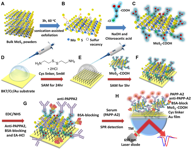
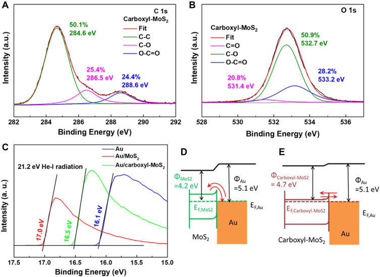
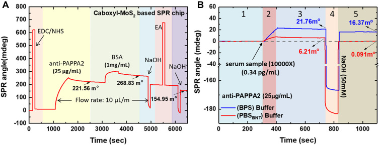
Methods: We developed an effective, highly sensitive, surface plasmon resonance (SPR) biosensor with biochemically amplified responses using carboxyl-molybdenum disulfide (MoS2) film. The use of carboxylic acid as a surface modifier of MoS2 promoted dispersion and formed specific three-dimensional coordination sites. The carboxylic acid immobilized unmodified antibodies in a way that enhanced the bioaffinity of MoS2 and preserved biorecognition properties of the SPR sensor surface. Complete antigen pregnancy-associated plasma protein-A2 (PAPP-A2) conjugated with the carboxyl-MoS2-modified gold chip to amplify the signal and improve detection sensitivity. This heterostructure interface had a high work function, and thus improved the efficiency of the electric field energy of the surface plasmon. These results provide evidence that the interface electric field improved performance of the SPR biosensor.

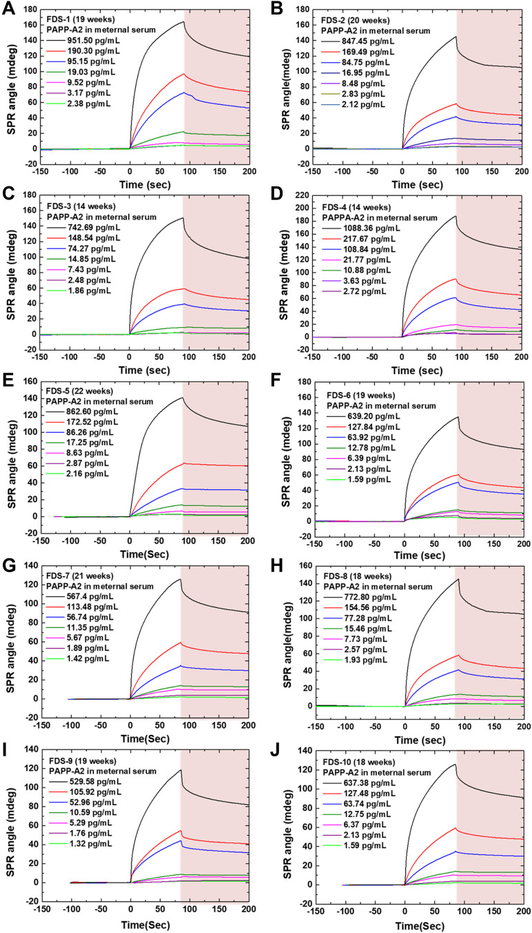
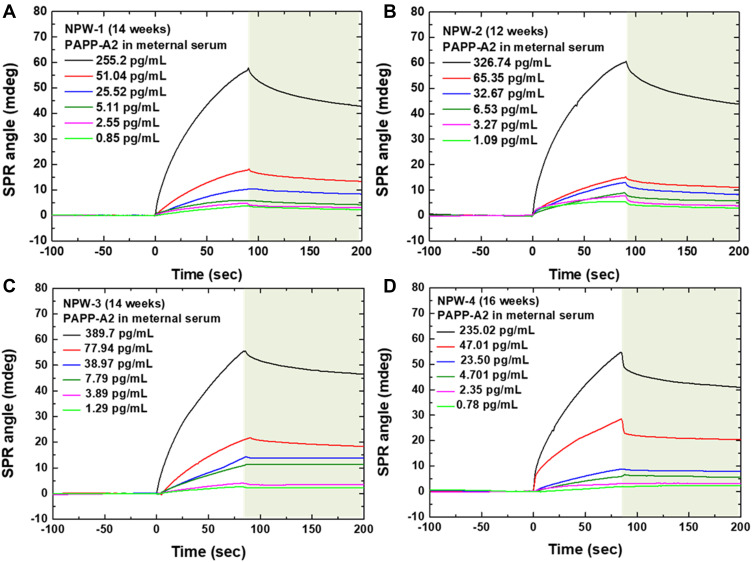
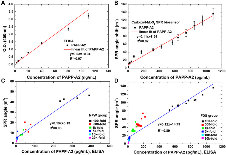
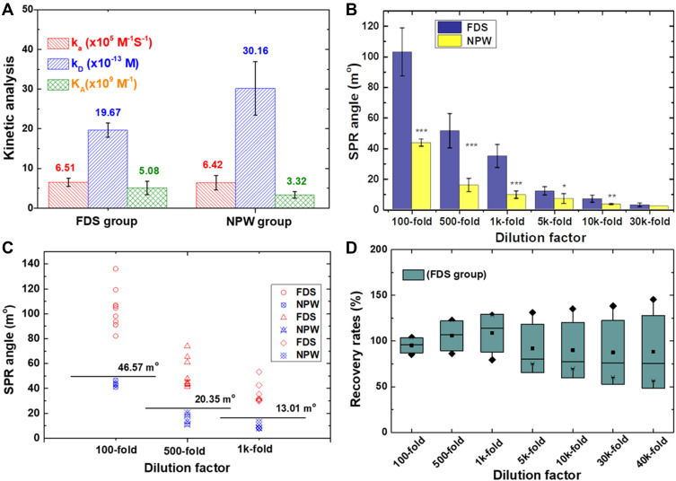
Results: The carboxyl-MoS2-based SPR biosensor was used successfully to evaluate PAPP-A2 level for fetal Down's syndrome screening in maternal serum samples. The detection limit was 0.05 pg/mL, and the linear working range was 0.1 to 1100 pg/mL. The women with an SPR angle >46.57 m° were more closely associated with fetal Down's syndrome. Once optimized for serum Down's syndrome screening, an average recovery of 95.2% and relative standard deviation of 8.5% were obtained. Our findings suggest that carboxyl-MoS2-based SPR technology may have advantages over conventional ELISA in certain situations.

Conclusion: Carboxyl-MoS2-based SPR biosensors can be used as a new diagnostic technology to respond to the increasing need for fetal Down's syndrome screening in maternal serum samples. Our results demonstrated that the carboxyl-MoS2-based SPR biosensor was capable of determining PAPP-A2 levels with acceptable accuracy and recovery. We hope that this technology will be investigated in diverse clinical trials and in real case applications for screening and early diagnosis in the future.

Keywords: DS; Down’s syndrome; FDS; PAPP-A2; SPR; carboxyl-MoS2; carboxyl-functionalized molybdenum disulfide; fetal Down’s syndrome; pregnancy-associated plasma protein A2; surface plasmon resonance.


Conflict of interest statement

Prof. Dr. Nan-Fu Chiu reports a patent US10634613B2 issued, a patent US10815259B2. The authors declare that the research was conducted in the absence of any other commercial or financial relationships that could be construed as a potential conflict of interest.

Figures

Figure 1
Figure 1
Carboxyl-MoS2 nanocomposite synthesis and the sensing mechanism of the carboxyl-MoS2-based SPR biosensor to detect PAPP-A2 protein in maternal serum samples. (A) Schematic diagram of sonication-assisted liquid-phase exfoliation (LPE) for the preparation of MoS2 sheets. (B) MoS2 exhibits a layered structure with planar Mo–S bonds and sulfur vacancies on the MoS2 surface. (C) The carboxyl-MoS2 nanocomposites were successfully modified by chloroacetic acid. Carboxyl-MoS2-based SPR chip immobilization steps and containing. (D) Au film substrate. (E) Cys linker containing a thiol-group to allow for assembly on the Au film substrate. (F) Covalent binding of carboxyl-MoS2 on the Cys-linker. (G) EDC/NHS activated carboxyl groups resulting in a higher density of immobilized antibodies. (H) To detect PAPP-A2 protein to screen for Down’s syndrome in maternal serum samples.
Figure 2
Figure 2
(A) SEM image of carboxyl-MoS2 sheets. (B) Cross-sectional SEM image of lateral flake thickness of carboxyl-MoS2 sheets. (C) TEM image of the carboxyl-MoS2 sheets. (D) TEM image of the MoS2 sheets. (E) EDS analysis of the carboxyl-MoS2 sheets (insert shows the carboxyl-MoS2 sheet for the EDS analysis).
Figure 3
Figure 3
The XPS survey spectra of (A) MoS2 sheets and (B) carboxyl-MoS2 sheets. The high-resolution XPS spectra of (C) C1 2p, (D) Mo 3d, (E) S 2p for MoS2 and carboxyl-MoS2 sheets. (F) Analysis of XPS surface atomic intensity ratios of C1s/Mo3d and O1s/Mo3d on MoS2 and carboxyl-MoS2 sheets.
Figure 4
Figure 4
High energy resolution XPS spectra of (A) C1s and (B) O1s regions on carboxyl-MoS2 sheets. (C) UPS spectra of different interfaces of Au, Au/MoS2 and Au/carboxyl-MoS2 film. Band diagram of (D) Au/MoS2 and (E) Au/carboxyl-MoS2 heterojunctions obtained from UPS measurements. The band-gap energies used in the diagram are optical gaps.
Figure 5
Figure 5
(A) Optimization of the immobilization process of antibodies in carboxyl-MoS2-based SPR chips. (B) Non-specific molecular dissociation reactions were tested using PBS and PBSBNT buffer, respectively.
Figure 6
Figure 6
Sensorgrams showing the SPR responses generated with a flow rate of 60 μL/min and different dilution factors of 10 FDS maternal serum samples. The 10 FDS samples were derived from blood drawn at a gestational age of (A) 19, (B) 20, (C) 14, (D) 14, (E) 22, (F) 19, (G) 21, (H) 18, (I) 19, and (J) 18 weeks, respectively.
Figure 7
Figure 7
Sensorgrams showing the SPR responses generated with a flow rate of 60 μL/min and different dilution factors of maternal serum samples from four healthy women (NPW group). The NPW samples were derived from blood drawn at a gestational age of (A) 14, (B) 12, (C) 14, and (D) 16 weeks, respectively.
Figure 8
Figure 8
Calibration curves obtained with (A) ELISA kit assays for PAPP-A2 quantification ranging from 2.5 to 108 ng/mL. (B) Calibration curve of the average SPR response to various PAPP-A2 concentrations ranging from 0.1 to 1100 pg/mL with the carboxyl-MoS2-based SPR chip. Error bars indicate standard deviations of means obtained from three replicates using SPR and three replicates using ELISA. Correlation between the concentration of PAPP-A2 in serum obtained by ELISA and signal responses by SPR. (C) NPW group (n = 24, R2 = 0.97) and (D) FDS group (n = 60, R2 = 0.91) samples.
Figure 9
Figure 9
SPR responses of serum samples of the FDS and NPW groups. (A) Kinetic analysis of the FDS and NPW groups on the carboxyl-MoS2-based SPR biosensor for affinity binding analysis of the PAPP-A2 interaction. (B) Correlation between two variables of the FDS and NPW groups to detect PAPP-A2 in serum samples. Data expressed as mean ± SD for triplicate tests. Asterisks indicate statistical significance using a generalized mixed effects model (highly significant recognition (***) (p < 0.001); very significant recognition (**) (0.001 < p < 0.01); significant recognition (*) (0.01 < p < 0.05). p: probability value). (C) The dot plot shows the distribution of SPR responses at the dilution rate (100-, 500-, and 1k-fold) of different serum samples and the difference between the FDS and NPW groups at the cut-off limit for screening. (D) Recovery rates of PAPP-A2 protein assay in the FDS group using the carboxyl-MoS2-based SPR chip.

Similar articles

Cited by

References

    1. Heffner LJ. Advanced maternal age – how old is too old? N Engl J Med. 2004;351:1927–1929. doi:10.1056/NEJMp048087 - DOI - PubMed
    1. Resta RG. Changing demographics of advanced maternal age (AMA) and the impact on the predicted incidence of Down syndrome in the United States: implications for prenatal screening and genetic counseling. Am J Med Genet A. 2005;133A(1):31–36. doi:10.1002/ajmg.a.30553 - DOI - PubMed
    1. Bayrampour H, Heaman M, Duncan KA, Tough S. Advanced maternal age and risk perception: a qualitative study. BMC Pregnancy Childbirth. 2012;12(1):100–113. doi:10.1186/1471-2393-12-100 - DOI - PMC - PubMed
    1. Kahveci B, Melekoglu R, Evruke IC, Cetin C. The effect of advanced maternal age on perinatal outcomes in nulliparous singleton pregnancies. BMC Pregnancy Childbirth. 2018;18(1):343–349. doi:10.1186/s12884-018-1984-x - DOI - PMC - PubMed
    1. Carr J, Collins S. 50 years with Down syndrome: a longitudinal study. J Appl Res Intellect Disabil. 2018;31:743–750. doi:10.1111/jar.12438 - DOI - PubMed