. 2021 Apr 9:16:2735-2749.
doi: 10.2147/IJN.S298093. eCollection 2021.

Anti-Cancer Activity Based on the High Docetaxel Loaded Poly(2-Oxazoline)s Micelles

Affiliations

Anti-Cancer Activity Based on the High Docetaxel Loaded Poly(2-Oxazoline)s Micelles

Min Xu et al. Int J Nanomedicine. .

Erratum in

Abstract

Purpose: Nanocarriers, with a high drug loading content and good safety, to achieve desirable therapeutic effect are always the goals for industry and research.

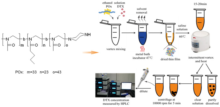
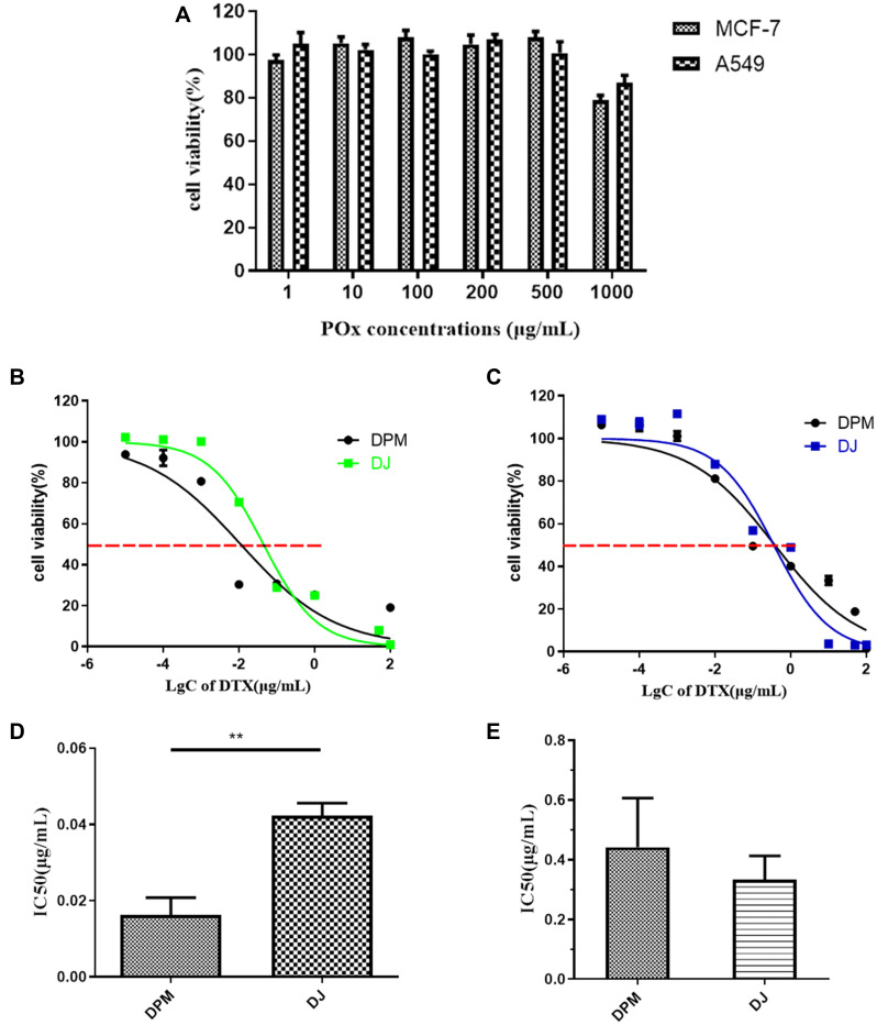
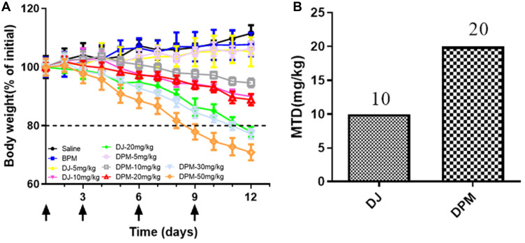
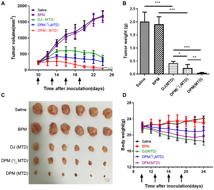
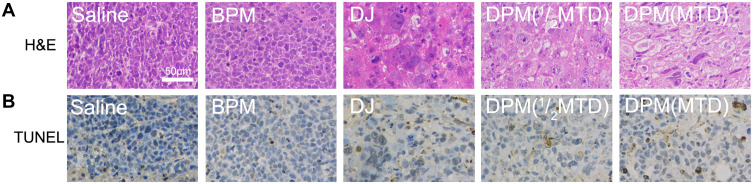
Methods and results: In the present study, we developed a docetaxel loaded poly-2-oxazoline polymer micellar system which employed poly-2-butyl-2 oxazoline and poly-2-methyl-2 oxazoline as the hydrophobic chain and hydrophilic chain, respectively. This micellar system achieves a high load up to 25% against the docetaxel, and further demonstrates an IC50 as low as 40% of the commercialized docetaxel injection in vitro and a double maximum tolerated dose in MCF-7 cells in vivo.

Conclusion: The high drug loading content, superior safety, and considerable anti-cancer activity make this newly developed docetaxel loaded poly(2-oxazoline) micelle go further in future clinical research.

Keywords: anti-cancer activity; docetaxel; high loading; micelle; poly(2-oxazoline)s.


Conflict of interest statement

The authors report no conflicts of interest in this work.

Figures

Figure 1
Figure 1
Chemical structure of amphiphilic poly(2-oxazoline)s (POx) triblock copolymers and the standard operating procedure for small scale micelle production (1–5mg) (blue: ethanol solution, yellow: DI water solution, black: undissolved drug).
Figure 2
Figure 2
Stability of the DTX loaded micelles determined by the size (A) and PDI (B) measurements over time.
Figure 3
Figure 3
Drug release profiles of DTX from different formulations at 37°C in PBS buffer at pH 5.5 and pH 7.4. Data are expressed as mean ± SD (n= 3).
Figure 4
Figure 4
The cell uptake of C6-POx towards MCF-7 and A549 cells by laser scanning confocal microscopy. ((A) MCF-7 cells, treated for 2h; (B) MCF-7 cells, treated for 4h; (C) A549 cells, treated for 2h; (D) A549 cells, treated for 4h).
Figure 5
Figure 5
In vitro cytotoxicity evaluation. (A) Blank POx micelles, (B) DPM and DJ in MCF-7 cells, (C) DPM and DJ in A549 cells, (D) IC50 in MCF-7 cells, (E) IC50 in A549 cells. Data are represented as mean ± SD (n=3). **p < 0.01.
Figure 6
Figure 6
Cell apoptotic assay. MCF-7 cells (A and C) and A549 cells (B and D) were treated with different formulations for 48h and measured by flow cytometry using V-APC kit and PI staining. Data are represented as mean ± SD (n=3). **p<0.01, ***p<0.001.
Figure 7
Figure 7
The evaluation of the distribution of DiR loaded micelles in MCF-7 tumor-bearing nude mice after tail vein injection of the formulations. (A) Images taken at 2, 4, 6, 8, 12, 24h after administration of DiR and DiR loaded micelles, respectively. (B) Ex vivo fluorescence images of organs and tumors were collected at 24h post-injection of formulations. (C) Total fluorescence signals of the ex vivo organs and tumors uptake DiR by Imaging Software. Data are expressed as mean ± SD (n=3). ***p<0.001,**p<0.01, nsIndicates p>0.05.
Figure 8
Figure 8
MTD in tumor-bearing nude mice. (A) Mice body weight change (% of initial) after repeated administration of different DTX formulation. (B) The maximum tolerated dose of BPM and DJ. Data are expressed as mean ± SD (n=4).
Figure 9
Figure 9
Anti-tumor efficacy of DTX formulations in MCF-7 tumor-bearing nude mice. (A) Tumor growth curve of mice administrated every three days for four times via tail vein. (B) The weight of the excised tumors from all groups. **p<0.01 and *p<0.05. (C) The images of excised tumor, scale bar 1cm. (D) Changes in body weight of tumor-bearing nude mice in each group during anti-tumor efficacy study. Data are expressed as mean ± SD (n= 6). ***p<0.001, vs saline; Δp<0.001, vs DJ(MTD); p<0.01, vs DPM (1/2MTD).
Figure 10
Figure 10
H&E and TUNEL assay. (A) Histological examination of tumors by hematoxylin and eosin (H&E) staining. (B) TUNEL staining from mice treated with different formulations. Images were taken with 200 × magnification, scale bar 50 μm.

References

    1. Bray F, Ferlay J, Soerjomataram I, Siegel RL, Torre LA, Jemal A. Global cancer statistics 2018: GLOBOCAN estimates of incidence and mortality worldwide for 36 cancers in 185 countries. CA Cancer J Clin. 2018;68(6):394–424. doi:10.3322/caac.21492 - DOI - PubMed
    1. Jordan MA, Wilson L. Microtubules as a target for anticancer drugs. Nat Rev Cancer. 2004;4(4):253. doi:10.1038/nrc1317 - DOI - PubMed
    1. Ojima I, Lichtenthal B, Lee S, Wang C, Wang X. Taxane anticancer agents: a patent perspective. Expert Opin Ther Pat. 2016;26(1):1–20. doi:10.1517/13543776.2016.1111872 - DOI - PMC - PubMed
    1. Cragg GM, Kingston DG, Newman DJ. Anticancer Agents from Natural Products. CRC press; 2011.
    1. Tan Q, Liu X, Fu X, Li Q, Dou J, Zhai G. Current development in nanoformulations of docetaxel. Expert Opin Drug Deliv. 2012;9(8):975–990. doi:10.1517/17425247.2012.696606 - DOI - PubMed

MeSH terms