

D-Chiro-Inositol Regulates Insulin Signaling in Human Adipocytes
- PMID: 33859622
- PMCID: PMC8042392
- DOI: 10.3389/fendo.2021.660815
D-Chiro-Inositol Regulates Insulin Signaling in Human Adipocytes
Abstract
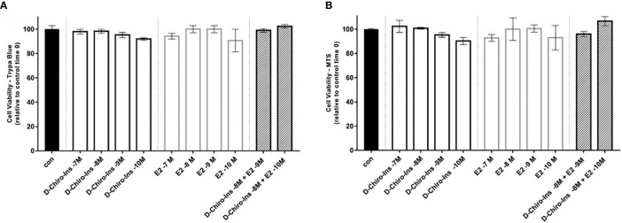
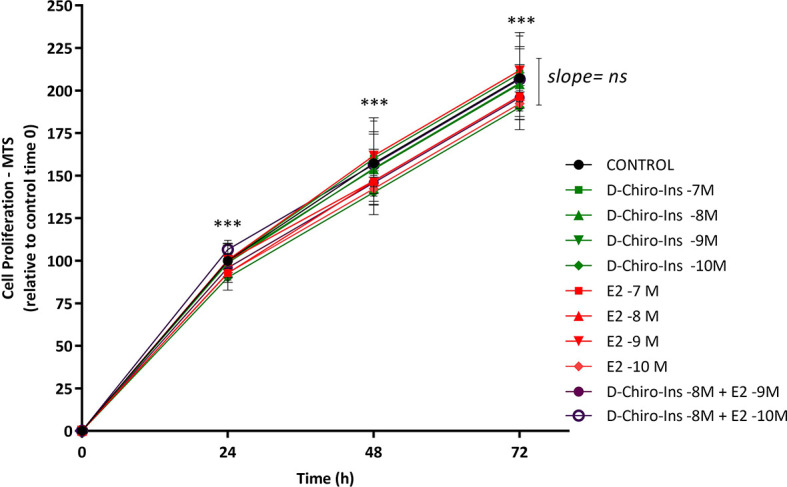
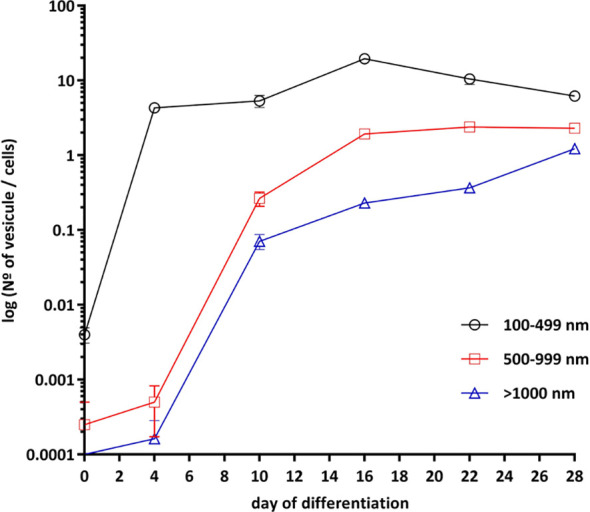
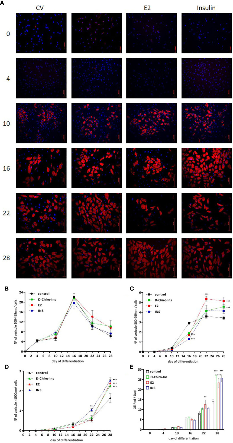
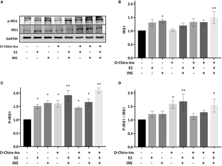
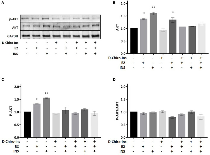
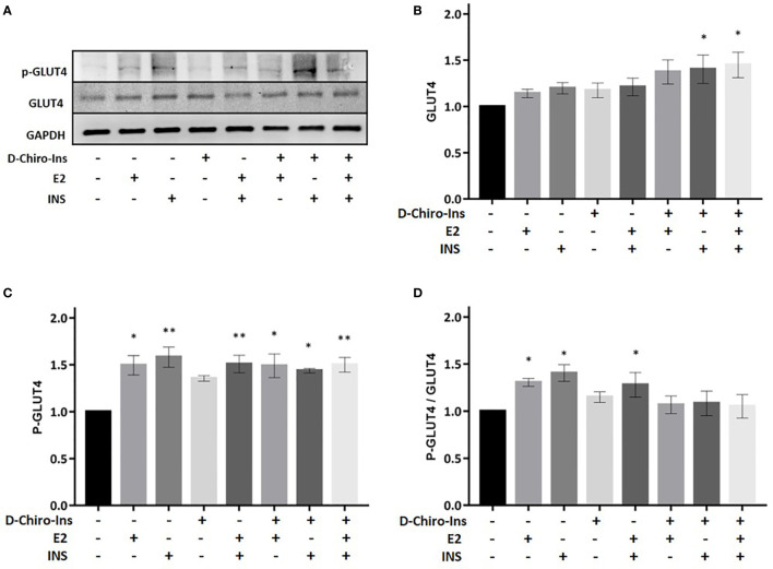
D-Chiro-Inositol (D-Chiro-Ins) is a secondary messenger in the insulin signaling pathway. D-Chiro-Ins modulates insulin secretion, the mitochondrial respiratory chain, and glycogen storage. Due to these actions D-Chiro-Ins has been proposed to correct defective insulin function in a variety of conditions characterized by metabolic dysfunction, such as polycystic ovary syndrome (PCOS), obesity, gestational diabetes and fat accumulation at menopause. Since it is unclear whether D-Chiro-Ins directly acts on adipocytes, we aimed to study D-Chiro-Ins's actions on adipocyte viability, proliferation, differentiation, and insulin-related protein expression using a human adipocyte cell line derived from Simpson-Golabi-Behmel Syndrome (SGBS) which fully differentiates to mature adipocytes. Throughout differentiation, cells were treated with D-Chiro-Ins, 17β-estradiol (E2) or Insulin. Cell viability and proliferation were not affected by D-Chiro-Ins, then D-Chiro-Ins promoted cell differentiation only during the final days of the process, while E2 enhanced it from the first phases. D-Chiro-Ins stimulated lipid storage and the production of big lipid droplets, thus reducing the content of free fatty acids. We also found that D-Chiro-Ins, either alone or in combination with insulin and E2 increased the expression and activation of insulin receptor substrate-1 (IRS1) and glucose transporter type 4 (GLUT4). In conclusion, this work shows that D-Chiro-Ins plays a direct role in the differentiation and in the function of human adipocytes, where it synergizes with insulin and estrogen through the recruitment of signal transduction pathways involved in lipid and glucose storage. These findings give clear insights to better understand the actions of D-Chiro-Ins on fat metabolism in women in physiology and in a variety of diseases.
Keywords: 17β-estradiol (E2); D-Chiro-Inositol (D-Chiro-Ins); Simpson–Golabi–Behmel syndrome cells (SGBS); insulin pathway; insulin receptor substrate (IRS1).
Copyright © 2021 Montt-Guevara, Finiguerra, Marzi, Fidecicchi, Ferrari, Genazzani and Simoncini.
Conflict of interest statement
The authors declare that the research was conducted in the absence of any commercial or financial relationships that could be construed as a potential conflict of interest.
Figures
Similar articles
-
Phytic acid and myo-inositol support adipocyte differentiation and improve insulin sensitivity in 3T3-L1 cells.Nutr Res. 2014 Aug;34(8):723-31. doi: 10.1016/j.nutres.2014.07.015. Epub 2014 Jul 30. Nutr Res. 2014. PMID: 25174657
-
Inositols: From Established Knowledge to Novel Approaches.Int J Mol Sci. 2021 Sep 30;22(19):10575. doi: 10.3390/ijms221910575. Int J Mol Sci. 2021. PMID: 34638926 Free PMC article. Review.
-
Interferon gamma attenuates insulin signaling, lipid storage, and differentiation in human adipocytes via activation of the JAK/STAT pathway.J Biol Chem. 2009 Nov 13;284(46):31936-44. doi: 10.1074/jbc.M109.061655. Epub 2009 Sep 23. J Biol Chem. 2009. PMID: 19776010 Free PMC article.
-
Hypoglycemic effect of D-chiro-inositol in type 2 diabetes mellitus rats through the PI3K/Akt signaling pathway.Mol Cell Endocrinol. 2016 Sep 15;433:26-34. doi: 10.1016/j.mce.2016.05.013. Epub 2016 May 20. Mol Cell Endocrinol. 2016. PMID: 27212205
-
The Role of Inositols in the Hyperandrogenic Phenotypes of PCOS: A Re-Reading of Larner's Results.Int J Mol Sci. 2023 Mar 27;24(7):6296. doi: 10.3390/ijms24076296. Int J Mol Sci. 2023. PMID: 37047265 Free PMC article. Review.
Cited by
-
Novel Chemical and Biological Insights of Inositol Derivatives in Mediterranean Plants.Molecules. 2022 Feb 24;27(5):1525. doi: 10.3390/molecules27051525. Molecules. 2022. PMID: 35268625 Free PMC article. Review.
-
D-Chiro-Inositol and Myo-Inositol Induce WAT/BAT Trans-Differentiation in Two Different Human Adipocyte Models (SGBS and LiSa-2).Int J Mol Sci. 2023 Apr 18;24(8):7421. doi: 10.3390/ijms24087421. Int J Mol Sci. 2023. PMID: 37108582 Free PMC article.
-
Preventing Adipogenesis and Preserving Mitochondria and GLUT-4 Functions by Extracts and Isolated Compounds of Australian Acacia saligna.Molecules. 2023 Sep 18;28(18):6677. doi: 10.3390/molecules28186677. Molecules. 2023. PMID: 37764453 Free PMC article.
-
New Insights into the Activities of D-Chiro-Inositol: A Narrative Review.Biomedicines. 2021 Oct 2;9(10):1378. doi: 10.3390/biomedicines9101378. Biomedicines. 2021. PMID: 34680494 Free PMC article. Review.
-
Effectiveness of a Food Supplement Based on Glucomannan, D-Chiro-Inositol, Cinnamomum zeylanicum Blume and Inulin in Patients with Metabolic Syndrome.Nutrients. 2024 Jan 12;16(2):249. doi: 10.3390/nu16020249. Nutrients. 2024. PMID: 38257142 Free PMC article. Clinical Trial.
References
Publication types
MeSH terms
Substances
LinkOut - more resources
Full Text Sources
Other Literature Sources
Medical
Miscellaneous