

Gyogamdan, a Traditional Medicine Prescription, Ameliorated Dermal Inflammation and Hyperactive Behavior in an Atopic Dermatitis Mouse Model Exposed to Psychological Stress
- PMID: 33859711
- PMCID: PMC8026289
- DOI: 10.1155/2021/6687513
Gyogamdan, a Traditional Medicine Prescription, Ameliorated Dermal Inflammation and Hyperactive Behavior in an Atopic Dermatitis Mouse Model Exposed to Psychological Stress
Abstract
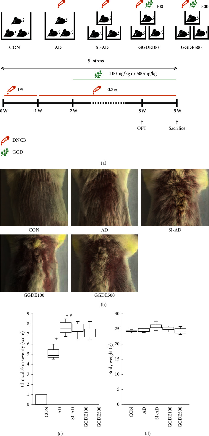
Psychological stress (PS) plays a significant role as an aggravating factor in atopic dermatitis (AD). The traditional medicine prescription, Gyogamdan, has been used to treat chest discomfort and mood disorders caused by PS. This study investigated the effects of an ethanolic extract of Gyogamdan (GGDE) on stress-associated AD models and the underlying mechanisms. 2,4-Dinitrochlorobenzene- (DNCB-) treated BALB/c mice were exposed to social isolation (SI) stress. The effects of orally administered GGDE (100 or 500 mg/kg) were evaluated by ELISA, western blotting, and an open field test (OFT). SI stress exaggerated the skin inflammation and induced locomotor hyperactivity in the AD mouse model. GGDE reduced the levels of IgE, TNF-α, IL-13, eotaxin, and VEGF and mast cell/eosinophil infiltration and prevented the decreases in the levels of involucrin and loricrin in the skin. GGDE also suppressed the SI-induced increases in corticotropin-releasing hormone (CRH), adrenocorticotropic hormone (ACTH), and corticosterone (CORT) in socially isolated AD mice. Furthermore, GGDE reduced traveling distances and mean speed significantly in the OFT. The in vitro experiments were performed using HaCaT, HMC-1, PC12, and BV2 cells. In the TNF-α/IFN-γ- (TI-) stimulated HaCaT cells, GGDE decreased the thymus and activation-regulated chemokine (TARC) and macrophage-derived chemokine (MDC) production significantly by inhibiting p-STAT1 and NF-κB signaling. GGDE also reduced VEGF production in HMC-1 cells stimulated with CRH/substance P (SP) by inhibiting p-ERK signaling pathway. GGDE increased the cell viability significantly and suppressed apoptosis in CORT-stimulated PC12 cells. Moreover, GGDE suppressed the LPS-induced production of NO, TNF-α, IL-1β, and IL-6 in BV2 cells. These results suggest that GGDE might be useful in patients with AD, which is exacerbated by PS.
Copyright © 2021 Ly Thi Huong Nguyen et al.
Conflict of interest statement
The authors have no conflicts of interest to declare.
Figures


Similar articles
-
Effect of Sopoongsan on Skin Inflammation and Hyperlocomotion in Socially Isolated Mice with Atopic Dermatitis.Evid Based Complement Alternat Med. 2022 Sep 16;2022:3323201. doi: 10.1155/2022/3323201. eCollection 2022. Evid Based Complement Alternat Med. 2022. PMID: 36159556 Free PMC article.
-
Inhibitory effect of 5,6-dihydroergosteol-glucoside on atopic dermatitis-like skin lesions via suppression of NF-κB and STAT activation.J Dermatol Sci. 2015 Sep;79(3):252-61. doi: 10.1016/j.jdermsci.2015.06.005. Epub 2015 Jun 15. J Dermatol Sci. 2015. PMID: 26100037
-
A Natural Compound Mixture Containing Arctigenin, Hederagenin, and Baicalein Alleviates Atopic Dermatitis in Mice by Regulating HPA Axis and Immune Activity.Evid Based Complement Alternat Med. 2020 Jun 29;2020:1970349. doi: 10.1155/2020/1970349. eCollection 2020. Evid Based Complement Alternat Med. 2020. PMID: 32714398 Free PMC article.
-
Cultivated ginseng inhibits 2,4-dinitrochlorobenzene-induced atopic dermatitis-like skin lesions in NC/Nga mice and TNF-α/IFN-γ-induced TARC activation in HaCaT cells.Food Chem Toxicol. 2013 Jun;56:195-203. doi: 10.1016/j.fct.2013.02.037. Epub 2013 Feb 27. Food Chem Toxicol. 2013. PMID: 23454147
-
Seungmagalgeun-Tang, a Traditional Herbal Formula, Alleviates Skin Inflammation and Depression-Like Behavior in Atopic Dermatitis Mice under Sleep Deprivation Conditions.Evid Based Complement Alternat Med. 2022 Mar 23;2022:1307173. doi: 10.1155/2022/1307173. eCollection 2022. Evid Based Complement Alternat Med. 2022. PMID: 35368752 Free PMC article.
Cited by
-
Effect of Sopoongsan on Skin Inflammation and Hyperlocomotion in Socially Isolated Mice with Atopic Dermatitis.Evid Based Complement Alternat Med. 2022 Sep 16;2022:3323201. doi: 10.1155/2022/3323201. eCollection 2022. Evid Based Complement Alternat Med. 2022. PMID: 36159556 Free PMC article.
References
-
- Sur B., Lee B., Yoon Y. S., et al. Extract of polygala tenuifolia alleviates stress-exacerbated atopy-like skin dermatitis through the modulation of protein kinase A and p38 mitogen-activated protein kinase signaling pathway. International Journal of Molecular Sciences. 2017;18(1) doi: 10.3390/ijms18010190. - DOI - PMC - PubMed
LinkOut - more resources
Full Text Sources
Other Literature Sources
Research Materials
Miscellaneous
