. 2018;14(7):658-669.
doi: 10.1038/s41567-018-0186-9. Epub 2018 Jul 4.

Physical biology of the cancer cell glycocalyx

Affiliations

Physical biology of the cancer cell glycocalyx

Joe Chin-Hun Kuo et al. Nat Phys. 2018.

Abstract

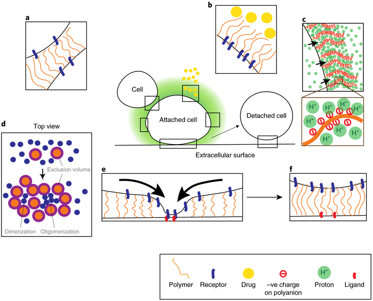
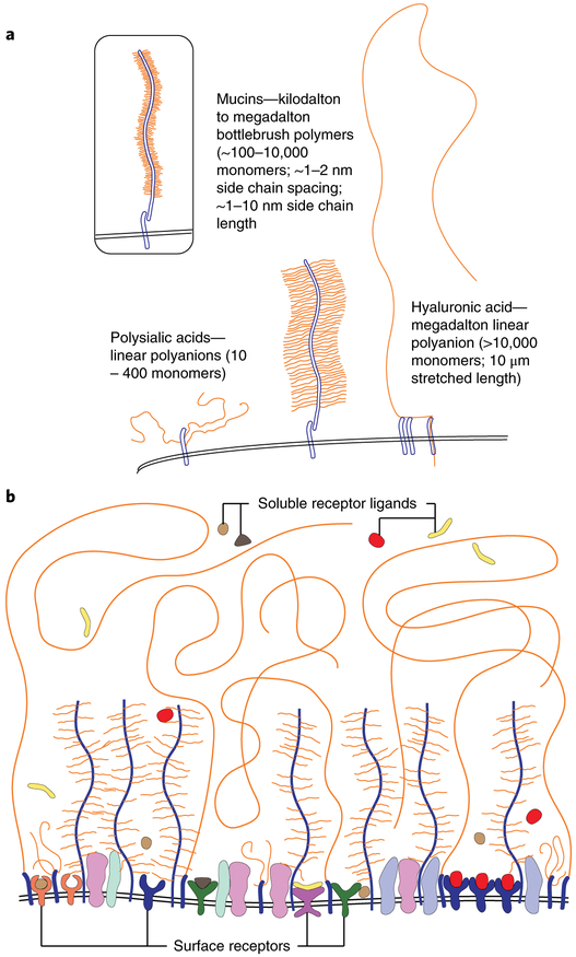
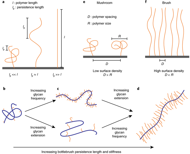
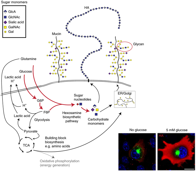
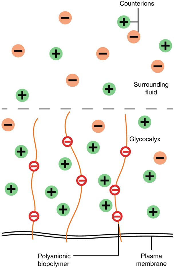
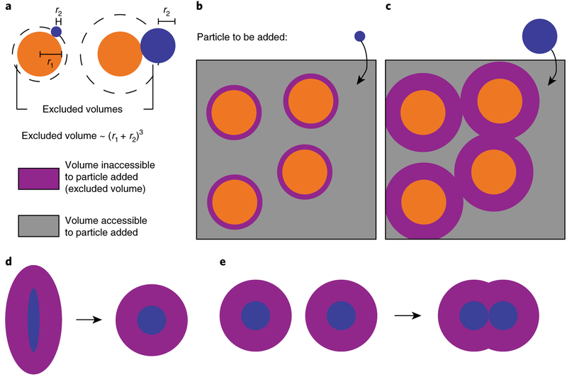
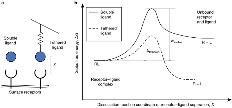
The glycocalyx coating the outside of most cells is a polymer meshwork comprising proteins and complex sugar chains called glycans. From a physical perspective, the glycocalyx has long been considered a simple 'slime' that protects cells from mechanical disruption or against pathogen interactions, but the great complexity of the structure argues for the evolution of more advanced functionality: the glycocalyx serves as the complex physical environment within which cell-surface receptors reside and operate. Recent studies have demonstrated that the glycocalyx can exert thermodynamic and kinetic control over cell signalling by serving as the local medium within which receptors diffuse, assemble and function. The composition and structure of the glycocalyx change markedly with changes in cell state, including transformation. Notably, cancer-specific changes fuel the synthesis of monomeric building blocks and machinery for production of long-chain polymers that alter the physical and chemical structure of the glycocalyx. In this Review, we discuss these changes and their physical consequences on receptor function and emergent cell behaviours.


Conflict of interest statement

Competing interests The authors declare no competing interests

Figures

Fig. 1
Fig. 1. Overview of large polymers on the cancer cell surface.
Cell surface receptors (blue) are communication devices that direct cancer cell behaviour. These receptors are usually of the order of 20–30 nm and are towered over by glycocalyx polymers (orange). a,b, The presence of large polymers can sterically block the interaction between receptors and their ligands to prevent cell-cell contacts (a) and impart barrier function (b) to block drug molecules (yellow). c, By charge attraction, the presence of polyanions may concentrate protons on the cell surface to decrease local pH. d, Top view of the cell surface. Glycocalyx polymers can occupy large volumes that exclude receptors as they avoid molecular overlaps (purple). This energetically favours the close-packing (oligomerization) of small receptors to reduce the excluded volume. e, A moderately dense glycocalyx can promote the clustering of cell surface receptors and enhance cell adhesion. The black arrows indicate the clustering of receptors towards and into adhesion sites. f, On the other hand, an overly dense glycocalyx can increase the spacing between receptors and their ligands. This can lead to cell detachment.
Fig. 2
Fig. 2. Glycocalyx architecture and communication interface on the cell surface.
a, Bottlebrush mucin polymers and polyanions such as HA and polysialic acid are common structures in the glycocalyx. In cancer, mucin polymers become decorated with a higher density of short glycan side chains on the polypeptide backbone (insert). b, The glycocalyx as a structural polymer brush and communication interface on the cell surface. A structural network composed of glycopolymers including mucins, HA and polysialic acid acts as a physically crowded layer shielding cell surface molecules. Large polymers are densely arrayed in the cancer cell glycocalyx, creating a crowded network in which embedded cell-surface receptors must operate. The glycocalyx may be ideally positioned to physically mediate or attenuate receptor function on a global level.
Fig. 3
Fig. 3. Physical aspects of polymers.
a, The persistence length, lp, of a polymer is the length scale over which the molecule does not bend. The persistence length is directly proportional to polymer bending stiffness. A flexible polymer with lpl adopts a coiled-up shape without any external forces. On the other hand, a polymer with lpl does not bend in the absence of external effects and thus is rod-like and stiff. Polymers with comparable length scales lpl fall in between, possessing intermediate stiffness and adopting partially deformed configurations. bd, Bottlebrush polymers are composed of side chains attached to a polymer backbone, and their structures are controlled by the density and length of the attached side chains. Bottlebrush polymers with short and infrequent side chains adopt flexible structures (b). Moderate increase in glycan frequency or glycan extension stretches out the bottlebrushes partially and enhances the persistence length (c). Heavily glycosylated bottlebrush polymers with high glycan frequency and extension are extended into stiff rod-like structures with a large persistence length (d). e,f, Polymers attached to a substrate with a characteristic polymer size, R, and a spacing between polymers of D exhibit two regimes of physical behaviour. Mushroom regime: at low densities such that D > R, polymers do not sense neighbouring molecules (e). Brush regime: at high surface concentrations, polymers experience steric repulsion with neighbours and, to reduce crowding, they extend out into a brush-like structure (f).
Fig. 4
Fig. 4. Cancer cell metabolism is rewired to produce large polymers in the glycocalyx.
Normal cells use nutrients such as glucose to run oxidative phosphorylation that efficiently produces energy (dotted grey line). In cancer cells, these nutrients are instead diverted into the hexosamine biosynthetic pathway to produce sugar nucleotide monomers and enhance the production of glycocalyx polymers such as mucins and HA. Cancer cells also produce large amounts of lactic acid and H+ that must be pumped out of the cell to avoid toxicity. Supplementing glucose can greatly increase the amount of HA (stained red in the inset) on the cell surface (fluorescence images reproduced from ref. , American Society for Biochemistry and Molecular Biology). ER, endoplasmic reticulum; TCA, tricarboxylic acid.
Fig. 5
Fig. 5. Donnan equilibrium between the glycocalyx and surrounding fluid.
Acting as a semi-permeable polyelectrolyte network, the glycocalyx attracts and traps cations from the surrounding fluid. This increases the osmotic pressure or concentration of ions in the glycocalyx and reduces the local pH.
Fig. 6
Fig. 6. Excluded volumes depict crowding in the glycocalyx.
a, Molecules have excluded volumes where the centre of another molecule cannot exist. The excluded volume of two spheres of size r1 and r2 is proportional to (r1 + r2)3. b,c, Addition of a particle (blue) to a box filled with other particles (orange) depicts the implications of excluded volume (purple) for molecular transport, as expected for diffusion of soluble ligands through the glycocalyx. A small particle encounters small excluded volumes, enjoying access to considerable space and several positional states, and thus requires less energy to diffuse to the cell surface (b). On the other hand, a large particle finds it difficult to enter and diffuse through a crowded environment owing to large excluded volumes and consequently requires higher energy for transport (c). d,e, A crowded environment energetically favours changes that decrease the total excluded volume of molecules, such as conformational shape changes in a protein (d) or the clustering and association of proteins (e) on the cell membrane.
Fig. 7
Fig. 7. Tethered ligand binding.
a, Whereas soluble ligands can bind receptors relatively freely, ligands tethered to stiff molecules and substrates in the cellular environment can be considerably impeded by a spring-like resistance from the tether. b, Typical energy curves for the dissociation of a receptor–ligand (RL) complex for soluble and tethered ligands, indicating the effects of the tether. Soluble ligands prefer binding receptors and encounter large activation energies for dissociation. Tethering the ligand tilts the energy landscape because of the force pulling the ligand. Tethered ligands thus encounter smaller activation energies and are more likely to dissociate from receptors.

References

    1. Sever R & Brugge JS Signal transduction in cancer. Cold Spring Harb. Perspect. Med 5, a006098 (2015). - PMC - PubMed
    1. Multhaupt HAB, Leitinger B, Gullberg D & Couchman JR Extracellular matrix component signaling in cancer. Adv. Drug Deliv. Rev 97, 28–40 (2016). - PubMed
    1. Casaletto JB & McClatchey AI Spatial regulation of receptor tyrosine kinases in development and cancer. Nat. Rev. Cancer 12, 387–400 (2012). - PMC - PubMed
    1. Twomey JD, Brahme NN & Zhang B Drug-biomarker co-development in oncology—20 years and counting. Drug Resist. Updat 30, 48–62 (2017). - PubMed
    1. Swain SM et al. Pertuzumab, trastuzumab, and docetaxel in HER2-positive metastatic breast cancer. N. Engl. J. Med 372, 724–734 (2015). - PMC - PubMed