. 2021 Mar 29:2021:6678276.
doi: 10.1155/2021/6678276. eCollection 2021.

Photobiomodulation Therapy Ameliorates Glutamatergic Dysfunction in Mice with Chronic Unpredictable Mild Stress-Induced Depression

Affiliations

Photobiomodulation Therapy Ameliorates Glutamatergic Dysfunction in Mice with Chronic Unpredictable Mild Stress-Induced Depression

Di Zhang et al. Oxid Med Cell Longev. .

Abstract

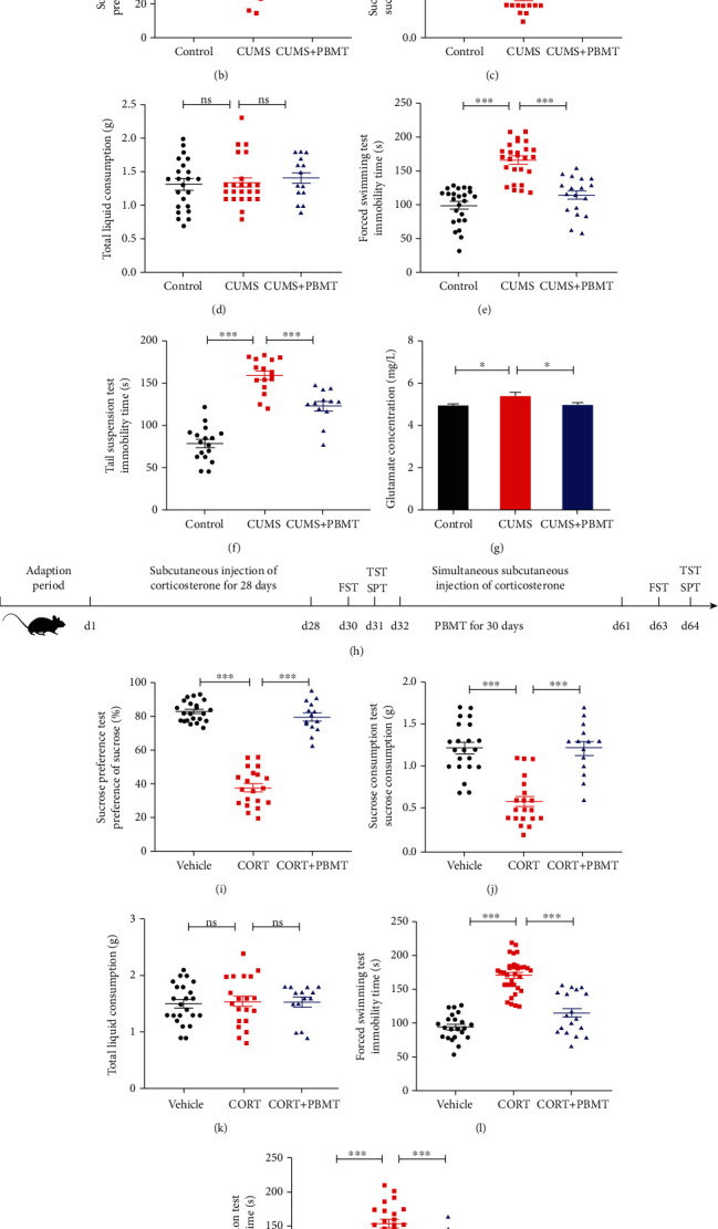
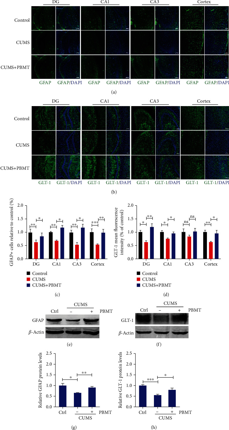
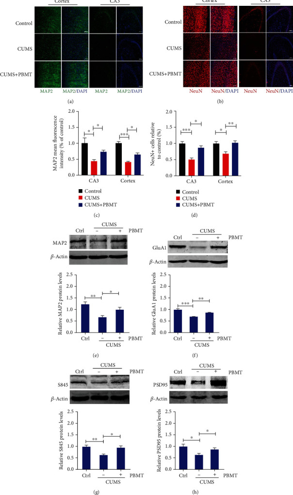
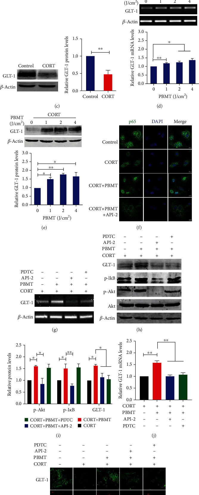
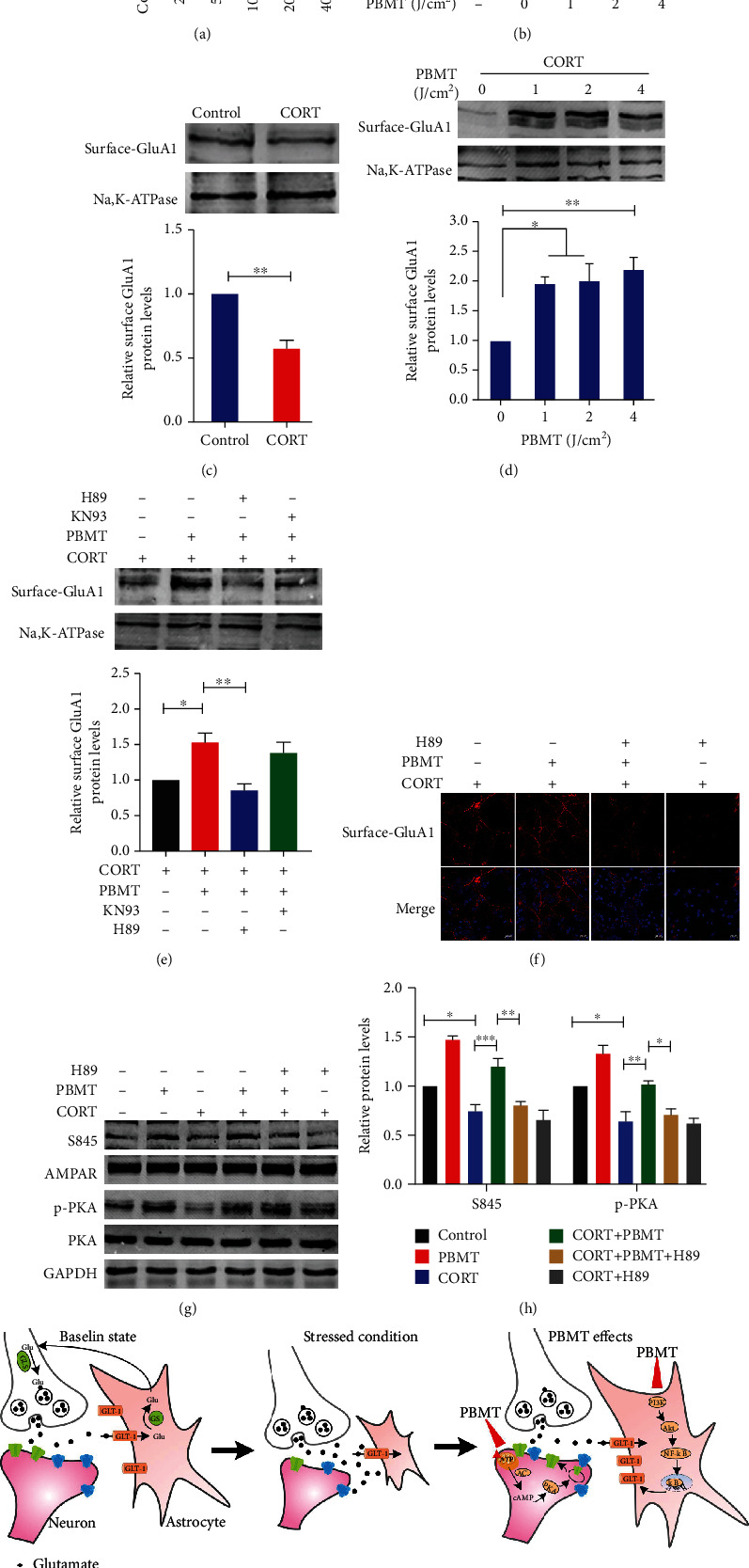
Accumulating evidence indicates that dysfunction of the glutamatergic neurotransmission has been widely involved in the pathophysiology and treatment of depression. Photobiomodulation therapy (PBMT) has been demonstrated to regulate neuronal function both in vitro and in vivo. Herein, we aim to investigate whether the antidepressant phenotype of PBMT is associated with the improvement of glutamatergic dysfunction and to explore the mechanisms involved. Results showed that PBMT decreased extracellular glutamate levels via upregulation of glutamate transporter-1 (GLT-1) and rescued astrocyte loss in the cerebral cortex and hippocampus, which also alleviated dendritic atrophy and upregulated the expression of AMPA receptors on the postsynaptic membrane, ultimately exhibiting behaviorally significant antidepressant effects in mice exposed to chronic unpredictable mild stress (CUMS). Notably, PBMT also obtained similar antidepressant effects in a depressive mouse model subcutaneously injected with corticosterone (CORT). Evidence from in vitro mechanistic experiments demonstrated that PBMT treatment significantly increased both the GLT-1 mRNA and protein levels via the Akt/NF-κB signaling pathway. NF-κB-regulated transcription was in an Akt-dependent manner, while inhibition of Akt attenuated the DNA-binding efficiency of NF-κB to the GLT-1 promoter. Importantly, in vitro, we further found that PKA activation was responsible for phosphorylation and surface levels of AMPA receptors induced by PBMT, which is likely to rescue excitatory synaptic transmission. Taken together, our research suggests that PBMT as a feasible therapeutic approach has great potential value to control the progression of depression.


Conflict of interest statement

The authors declare that they have no known competing financial interests or personal relationships that could have appeared to influence the work reported in this paper.

Figures

Figure 1
Figure 1
Antidepressant effects of PBMT in CUMS- and CORT-induced mouse models of depression. (a) The experimental design of CUMS, treatment schedule, and behavioral tests. (b–d) PBMT significantly attenuated the decreased sucrose preference induced by CUMS but did not affect total fluid consumption. (e, f) PBMT significantly attenuated the increased immobility time of FST and TST induced by CUMS. (g) Glutamate concentration in CUMS-exposed mice was significantly higher than that in control mice, and PBMT could restore glutamate levels to normal (n = 6 per group). (h) The experimental design of CORT, treatment schedule, and behavioral tests. (i–k) PBMT significantly attenuated the decreased sucrose preference induced by CORT but did not affect total fluid consumption. (l, m) PBMT significantly attenuated the increased immobility time of FST and TST induced by CORT. Number of mice used in behavior tests: control/vehicle, n = 23 mice; CUMS, n = 23 mice; CUMS+PBMT, n = 14 mice; CORT, n = 20 mice; and CORT+PBMT, n = 14 mice. Data represent mean ± SEM. ns: not significant. p < 0.05, ∗∗p < 0.01, and ∗∗∗p < 0.001, one-way ANOVA with Tukey's post hoc analysis. CUMS: chronic unpredictable mild stress; CORT: mice treated with corticosterone at a dose of 20 mg/kg for 28 days; PBMT: photobiomodulation therapy; SPT: sucrose preference test; FST: forced swimming test; TST: tail suspension test.
Figure 2
Figure 2
Effects of PBMT on the loss of astrocytes and expression of GLT-1 in the cortex and hippocampus of depressed mice. (a) Representative immunofluorescent images of GFAP in the hippocampal and cortex regions of each group. Nuclei were counterstained with DAPI (blue). Scale bar: 100 μm (n = 6 per group). (b) Representative immunofluorescent images of GLT-1 in the hippocampal and cortex regions of each group. Scale bar: 100 μm (n = 6 per group). (c) Quantification analysis of GFAP-positive cells in the hippocampal and cortex regions of different groups. (d) Quantification analysis of GLT-1 in the hippocampal and cortex regions of different groups. (e–h) GFAP and GLT-1 expression detected by western blot in the cortex and hippocampus lysates from mice exposed to CUMS with or without PBMT (n = 9 mice for each group). Data represent mean ± SEM. ns: not significant. p < 0.05, ∗∗p < 0.01, and ∗∗∗p < 0.001, one-way ANOVA with Tukey's post hoc analysis. DAPI: 4′,6-diamidino-2-phenylindole; GFAP: glial fibrillary acidic protein; GLT-1: glutamate transporter-1.
Figure 3
Figure 3
Effects of PBMT on dendritic atrophy and expression of GluA1 in the cortex and hippocampus of depressed mice. (a) Typical staining of MAP2 (green) in the cortex and hippocampal regions from CUMS mice with or without PBMT and the control group. Nuclei were counterstained with DAPI (blue). Scale bar, 100 μm (n = 6 per group). (b) Representative immunofluorescent image analysis of NeuN-positive cells (red) in the hippocampal and cortex regions of each group; scale bar represents 100 μm. (c) Quantification of MAP2 density in the hippocampal and cortex regions of different groups. (d) Quantitative analyses of the NeuN-positive cells in the hippocampal and cortex regions of different groups. (e) Western blot and quantification analysis of MAP2 from control vs. chronic stressed mice without or with PBMT treatment (n = 6-8 per group). (f) Western blot and quantification analysis of GluA1 from control vs. chronic stressed mice without or with PBMT treatment. (g) Representative western blot and quantification analysis of S845 in chronic stressed mice under the treatment with or without PBMT. (h) Western blot and quantification analysis of PSD95 from control vs. chronic stressed mice without or with PBMT treatment. All the data represent mean ± SEM. p < 0.05, ∗∗p < 0.01, and ∗∗∗p < 0.001, one-way ANOVA with Tukey's post hoc analysis. MAP2: microtubule-associated protein 2; PSD95: postsynaptic density 95.
Figure 4
Figure 4
PBMT upregulates GLT-1 expression by activating the PI3K/Akt/NF-κB signaling pathway in CORT-treated primary astrocytes. (a) CORT induced reduced viability of primary cultured astrocytes in dose-dependent manners as measured using the CCK-8 assay. The ordinate represents the percentage of cell survival compared with controls as measured by the CCK-8 assay. (b) Primary astrocytes were exposed to 200 μΜ corticosterone followed by irradiation with PBMT at 1 J/cm2, 2 J/cm2, or 4 J/cm2, respectively. Cell viability was assessed by the CCK-8 assay after 24 h. (c) Western blot and quantification analysis of GLT-1 expression in 200 μΜ CORT-treated primary astrocytes. (d, e) Representative PCR and western blot and quantification analysis that PBMT increases GLT-1 mRNA and protein levels in a dose-dependent manner. (f) Representative immunofluorescent images of p65 (green) in primary astrocytes under the indicated treatments. Staining with DAPI (blue) to visualize the nucleus. Scale bar: 10 μm. (g, j) GLT-1 mRNA levels were detected by PCR stimulated with CORT and/or PBMT in the preference of API-2 (6 μΜ) and PDTC (8 μΜ) in primary astrocytes. (h, i) Representative western blot and quantification analysis of GLT-1 stimulated with CORT and/or PBMT in the preference of API-2 (6 μΜ) and PDTC (8 μΜ) in primary astrocytes. (k) Representative immunofluorescent images of GLT-1 (green) in astrocytes (red) under the indicated treatments. Staining with DAPI (blue) to visualize the nucleus. Scale bar: 20 μm. All the data represent mean ± SEM. p < 0.05, ∗∗p < 0.01, and ∗∗∗p < 0.001. Significant differences were analyzed by the two-sided unpaired Student's t-test for two-group comparisons and one-way ANOVA followed by Tukey's post hoc test for multiple comparisons. CORT: primary astrocytes treated with corticosterone.
Figure 5
Figure 5
PBMT promotes AMPA receptor insertion through activation of PKA in CORT-treated primary neurons. (a) CORT induced reduced viability of primary cultured neurons in dose-dependent manners as measured using the CCK-8 assay. The ordinate represents the percentage of cell survival compared with controls. (b) Primary neurons were exposed to 100 μΜ CORT followed by irradiation with PBMT at 1 J/cm2, 2 J/cm2, or 4 J/cm2, respectively. Cell viability was assessed by the CCK-8 assay after 24 h. (c) Representative western blot and quantification analysis of surface levels of AMPA receptor subunit GluA1 in 100 μΜ CORT-treated primary neurons. (d) Representative western blot and quantification analysis of the dose-dependent effect of PBMT on surface levels of AMPA receptor subunit GluA1 expression after 24 h. (e) Representative western blot and quantification analysis of surface GluA1 stimulated with CORT and/or PBMT in the preference of H89 (20 μΜ) and KN93 (10 μΜ) in primary neurons. (f) Representative immunofluorescent images of surface GluA1 (red) in neurons under the indicated treatments. Staining with DAPI (blue) to visualize the nucleus. Scale bar: 20 μm. (g, h) Representative western blot and quantification analysis of S845, PKA, and p-PKA stimulated with CORT and/or PBMT in the preference of H89 (20 μΜ) and KN93 (10 μΜ) in primary neurons. (i) Schematic representation of the signaling pathway for PBMT ameliorates glutamatergic dysfunction. All the data represent mean ± SEM. p < 0.05, ∗∗p < 0.01, and ∗∗∗p < 0.001. Significant differences were analyzed by the two-sided unpaired Student's t-test for two-group comparisons and one-way ANOVA followed by Tukey's post hoc test for multiple comparisons. CORT: primary neurons treated with corticosterone.

Similar articles

Cited by

References

    1. Moussavi S., Chatterji S., Verdes E., Tandon A., Patel V., Ustun B. Depression, chronic diseases, and decrements in health: results from the World Health Surveys. The Lancet. 2007;370(9590):851–858. doi: 10.1016/S0140-6736(07)61415-9. - DOI - PubMed
    1. GBD 2015 Disease and Injury Incidence and Prevalence Collaborators. Global, regional, and national incidence, prevalence, and years lived with disability for 310 diseases and injuries, 1990-2015: a systematic analysis for the Global Burden of Disease Study 2015. The Lancet. 2016;388(10053):1545–1602. doi: 10.1016/S0140-6736(16)31678-6. - DOI - PMC - PubMed
    1. Stimmel G. L., Dopheide J. A., Stahl S. M. Mirtazapine: an antidepressant with noradrenergic and specific serotonergic effects. Pharmacotherapy the Journal of Human Pharmacology & Drug Therapy. 1997;17(1):10–21. doi: 10.1002/j.1875-9114.1997.tb03674.x. - DOI - PubMed
    1. Wong E. H. F., Sonders M. S., Amara S. G., et al. Reboxetine: a pharmacologically potent, selective, and specific norepinephrine reuptake inhibitor. Biological Psychiatry. 2000;47(9):818–829. doi: 10.1016/s0006-3223(99)00291-7. - DOI - PubMed
    1. Rush A. J., Trivedi M. H., ., Wisniewski S. R., ., et al. Acute and longer-term outcomes in depressed outpatients requiring one or several treatment steps: a STAR∗D report. The American Journal of Psychiatry. 2006;163(11):1905–1917. doi: 10.1176/ajp.2006.163.11.1905. - DOI - PubMed

LinkOut - more resources