Review
. 2021 Mar 11;13(3):e13816.
doi: 10.7759/cureus.13816.

Tumor Lysis Syndrome: Introduction of a Cutaneous Variant and a New Classification System

Affiliations
Review

Tumor Lysis Syndrome: Introduction of a Cutaneous Variant and a New Classification System

Philip R Cohen et al. Cureus. .

Abstract

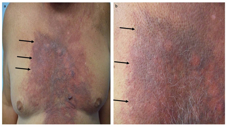
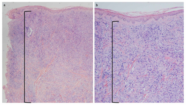
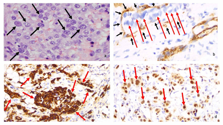
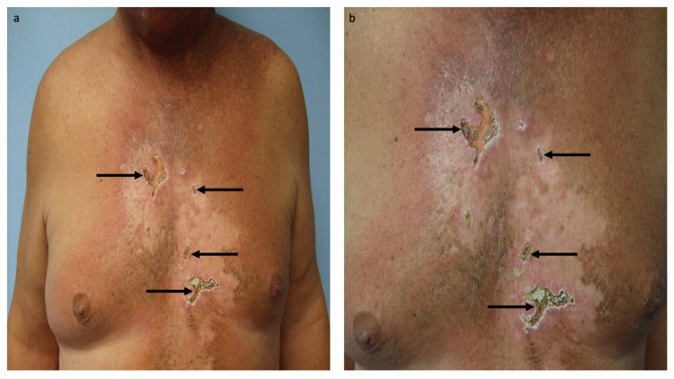
Tumor lysis syndrome, an oncological emergency, is characterized by laboratory parameters such as hyperuricemia, hyperkalemia, hyperphosphatemia, and hypocalcemia, as well as renal injury with an elevated creatinine. Tumor lysis syndrome is seen in patients with aggressive malignancies and high tumor burden. More frequently, it occurs in individuals with hematologic malignancies such as high-grade lymphomas (such as Burkitt lymphoma) and leukemia (such as acute lymphocytic leukemia). It also, albeit less commonly, can be seen in patients with widespread solid tumors that are rapidly proliferating and are markedly sensitivity to antineoplastic therapy. Tumor lysis syndrome is usually preceded by cancer-directed therapy; however, the syndrome can present spontaneously prior to the individual receiving malignancy-directed treatment. We reported a man with metastatic salivary duct carcinoma who had cutaneous metastases that presented as carcinoma hemorrhagiectoides. Microscopic examination demonstrated that the metastatic tumor cells had infiltrated and replaced the entire dermis. After the patient received his first dose of antineoplastic therapy, he had an excellent response and the cutaneous metastases developed into ulcers; we hypothesize that most of the dermis, which had been replaced by tumor cells, disappeared as a result of the therapeutic response, and the overlying epidermis became necrotic and shed, leaving an ulcer. His dramatic response to treatment prompted us to propose a new classification of tumor lysis syndrome, which should include the systemic form of the condition as well as the new variant: cutaneous tumor lysis syndrome. We anticipate that, with improvement in targeted therapies, there may be an increase in therapy-associated cutaneous tumor lysis syndrome.

Keywords: cancer; carcinoma; cutaneous; hematologic; lysis; malignancy; metastatic; solid; syndrome; tumor.


Conflict of interest statement

Funded in part by the Joan and Irwin Jacobs Fund and by the National Cancer Institute grant P30 CA016672 (RK).

Figures

Figure 1
Figure 1. Cutaneous metastatic salivary duct carcinoma-associated carcinoma hemorrhagiectoides presenting as the shield sign.
A 70-year-old man with metastatic salivary duct carcinoma appearing as shield sign-associated carcinoma hemorrhagiectoides presented 11 months earlier with a left preauricular parotid gland mass. Additional lesions developed in his left axilla and chest wall within two months. Both the parotid gland mass and left axillary lymph node showed salivary duct carcinoma. He was treated with five cycles of docetaxel followed by four weeks of chemoradiation (45 Gray in 20 fractions and weekly cetuximab). Two months after completing chemoradiation, he presented with progressive neoplastic disease not only involving lymph nodes of the mediastinum and both axillae but also cutaneous metastases. Distant (a) and closer (b) views of his cutaneous metastatic salivary duct carcinoma appeared as a hemorrhagic and erythematous confluent large violaceous and purpuric plaque (black arrows) that extended from his neck to his abdomen and across his chest, resembling a medieval knight’s shield. The images have not previously been published; however, the details of the patient’s skin lesions and clinical course have previously been described [3-5].
Figure 2
Figure 2. Microscopic examination of cutaneous metastatic salivary duct carcinoma.
Low (a) and higher (b) magnification views of the punch biopsy in the hemorrhagic area of the chest are shown. The depth of the biopsy was 10 mm and extended into the deep reticular dermis; the deep margin of the skin biopsy did not reach the subcutaneous fat. Microscopic examination of the tissue specimen shows a narrow zone of normal-appearing papillary dermis beneath the epidermis. Below this narrow zone, there is a confluent and dense infiltrate of metastatic carcinoma cells that are not only present in the dermal vessels but also extend to the base of the specimen in the deep reticular dermis (demonstrated by the area demarcated by the black open bracket (hematoxylin and eosin: a, ×20; b, ×40). The images have not previously been published; however, the details of the pathology of the patient’s skin lesions have previously been described [3-5].
Figure 3
Figure 3. Microscopic examination of cutaneous metastatic salivary duct carcinoma.
Microscopic examination at high magnification of the neoplastic salivary duct carcinoma cells shows large, irregular nuclei (black arrows) with visible nucleoli (a). Immunohistochemistry (brown staining) with anti-cluster of differentiation 31 (anti-CD31) highlights vascular endothelial cells (black arrows) containing neoplastic cells (red arrows), thereby demonstrating vascular invasion by the metastatic carcinoma (b). Anti-cytokeratin 7 (anti-CK7) and androgen receptor (both brown staining) label tumor cells (red arrows) (c and d, respectively) (a, hematoxylin and eosin, ×400; b, anti-CD31, diaminobenzidine immunoperoxidase and light hematoxylin, x 400; c, anti-CK7, diaminobenzidine immunoperoxidase and light hematoxylin, ×400; anti-androgen receptor, diaminobenzidine immunoperoxidase and light hematoxylin, ×400). The images have not previously been published; however, the details of the pathology of the patient’s skin lesions have previously been described [3-5].
Figure 4
Figure 4. Cutaneous tumor lysis syndrome following treatment with temsirolimus and bevacizumab.
After the patient had progressed on chemoradiation, he was treated with temsirolimus (at a dose of 25 mg intravenously on days one, eight, and 15) and bevacizumab (at a dose of 15 mg/kg on day one) on 21 day therapy cycles. Distant (a) and closer (b) views show therapy-associated cutaneous tumor lysis syndrome presenting as several areas of full thickness ulcers (black arrows) on his chest that he experienced after his first cycle of treatment; the other areas of his chest and abdomen also demonstrated the dramatic resolution of his cutaneous metastases. The images have not previously been published; however, the details of the patient’s skin lesions and clinical course have previously been described [3-5].

Similar articles

Cited by

References

    1. The tumor lysis syndrome. Howard SC, Jones DP, Pui CH. N Engl J Med. 2011;364:1844–1854. - PMC - PubMed
    1. Spontaneous tumour lysis syndrome. Kekre N, Djordjevic B, Touchie C. CMAJ. 2012;184:913–916. - PMC - PubMed
    1. Salivary duct carcinoma: targeting the phosphatidylinositol 3-kinase pathway by blocking mammalian target of rapamycin with temsirolimus. Piha-Paul SA, Cohen PR, Kurzrock R. J Clin Oncol. 2011;29:727–730. - PubMed
    1. The “shield sign” in two men with metastatic salivary duct carcinoma to the skin: cutaneous metastases presenting as carcinoma hemorrhagiectoides. Cohen PR, Prieto VG, Piha-Paul SA, Kurzrock R. https://www.ncbi.nlm.nih.gov/pubmed/23050032 J Clin Aesthet Dermatol. 2012;5:27–36. - PMC - PubMed
    1. Cutaneous metastatic cancer: carcinoma hemorrhagiectoides presenting as the shield sign. Cohen PR, Kurzrock R. Cureus. 2021;13:12627. - PMC - PubMed

LinkOut - more resources