

Magnetic Resonance Imaging Features in Different Types of Invasive Breast Cancer: A Systematic Review of the Literature
- PMID: 33859904
- PMCID: PMC8038870
- DOI: 10.7759/cureus.13854
Magnetic Resonance Imaging Features in Different Types of Invasive Breast Cancer: A Systematic Review of the Literature
Abstract
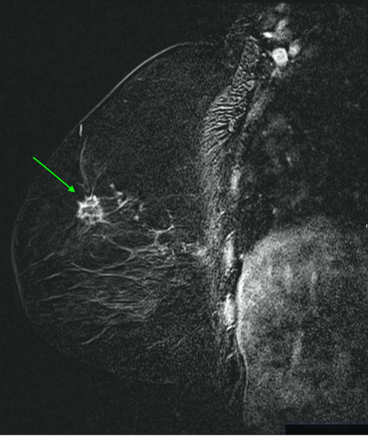
Breast cancer is the most common malignancy affecting women worldwide, and early diagnosis of breast cancer is the key to its successful and effective treatment. Traditional imaging techniques such as mammography and ultrasound are used to detect and configure breast abnormalities; unfortunately, these modalities have low sensitivity and specificity, particularly in young patients with dense breast tissue, breast implants, or post-surgical scar/architecture distortions. Therefore, breast magnetic resonance imaging (MRI) has been superior in the characterization and detection of breast cancer, especially that with invasive features. This review article explores the importance of breast MRI in the early detection of invasive breast cancer versus traditional tools, including mammography and ultrasound, while also analyzing the use of MRI as a screening tool for high-risk women. We will also discuss the different MRI features for invasive ductal carcinoma and lobular carcinoma and the role of breast MRI in the detection of ductal carcinoma in situ with a focus on the utilization of new techniques, including MR spectroscopy and diffusion-weighted imaging.
Keywords: breast cancer; dcis; idc; ilc; mammogram; mri breast; ultrasound.
Copyright © 2021, Alaref et al.
Conflict of interest statement
The authors have declared that no competing interests exist.
Figures


Similar articles
-
Diagnostic accuracy of mammography, clinical examination, US, and MR imaging in preoperative assessment of breast cancer.Radiology. 2004 Dec;233(3):830-49. doi: 10.1148/radiol.2333031484. Epub 2004 Oct 14. Radiology. 2004. PMID: 15486214
-
[Preoperative MRT of the breast in invasive lobular carcinoma in comparison with invasive ductal carcinoma].Rofo. 2004 Apr;176(4):544-9. doi: 10.1055/s-2004-813011. Rofo. 2004. PMID: 15088179 German.
-
Pathologic Tumor Size versus Mammography, Sonography, and MRI in Breast Cancer Based on Pathologic Subtypes.Am Surg. 2023 Aug;89(8):3652-3654. doi: 10.1177/00031348231174019. Epub 2023 May 4. Am Surg. 2023. PMID: 37140069
-
Magnetic resonance imaging in breast cancer: A literature review and future perspectives.World J Clin Oncol. 2014 May 10;5(2):61-70. doi: 10.5306/wjco.v5.i2.61. World J Clin Oncol. 2014. PMID: 24829852 Free PMC article. Review.
-
[New trends and novel possibilities in the diagnostic imaging of breast cancer].Magy Onkol. 2015 Mar;59(1):44-55. Epub 2014 Oct 13. Magy Onkol. 2015. PMID: 25763913 Review. Hungarian.
Cited by
-
Magnetic resonance imaging of the breast: Could it be used as a screening test?Saudi Med J. 2024 Aug;45(8):799-807. doi: 10.15537/smj.2024.45.8.20230748. Saudi Med J. 2024. PMID: 39074890 Free PMC article.
-
Diagnostic value of mammography combined with ultrasound shear wave elastography and magnetic resonance imaging in breast cancer.Oncol Lett. 2024 Dec 3;29(2):85. doi: 10.3892/ol.2024.14831. eCollection 2025 Feb. Oncol Lett. 2024. PMID: 39664614 Free PMC article.
-
Assessing Breast Cancer through Tumor Microenvironment Mapping of Collagen and Other Biomolecule Spectral Fingerprints─A Review.ACS Sens. 2024 Sep 27;9(9):4364-4379. doi: 10.1021/acssensors.4c00585. Epub 2024 Aug 22. ACS Sens. 2024. PMID: 39175278 Free PMC article. Review.
-
Differentiation of invasive ductal and lobular carcinoma of the breast using MRI radiomic features: a pilot study.F1000Res. 2024 Mar 14;13:91. doi: 10.12688/f1000research.146052.2. eCollection 2024. F1000Res. 2024. PMID: 38571894 Free PMC article.
-
Meta-analysis of the synergistic effect of magnetic resonance imaging and mammography in breast cancer detection in women with dense breasts.Am J Transl Res. 2025 Mar 15;17(3):1554-1567. doi: 10.62347/BMWY7899. eCollection 2025. Am J Transl Res. 2025. PMID: 40226002 Free PMC article. Review.
References
-
- Magnetic resonance imaging in patients with newly diagnosed breast cancer: a review of the literature. Pilewskie M, King TA. Cancer. 2014;120:2080–2089. - PubMed
-
- Magnetic resonance imaging of the breast: recommendations from the EUSOMA working group. Sardanelli F, Boetes C, Borisch B, et al. Eur J Cancer. 2010;46:1296–1316. - PubMed
-
- The effectiveness of MR imaging in the assessment of invasive lobular carcinoma of the breast. Mann RM. Magn Reson Imaging Clin N Am. 2010;18:259–276. - PubMed
-
- Magnetic resonance mammography of invasive lobular versus ductal carcinoma: systematic comparison of 811 patients reveals high diagnostic accuracy irrespective of typing. Dietzel M, Baltzer PA, Vag T, Gröschel T, Gajda M, Camara O, Kaiser W. J Comput Assist Tomogr. 2010;34:587–595. - PubMed
Publication types
LinkOut - more resources
Full Text Sources
Other Literature Sources