. 2021 Apr:22:100309.
doi: 10.1016/j.impact.2021.100309. Epub 2021 Mar 13.

STAT6-Dependent Exacerbation of House Dust Mite-Induced Allergic Airway Disease in Mice by Multi-Walled Carbon Nanotubes

Affiliations

STAT6-Dependent Exacerbation of House Dust Mite-Induced Allergic Airway Disease in Mice by Multi-Walled Carbon Nanotubes

Mark D Ihrie et al. NanoImpact. 2021 Apr.

Abstract

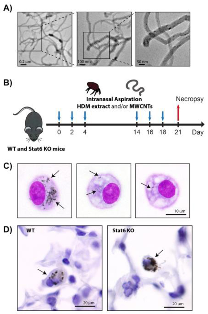
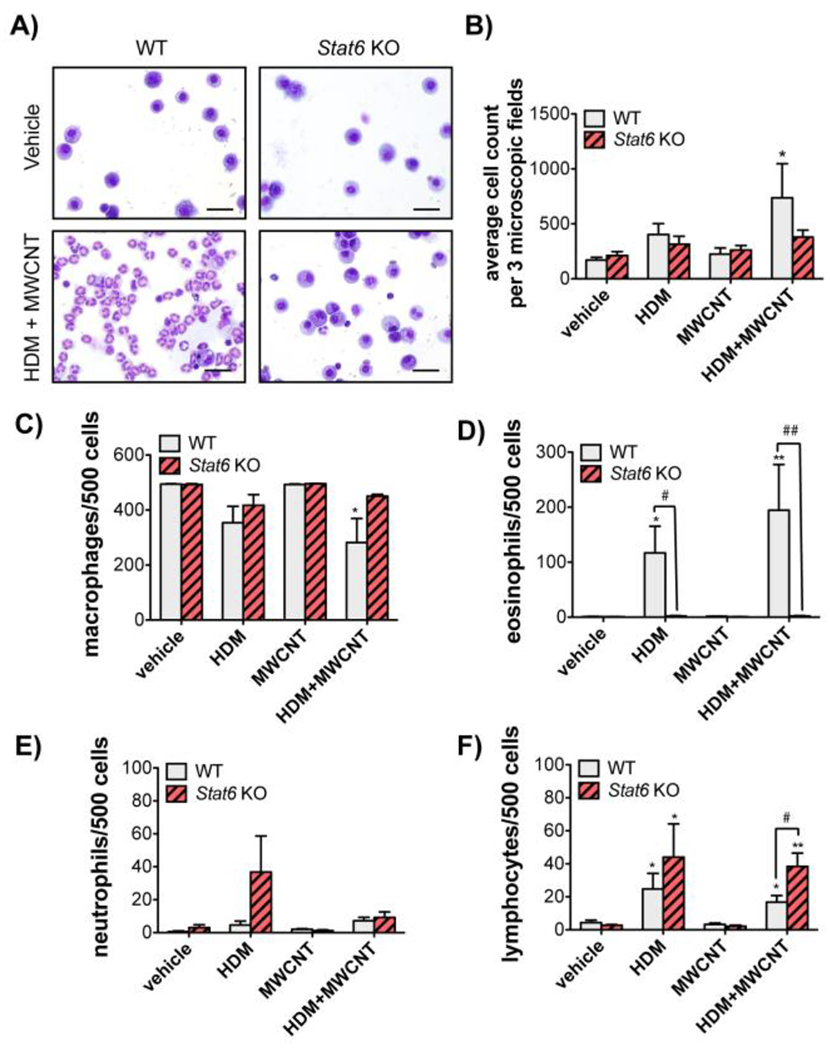
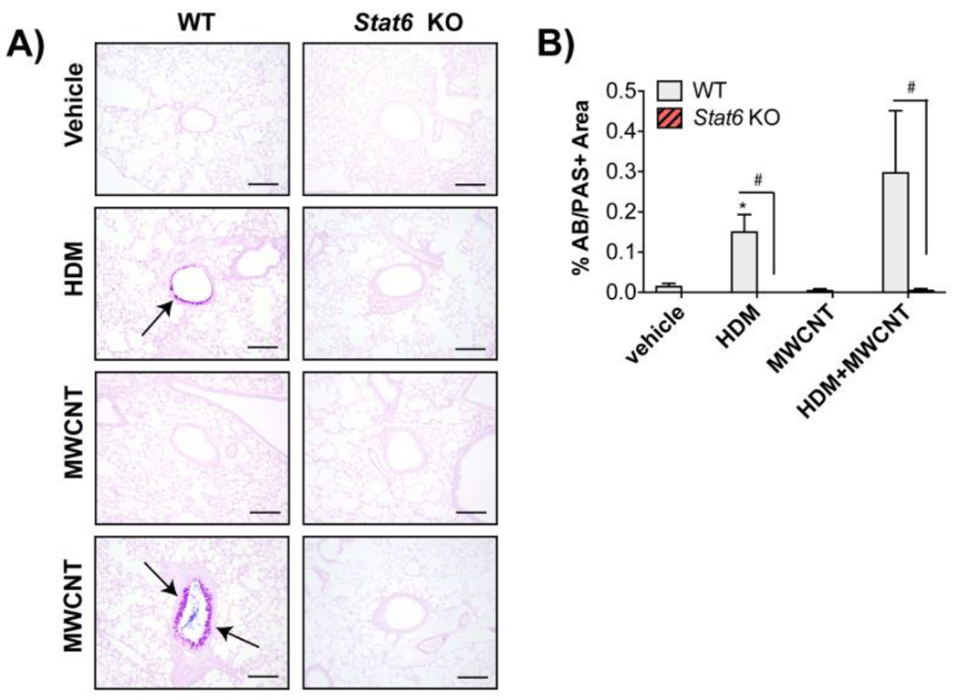
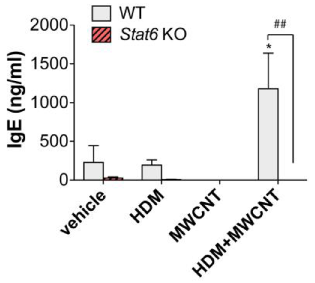
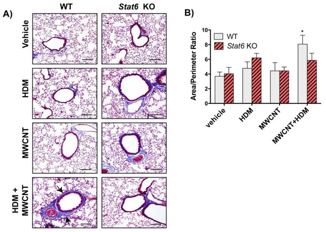
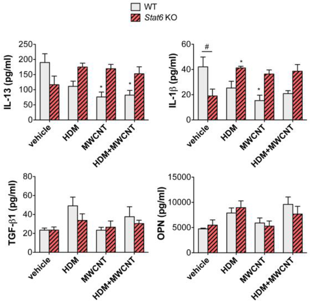
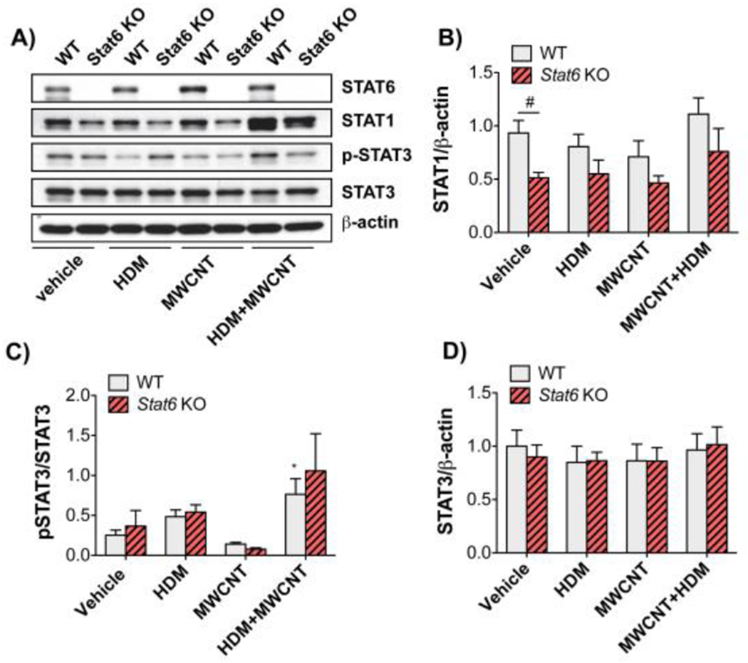
There is increasing evidence that inhaled multi-walled carbon nanotubes (MWCNTs) can have harmful effects on the respiratory system. Rodent studies suggest that individuals with asthma may be susceptible to the adverse pulmonary effects of MWCNTs. Asthma is an allergic lung disease characterized by a TH2 immune response that results in chronic airway disease characterized by eosinophilic lung inflammation, airway mucous cell metaplasia, and airway fibrosis. Signal transducer and activator of transcription 6 (STAT6) is a transcription factor with multiple roles in TH2 type inflammation. Herein we sought to examine the role of STAT6 in the exacerbation of house dust mite (HDM) allergen-induced allergic airway disease by MWCNTs. Male wild type (WT) and STAT6 knockout (Stat6 KO) mice were dosed via intranasal aspiration on days 0, 2, 4, 14, 16 and 18 with either vehicle, HDM extract, MWCNTs, or a combination of HDM and MWCNTs. Necropsy was performed on day 21 to collect bronchoalveolar lavage fluid (BALF), serum and lung tissue. MWCNTs exacerbated HDM-induced allergic endpoints, including eosinophilic lung inflammation, mucous cell metaplasia, and serum IgE levels. HDM-induced eosinophilic lung inflammation, mucous cell metaplasia, and serum IgE and exacerbation of these endpoints by MWCNTs were ablated in Stat6 KO mice. In addition, airway fibrosis was significantly increased by the combination of HDM and MWCNTs in WT mice but not in Stat6 KO mice. These findings provide new mechanistic insight by demonstrating a requirement for STAT6 in MWCNT-induced exacerbation of allergic respiratory disease.

Keywords: STAT6; allergens; asthma; carbon nanotubes; house dust mite.


Conflict of interest statement

Declaration of competing interests The authors declare that they have no known competing financial interests or relationships that could have appeared to influence the work reported in this paper.

Figures

Figure 1.
Figure 1.
A) TEM images of the MWCNTs used in this study. B) Illustration of exposure protocol timeline. Wild type (WT) and Stat6−/− mice were exposed repeatedly to HDM extract and/or MWCNTs by intranasal aspiration at days 0, 2 and 4, followed another round of repeated intranasal aspiration exposure at days 14, 16 and 18. Necropsy was performed at day 21 to collect BALF, lung tissue and serum. See the Methods section for details. C) Oil immersion (1000x) images of alveolar macrophages isolated by Cytospin centrifugation from the BALF of WT mice 21 days after exposure to MWCNTs using the protocol illustrated in panel B. Arrows indicate MWCNTs in the cytoplasm. D) Oil immersion (1000x) images of MWCNTs in the lung tissue of WT and Stat6 mice in situ. Arrows indicate macrophages with MWCNT inclusions.
Figure 2.
Figure 2.. Analysis of inflammatory cells in the BALF of WT and Stat6 KO mice after exposure to MWCNTs with or without HDM allergen.
A) Representative photomicrographs of BALF cells isolated by Cytospin centrifugation from WT and Stat6 KO mice after exposure to vehicle or HDM extract and MWCNTs (Bars = 20 μm). B) Total cell counts from Cytospin slides in each treatment group from WT and Stat6 KO mice. Three microscopic frames per slide were counted at 100x magnification for each animal. *p<0.05 vs. vehicle, Mann-Whitney test. C) Numbers of macrophages per 500 cells in the BALF of animals from each treatment group. *p<0.05 vs. vehicle of same genotype. D) Numbers of eosinophils per 500 cells. *p<0.05 or **p<0.01 compared to vehicle in WT mice. #p<0.05 or ##p<0.01 between genotypes. One-way ANOVA. E) Numbers of neutrophils per 500 cells. F) Numbers of lymphocytes per 500 cells. *p<0.05 or **p<0.01 vs. vehicle of same genotype. #p<0.05 between genotypes, one-way ANOVA. N=4 to 6 animals per group for panels B-F.
Figure 3.
Figure 3.. Requirement of STAT6 for HDM-induced mucous cell metaplasia and exacerbation by MWCNTs in mice.
A) Representative photomicrographs of Alcian blue periodic acid Schiff (AB/PAS) stained lung sections in WT and Stat6KO mice after exposure to vehicle, HDM, MWCNTs or MWCNTs with HDM allergen (Bars = 200 μm). B) Quantification of AB/PAS-positive mucosubstances in the airways of WT and Stat6 KO mice. N=4 to 6 animals per group. *p<0.05 vs. vehicle of same genotype, Student’s t-test. #p< 0.05 between WT and Stat6 KO mice, Mann-Whitney test.
Figure 4.
Figure 4.. Serum IgE is increased by combined HDM and MWCNT exposure in the lungs of WT mice but not in Stat6 KO mice.
Blood was collected from mice at necropsy and serum IgE was measured by ELISA. N=4 to 6 animals per group. *p<0.05 vs. vehicle of same genotype, ##p< 0.01 between WT and Stat6 KO mice, one-way ANOVA.
Figure 5.
Figure 5.. Airway fibrosis is increased by combined HDM and MWCNT exposure in the lungs of WT mice but not in Stat6 KO mice.
A) Representative photomicroscopic images of Gomori’s trichrome-stained lung sections from animals in each of the treatment groups in each genotype (Bars = 200 μm). B) Area to perimeter analysis of trichrome stained lung sections showed a significant increase in airway fibrosis caused by the combination of MWCNTs and HDM extract in WT mice. N=4 to 6 animals per group for each genotype. *p<0.05 vs. vehicle, one-way ANOVA.
Figure 6.
Figure 6.. Cytokine levels in the BALF of WT and Stat6 KO mice after exposure to HDM extract in the absence or presence of MWCNTs.
BALF was collected from mice at necropsy. Cytokines in BALF (IL-13, IL-1β, TGF-β1, OPN) were measured by ELISA as described in the Methods section. N=4 to 6 animals per group. *p<0.05 vs. vehicle of same genotype, #p< 0.05 between WT and Stat6 KO mice, one-way ANOVA.
Figure 7.
Figure 7.. STAT protein levels and activation in lung tissue from WT and Stat6 KO mice after treatment with HDM extract in the absence or presence of MWCNTs.
Protein lysates of right lung lobe tissue collected from mice at necropsy were assayed by Western blotting for STAT proteins and β-actin. A) Representative Western blots of lung tissue from WT or Stcit6 KO mice exposed to vehicle, HDM, MWCNTs, or HDM and MWCNTs. B)Densitometry of STAT1 signal normalized against the β-actin signal. #P < 0.05 between genotypes, one-way ANOVA. N=5 animals per group. C)Densitometry of p-STAT3 normalized against total STAT3. *P < 0.05 compared to vehicle, Mann-Whitney test. D)Densitometry of total STAT3 normalized against β-actin. N=4 to 6 animals per group.

References

    1. Akimoto T, Numata F, Tamura M, Takata Y, Higashida N, Takashi T, Takeda K, Akira S, 1998. Abrogation of bronchial eosinophilic inflammation and airway hyperreactivity in signal transducers and activators of transcription (STAT)6-deficient mice. J. Exp. Med 187, 1537–42. - PMC - PubMed
    1. Bhattacharya K, Mukherjee SP, Gallud A, Burkert SC, Bistarelli S, Bellucci S, Bottini M, Star A, Fadeel B, 2016. Biological interactions of carbon-based nanomaterials: From coronation to degradation. Nanomedicine Nanotechnology, Biol. Med 10.1016/j.nano.2015.11.011 - DOI - PMC - PubMed
    1. Bonner JC, 2010. Mesenchymal cell survival in airway and interstitial pulmonary fibrosis. Fibrogenes. tissue repair 10.1186/1755-1536-3-15 - DOI - PMC - PubMed
    1. Castañeda AR, Pinkerton KE, 2016. Investigating the Effects of Particulate Matter on House Dust Mite and Ovalbumin Allergic Airway Inflammation in Mice . Curr. Protoc. Toxicol 68, 18.18.1–18.18.18. 10.1002/cptx.5 - DOI - PMC - PubMed
    1. Chapoval SP, Dasgupta P, Smith EP, DeTolla LJ, Lipsky MM, Kelly-Welch AE, Keegan AD, 2011. STAT6 expression in multiple cell types mediates the cooperative development of allergic airway disease. J. Immunol 186, 2571–83. 10.4049/jimmunol.1002567 - DOI - PMC - PubMed

Publication types