Review
. 2021 Mar 3;4(2):589-612.
doi: 10.1021/acsptsci.0c00212. eCollection 2021 Apr 9.

Ultrasound-Responsive Nanocarriers in Cancer Treatment: A Review

Affiliations
Review

Ultrasound-Responsive Nanocarriers in Cancer Treatment: A Review

Nahid S Awad et al. ACS Pharmacol Transl Sci. .

Abstract

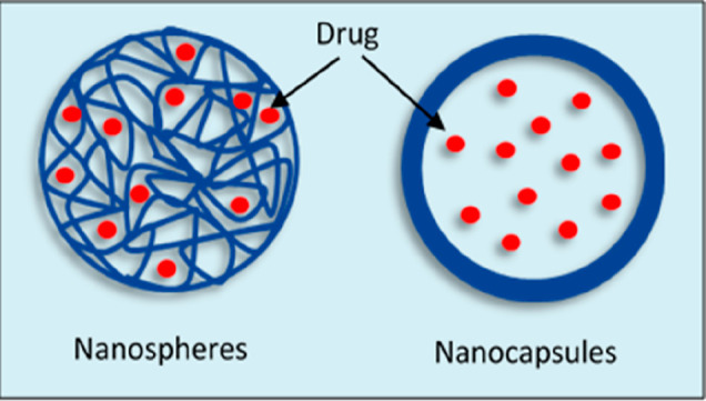
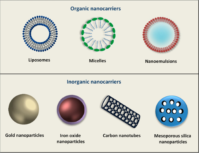
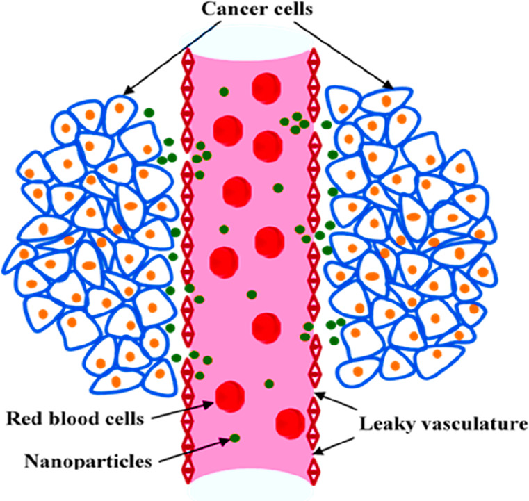
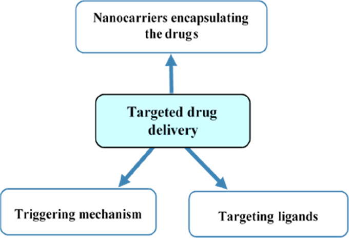
The safe and effective delivery of anticancer agents to diseased tissues is one of the significant challenges in cancer therapy. Conventional anticancer agents are generally cytotoxins with poor pharmacokinetics and bioavailability. Nanocarriers are nanosized particles designed for the selectivity of anticancer drugs and gene transport to tumors. They are small enough to extravasate into solid tumors, where they slowly release their therapeutic load by passive leakage or biodegradation. Using smart nanocarriers, the rate of release of the entrapped therapeutic(s) can be increased, and greater exposure of the tumor cells to the therapeutics can be achieved when the nanocarriers are exposed to certain internally (enzymes, pH, and temperature) or externally (light, magnetic field, and ultrasound) applied stimuli that trigger the release of their load in a safe and controlled manner, spatially and temporally. This review gives a comprehensive overview of recent research findings on the different types of stimuli-responsive nanocarriers and their application in cancer treatment with a particular focus on ultrasound.


Conflict of interest statement

The authors declare no competing financial interest.

Figures

Figure 1
Figure 1
Schematic representation of nanocarriers encapsulating drugs.
Figure 2
Figure 2
Nanocarriers can be composed of organic and/or inorganic materials. This figure shows the types of organic and inorganic nanoparticles discussed in this paper.
Figure 3
Figure 3
Leaky blood vessels surrounding the tumor with retarded lymphatic drainage (EPR effect) allow nanoparticles to extravasate through the blood vessels and accumulate inside the tumor.
Figure 4
Figure 4
Key components of responsive targeted nanocarriers in drug delivery.
Figure 5
Figure 5
Schematic representation of the acoustic pressure waves (A) and stable and collapsed cavitation of microbubbles (B).
Figure 6
Figure 6
Different mechanisms by which oscillating microbubbles may disrupt the integrity of cell membranes. (A) Stable cavitation produces shear forces that can perturb membrane structure. (B) Stable cavitation generates radiation forces that push microbubbles toward the cell and possibly through the cell membrane. (C) The mechanical stress created by microstreaming around microbubbles during stable cavitation causes pore formation. (D) During inertial cavitation, the shock waves formed by the collapsing microbubbles generate high pressure which disrupts the cell membrane. (E) The asymmetrical collapse of microbubble accelerates a microjet toward the cell membrane, forming a pore. (F) During inertial cavitation, ROS can be formed. This leads to disruption of the cell membrane through lipid peroxidation.
Figure 7
Figure 7
Ultrasound-triggered drug release from targeted nanoparticles. The controlled ultrasound beam is focused on the tumor tissue; nanocarriers passing through the high intensity focused beam are disrupted or activated.
Figure 8
Figure 8
Microbubble-conjugated liposomes (adapted from ref (240)).
Figure 9
Figure 9
Considerations for clinical translation of nanocarriers.

Similar articles

Cited by

References

    1. Cancer Research UK . Worldwide cancer statistics. https://www.cancerresearchuk.org/health-professional/cancer-statistics/w... (accessed September 8, 2020).
    1. Kaplun A. P.; Bezrukov D. A.; Shvets V. I. (2011) Rational Design of Nano- and Micro-Size Medicinal Forms of Biologically Active Substances. Appl. Biochem. Microbiol. 47 (8), 711–717. 10.1134/S0003683811080072. - DOI
    1. Zhang L.; Gu F. X.; Chan J. M.; Wang A. Z.; Langer R. S.; Farokhzad O. C. (2008) Nanoparticles in Medicine: Therapeutic Applications and Developments. Clin. Pharmacol. Ther. 83 (5), 761–769. 10.1038/sj.clpt.6100400. - DOI - PubMed
    1. Din F. U.; Aman W.; Ullah I.; Qureshi O. S.; Mustapha O.; Shafique S.; Zeb A. (2017) Effective Use of Nanocarriers as Drug Delivery Systems for the Treatment of Selected Tumors. Int. J. Nanomed. 12, 7291–7309. 10.2147/IJN.S146315. - DOI - PMC - PubMed
    1. Mishra B.; Patel B. B.; Tiwari S. (2010) Colloidal Nanocarriers: A Review on Formulation Technology, Types and Applications toward Targeted Drug Delivery. Nanomedicine 6 (1), 9–24. 10.1016/j.nano.2009.04.008. - DOI - PubMed