

COVID-19-related refractory status epilepticus with the presence of SARS-CoV-2 (RNA) in the CSF: a case report
- PMID: 33860395
- PMCID: PMC8049392
- DOI: 10.1007/s10072-021-05239-6
COVID-19-related refractory status epilepticus with the presence of SARS-CoV-2 (RNA) in the CSF: a case report
Abstract
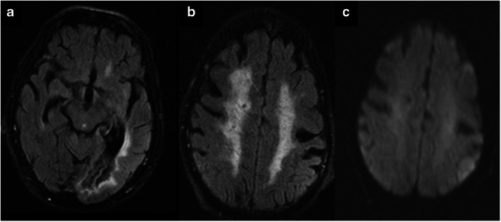
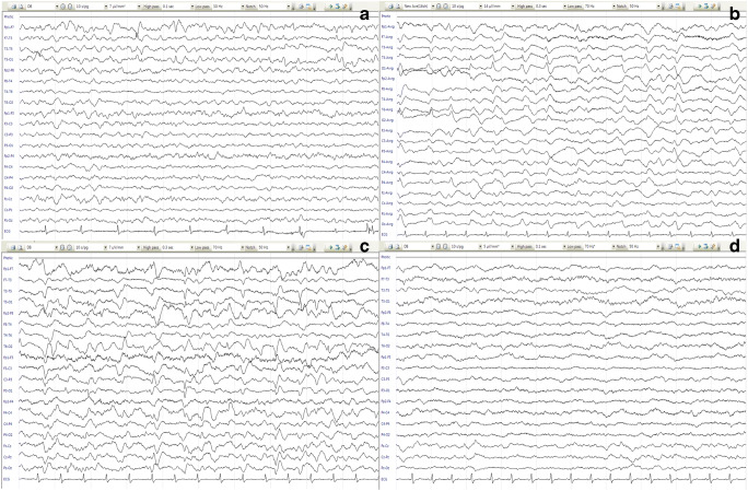
Amongst the neurologic complications of COVID-19 disease, very few reports have shown the presence of the virus in the cerebrospinal fluid (CSF). Seizure and rarely status epilepticus can be associated with COVID-19 disease. Here we present a 73-year-old male with prior history of stroke who has never experienced seizure before. He had no systemic presentation of COVID-19 disease. The presenting symptoms were two consecutive generalized tonic-clonic seizures that after initial resolution turned into a nonconvulsive status epilepticus despite antiepileptic treatment (a presentation similar to NORSE (new-onset refractory status epilepticus)). There was no new lesion in the brain magnetic resonance imaging (MRI). The CSF analysis only showed an increased protein levels and positive reverse transcription polymerase chain reaction (RT-PCR) of 2019-nCoV. Patient recovered partially after anesthetic, IVIG, steroid, and remdesivir. To our knowledge, this is the first report of a refractory status epilepticus with the presence of SARS-CoV-2 ribonucleic acid (RNA) in the CSF.
Keywords: COVID-19; CSF; Status epilepticus.
Conflict of interest statement
The authors declare no competing interests.
Figures
References
-
- Carod-Artal FJ. Neurological complications of coronavirus and COVID-19. Rev Neurol. 2020;70(9):311–322. - PubMed
Publication types
MeSH terms
Substances
LinkOut - more resources
Full Text Sources
Other Literature Sources
Medical
Miscellaneous
