

Smartphone-based imaging systems for medical applications: a critical review
- PMID: 33860648
- PMCID: PMC8047775
- DOI: 10.1117/1.JBO.26.4.040902
Smartphone-based imaging systems for medical applications: a critical review
Abstract
Significance: Smartphones come with an enormous array of functionality and are being more widely utilized with specialized attachments in a range of healthcare applications. A review of key developments and uses, with an assessment of strengths/limitations in various clinical workflows, was completed.
Aim: Our review studies how smartphone-based imaging (SBI) systems are designed and tested for specialized applications in medicine and healthcare. An evaluation of current research studies is used to provide guidelines for improving the impact of these research advances.
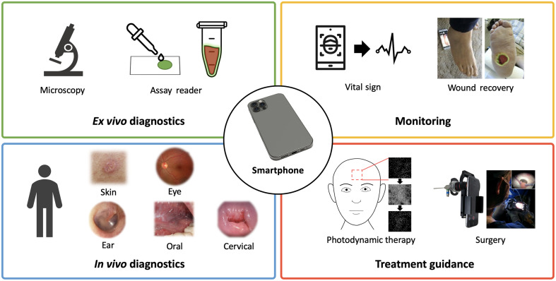
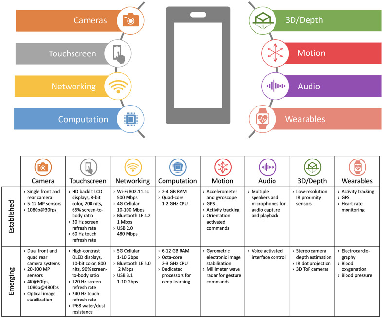
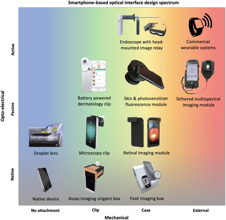
Approach: First, the established and emerging smartphone capabilities that can be leveraged for biomedical imaging are detailed. Then, methods and materials for fabrication of optical, mechanical, and electrical interface components are summarized. Recent systems were categorized into four groups based on their intended application and clinical workflow: ex vivo diagnostic, in vivo diagnostic, monitoring, and treatment guidance. Lastly, strengths and limitations of current SBI systems within these various applications are discussed.
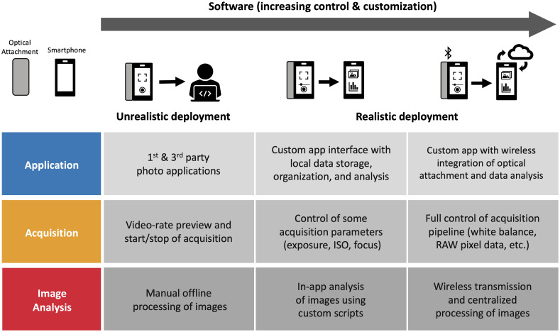
Results: The native smartphone capabilities for biomedical imaging applications include cameras, touchscreens, networking, computation, 3D sensing, audio, and motion, in addition to commercial wearable peripheral devices. Through user-centered design of custom hardware and software interfaces, these capabilities have the potential to enable portable, easy-to-use, point-of-care biomedical imaging systems. However, due to barriers in programming of custom software and on-board image analysis pipelines, many research prototypes fail to achieve a prospective clinical evaluation as intended. Effective clinical use cases appear to be those in which handheld, noninvasive image guidance is needed and accommodated by the clinical workflow. Handheld systems for in vivo, multispectral, and quantitative fluorescence imaging are a promising development for diagnostic and treatment guidance applications.
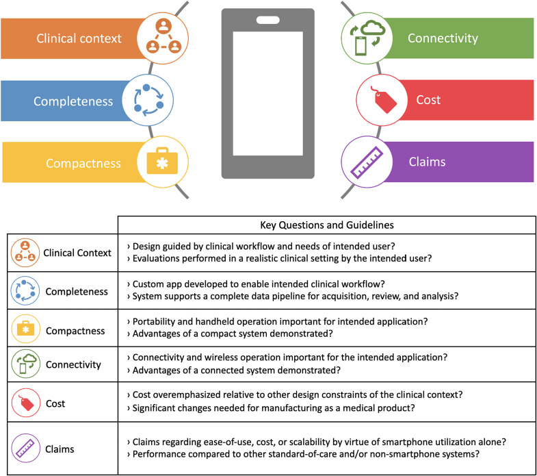
Conclusions: A holistic assessment of SBI systems must include interpretation of their value for intended clinical settings and how their implementations enable better workflow. A set of six guidelines are proposed to evaluate appropriateness of smartphone utilization in terms of clinical context, completeness, compactness, connectivity, cost, and claims. Ongoing work should prioritize realistic clinical assessments with quantitative and qualitative comparison to non-smartphone systems to clearly demonstrate the value of smartphone-based systems. Improved hardware design to accommodate the rapidly changing smartphone ecosystem, creation of open-source image acquisition and analysis pipelines, and adoption of robust calibration techniques to address phone-to-phone variability are three high priority areas to move SBI research forward.
Keywords: handheld; mobile; point-of-care; smartphone; smartphone imaging; smartphone systems.
Figures
Similar articles
-
Design of a 3D printed smartphone microscopic system with enhanced imaging ability for biomedical applications.J Microsc. 2019 Oct;276(1):13-20. doi: 10.1111/jmi.12829. Epub 2019 Sep 22. J Microsc. 2019. PMID: 31498428
-
Current progress in the utilization of smartphone-based imaging for quality assessment of food products: a review.Crit Rev Food Sci Nutr. 2022;62(13):3631-3643. doi: 10.1080/10408398.2020.1867820. Epub 2020 Dec 30. Crit Rev Food Sci Nutr. 2022. PMID: 33377398 Review.
-
Multiplex Smartphone Diagnostics.Methods Mol Biol. 2017;1546:295-302. doi: 10.1007/978-1-4939-6730-8_26. Methods Mol Biol. 2017. PMID: 27896779
-
Mobile healthcare applications: system design review, critical issues and challenges.Australas Phys Eng Sci Med. 2015 Mar;38(1):23-38. doi: 10.1007/s13246-014-0315-4. Epub 2014 Dec 5. Australas Phys Eng Sci Med. 2015. PMID: 25476753 Review.
-
Multi-Contrast Imaging and Digital Refocusing on a Mobile Microscope with a Domed LED Array.PLoS One. 2015 May 13;10(5):e0124938. doi: 10.1371/journal.pone.0124938. eCollection 2015. PLoS One. 2015. PMID: 25969980 Free PMC article.
Cited by
-
Advances in Portable Optical Microscopy Using Cloud Technologies and Artificial Intelligence for Medical Applications.Sensors (Basel). 2024 Oct 17;24(20):6682. doi: 10.3390/s24206682. Sensors (Basel). 2024. PMID: 39460161 Free PMC article. Review.
-
Smartphone-based dual radiometric fluorescence and white-light imager for quantification of protoporphyrin IX in skin.J Biomed Opt. 2023 Aug;28(8):086003. doi: 10.1117/1.JBO.28.8.086003. Epub 2023 Aug 26. J Biomed Opt. 2023. PMID: 37638107 Free PMC article.
-
Paper-based analytical devices for virus detection: Recent strategies for current and future pandemics.Trends Analyt Chem. 2021 Nov;144:116424. doi: 10.1016/j.trac.2021.116424. Epub 2021 Aug 26. Trends Analyt Chem. 2021. PMID: 34462612 Free PMC article. Review.
-
Mobile-linked point-of-care diagnostics in community-based healthcare: a scoping review of user experiences.Arch Public Health. 2024 Aug 27;82(1):139. doi: 10.1186/s13690-024-01376-4. Arch Public Health. 2024. PMID: 39192369 Free PMC article.
-
FluoroPi Device With SmartProbes: A Frugal Point-of-Care System for Fluorescent Detection of Bacteria From a Pre-Clinical Model of Microbial Keratitis.Transl Vis Sci Technol. 2023 Jul 3;12(7):1. doi: 10.1167/tvst.12.7.1. Transl Vis Sci Technol. 2023. PMID: 37395707 Free PMC article.
References
-
- Roda A., et al. , “Smartphone-based biosensors: a critical review and perspectives,” TRAC Trends Anal. Chem. 79, 317–325 (2016).10.1016/j.trac.2015.10.019 - DOI
Publication types
MeSH terms
Grants and funding
LinkOut - more resources
Full Text Sources
Other Literature Sources
