. 2021 Jun;32(11-12):599-615.
doi: 10.1089/hum.2020.301. Epub 2021 Jun 1.

Intracerebroventricular Administration of AAV9-PHP.B SYN1-EmGFP Induces Widespread Transgene Expression in the Mouse and Monkey Central Nervous System

Affiliations

Intracerebroventricular Administration of AAV9-PHP.B SYN1-EmGFP Induces Widespread Transgene Expression in the Mouse and Monkey Central Nervous System

Adriana Galvan et al. Hum Gene Ther. 2021 Jun.

Abstract

Viral vectors made from adeno-associated virus (AAV) have emerged as preferred tools in basic and translational neuroscience research to introduce or modify genetic material in cells of interest. The use of viral vectors is particularly attractive in nontransgenic species, such as nonhuman primates. Injection of AAV solutions into the cerebrospinal fluid is an effective method to achieve a broad distribution of a transgene in the central nervous system. In this study, we conducted injections of AAV9-PHP.B, a recently described AAV capsid mutant, in the lateral ventricle of mice and rhesus macaques. To enhance the expression of the transgene (the tag protein emerald green fluorescent protein [EmGFP]), we used a gene promoter that confers high neuron-specific expression of the transgene, the human synapsin 1 (SYN1) promoter. The efficacy of the viral vector was first tested in mice. Our results show that intracerebroventricular injections of AAV9-PHP.B SYN1-EmGFP-woodchuck hepatitis virus posttranscriptional regulatory element resulted in neuronal EmGFP expression throughout the mice and monkey brains. We have provided a thorough characterization of the brain regions expressing EmGFP in both species. EmGFP was observed in neuronal cell bodies over the whole cerebral cortex and in the cerebellum, as well as in some subcortical regions, including the striatum and hippocampus. We also observed densely labeled neuropil in areas known to receive projections from these regions. Double fluorescence studies demonstrated that EmGFP was expressed by several types of neurons throughout the mouse and monkey brain. Our results demonstrate that a single injection in the lateral ventricle is an efficient method to obtain transgene expression in many cortical and subcortical regions, obviating the need of multiple intraparenchymal injections to cover large brain areas. The use of intraventricular injections of AAV9-PHP.B SYN1-EmGFP could provide a powerful approach to transduce widespread areas of the brain and may contribute to further development of methods to genetically target-specific populations of neurons.

Keywords: ICV; adeno-associated virus; gene therapy; primate; synapsin.


Conflict of interest statement

No competing financial interests exist.

Figures

Figure 1.
Figure 1.
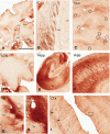
Widespread expression of SYN1-EmGFP after ICV injection of AAV9-PHP.B into the lateral ventricle of mice. (A) Low-power view of GFP immunostaining in the mouse forebrain at the level of the rostral Str. (B) Higher magnification of the area outlined by box 1 in (A), demonstrating GFP-positive axonal bundles projecting from the cortex. (C–E) Double immunostaining of cells in the Str (box 2 in A) for GFP and DARPP32 (a marker for medium spiny neurons) demonstrates a high degree of co-labeled cells (arrowheads in E). (F) Low-power view of GFP immunostaining at the level of the rostral Hipp and thalamus. (G) Higher magnification of the area outlined by box 1 in (F), demonstrating GFP immunoreactivity in the so and the sr of the CA1 region. Few pyramidal neuron cell bodies were GFP-positive. (H–J) Double immunostaining of cells in the S1 cortex (box 2 in F) for GFP and PV demonstrates the GABAergic phenotype of many nonpyramidal transduced cells (arrowheads in J). Large GFP-positive pyramidal neurons are also apparent (arrows in J). (K) Low-power view of GFP immunostaining in the mouse midbrain. (L) Higher magnification of area outlined in the box in (K), demonstrating the SN labeled with TH (a marker of catecholaminergic cells, red) and GFP (green). (M–O) Double immunostaining of cells in the SN for GFP and TH demonstrates the catecholaminergic phenotype of a subset of GFP-positive cells in this region (arrowheads in O). (P) Low-power view of GFP immunostaining in the Cer and brainstem. (Q) Higher magnification of the area outlined in the box in (P), demonstrating sparse GFP immunoreactivity in the Gl, the Mol, and the PCL of the cerebellar cortex. (R–T) Double immunostaining of cells for GFP and CB (a marker of PC, red) demonstrates occasional co-labeled cells (arrowheads in T). Scale bar is 1 mm in (A, F, K, and P); scale bar is 50 μm in (R, S, and T); scale bar is 100 μm in all other panels. All images are representative of at least two sections each from three mice where results were similar in all images examined. Boxed areas are representative of where higher magnification images were taken, but not all images shown are from the same section. DAPI (blue) is used as a counterstain in lower magnification panels and overview images. AAV9, adeno-associated virus 9; AC, anterior commissure; CB, calbindin; CC, corpus callosum; Cer, cerebellum; DAPI, 4′,6-diamidino-2-phenylindole; DARPP32, dopamine and cAMP-regulated phospho = protein Mr 32 kDa; EmGFP, emerald GFP; GABA, gamma aminobutyric acid; GFP, green fluorescent protein; Gl, granule cell layer; Hipp, hippocampus; ICV, intracerebroventricular; Mol, molecular layer; PC, Purkinje cells; PCL, Purkinje cell layer; PV, parvalbumin; SN, substantia nigra; so, stratum oriens; sr, stratum radiatum; Str, striatum; SYN1, synapsin 1; TH, tyrosine hydroxylase; ZI, zona incerta. Color images are available online.
Figure 1.
Figure 1.
Widespread expression of SYN1-EmGFP after ICV injection of AAV9-PHP.B into the lateral ventricle of mice. (A) Low-power view of GFP immunostaining in the mouse forebrain at the level of the rostral Str. (B) Higher magnification of the area outlined by box 1 in (A), demonstrating GFP-positive axonal bundles projecting from the cortex. (C–E) Double immunostaining of cells in the Str (box 2 in A) for GFP and DARPP32 (a marker for medium spiny neurons) demonstrates a high degree of co-labeled cells (arrowheads in E). (F) Low-power view of GFP immunostaining at the level of the rostral Hipp and thalamus. (G) Higher magnification of the area outlined by box 1 in (F), demonstrating GFP immunoreactivity in the so and the sr of the CA1 region. Few pyramidal neuron cell bodies were GFP-positive. (H–J) Double immunostaining of cells in the S1 cortex (box 2 in F) for GFP and PV demonstrates the GABAergic phenotype of many nonpyramidal transduced cells (arrowheads in J). Large GFP-positive pyramidal neurons are also apparent (arrows in J). (K) Low-power view of GFP immunostaining in the mouse midbrain. (L) Higher magnification of area outlined in the box in (K), demonstrating the SN labeled with TH (a marker of catecholaminergic cells, red) and GFP (green). (M–O) Double immunostaining of cells in the SN for GFP and TH demonstrates the catecholaminergic phenotype of a subset of GFP-positive cells in this region (arrowheads in O). (P) Low-power view of GFP immunostaining in the Cer and brainstem. (Q) Higher magnification of the area outlined in the box in (P), demonstrating sparse GFP immunoreactivity in the Gl, the Mol, and the PCL of the cerebellar cortex. (R–T) Double immunostaining of cells for GFP and CB (a marker of PC, red) demonstrates occasional co-labeled cells (arrowheads in T). Scale bar is 1 mm in (A, F, K, and P); scale bar is 50 μm in (R, S, and T); scale bar is 100 μm in all other panels. All images are representative of at least two sections each from three mice where results were similar in all images examined. Boxed areas are representative of where higher magnification images were taken, but not all images shown are from the same section. DAPI (blue) is used as a counterstain in lower magnification panels and overview images. AAV9, adeno-associated virus 9; AC, anterior commissure; CB, calbindin; CC, corpus callosum; Cer, cerebellum; DAPI, 4′,6-diamidino-2-phenylindole; DARPP32, dopamine and cAMP-regulated phospho = protein Mr 32 kDa; EmGFP, emerald GFP; GABA, gamma aminobutyric acid; GFP, green fluorescent protein; Gl, granule cell layer; Hipp, hippocampus; ICV, intracerebroventricular; Mol, molecular layer; PC, Purkinje cells; PCL, Purkinje cell layer; PV, parvalbumin; SN, substantia nigra; so, stratum oriens; sr, stratum radiatum; Str, striatum; SYN1, synapsin 1; TH, tyrosine hydroxylase; ZI, zona incerta. Color images are available online.
Figure 2.
Figure 2.
General pattern of expression of SYN1-EmGFP after ICV injection of AAV9-PHP.B in the monkey lateral ventricle. Schematics of coronal sections through the rostrocaudal axis of the rhesus monkey brain to map the distribution and relative density of GFP-immunoreactive neuronal cell bodies and axonal processes in the two monkeys (MR322 and MR325) that received ICV injections of AAV9-PHP.B SYN1-EmGFP-WPRE. For each animal, the schematics are based on GFP-immunostained sections from the hemisphere ipsilateral to the ICV injection. The interaural level of each section, based on the stereotaxic atlas of Paxinos et al., is indicated in the lower right corner of the schematics. The relative abundance of GFP-positive neuronal cell bodies is indicated in the maps as green circles, while areas with GFP-positive neuropil are represented with orange and brown shadings. Color images are available online.
Figure 3.
Figure 3.
Expression of EmGFP in monkey PFC and forebrain. (A–C) Low-power views of GFP-immunoreactive neuronal cell bodies and axonal processes in the PFC and basal forebrain structures of monkey MR322. (D, E) Medium- and high-power views of labeled neurons in the PFC. Both pyramidal (black arrows) and nonpyramidal (non-filled arrows) cells were immunostained in deep and superficial cortical layers, respectively (one in A). (F) Dense aggregates of immunoreactive neuronal cell bodies within a strongly immunostained neuropil in the periventricular region of the dorsomedial CD. (G) Immunoreactive axonal processes in the PU. (H) Dense immunostained neuropil and a large number of immunoreactive neuronal cell bodies in the BLA and BMA regions of the amygdala. (I) Immunoreactive neurons in the NBM/SI region. (J) Labeled axonal processes (arrows) and a few immunoreactive cell bodies (marked with circles) in the dorsal part of the GPe. Scale bar shown in (A) represents 5 mm in (A–C); 1 mm in (F–J); 0.5 mm in (D); 0.1 mm in (E). BLA, basolateral; BMA, basomedial; CD, caudate nucleus; GPe, external globus pallidus; NBM, nucleus basalis of Meynert; PFC, prefrontal cortex; PU, putamen; sep, septum; SI, substantia innominata. Color images are available online.
Figure 4.
Figure 4.
Expression of EmGFP in monkey midbrain. (A) Low-power view of GFP-immunostained neuronal elements at the level of the midbrain in monkey MR322. (B) Bundle of immunostained axons traveling through the IC. Note paucity of labeling in the neighboring RTN and VP thalamic nuclei. (C) Patches of GFP-immunostained axons and terminals with sparsely distributed immunoreactive neuronal cell bodies (open circles) throughout the medial VLm of the thalamus. (D) GFP-immunoreactive cell bodies in superficial layers of the LGN. (E, F) Low- and high-power views of GFP labeling throughout the Hipp. Rich plexuses of immunoreactive pyramidal cell bodies and dendritic processes within a dense immunostained neuropil are found throughout CA1-3 regions (F). In contrast, the DG is almost completely devoid of immunoreactivity. (G, H) GFP immunoreactivity in the SNc and SNr. In both regions, the GFP labeling is largely confined to rich plexuses of axons and terminals with few immunoreactive neuronal cell bodies (H). (I) Strong labeling of pyramidal (arrows) and nonpyramidal (boxed area) cells in the insular somatosensory cortices. Scale bar shown in (A) represents 10 mm in (A), 5 mm in (G), 3 mm in (C–E); 2 mm in (B); 1.5 mm in (H, I); and 1 mm in (F). DG, dentate gyrus; IC, internal capsule; LGN, lateral geniculate nucleus; RTN, reticular thalamic nucleus; SNc, substantia nigra pars compacta; SNr, substantia nigra reticulata; VLm, ventrolateral nucleus; VP, ventroposterior. Color images are available online.
Figure 5.
Figure 5.
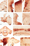
Expression of EmGFP in the monkey brainstem and Cer. (A) Low-power view of GFP immunostaining in the upper brainstem in monkey MR322. (B–D) High-power views of GFP-immunoreactive cell bodies and fibers in the PAG (B), SC (C), PBG (D) and PPN (D). The inset in (B) is a high-power view of the area in the rectangle showing GFP-positive periventricular ependymal cells (arrows). (E) Low-power view of GFP labeling in the monkey upper medulla. The DR and LDT nuclei contain labeled cell bodies, whereas the Py display dense axonal labeling. (F) High-power views of GFP-labeled cells in the DR, the LDT, and the LC. Note the stronger neuropil immunoreactivity in the DR compared with other regions surrounding the 4v. (G) GFP-immunoreactive cell bodies in the POI surrounding the IO devoid of GFP immunolabeling. (H) GFP immunostaining in the Cer and RF at the level of the central medulla. Areas of strong cellular labeling at this level include the CoN and the cerebellar cortex. Py are enriched in GFP-labeled axons. Sparsely distributed neuronal cell bodies are found throughout the RF. (I) Dense clusters of GFP-positive GC and Pf within the cerebellar cortex. The dendrites of some PC are also seen. (J) High-power view of the dense cell body and fibers labeling in the CoN. (K) GFP labeling in the lower medulla at the level of the PYx. Some cellular labeling can be found in the SpT and Cu/Gr nuclei. The PYx is enriched in GFP-labeled axons. (L) Clusters of GFP-positive PC and their dendritic processes that extend in the Mol of the cerebellar cortex. Scale bar shown in (A) represents 10 mm in (A, K); 5 mm in (E, H); 2 mm in (I, J, L); 1 mm in (B, D, F, G); 0.7 mm in (C). 4v, 4th ventricle; AQ, aqueduct; CoN, cochlear nucleus; Cu/Gr, cuneate/gracile; DR, dorsal raphe; GC, granule cells; IO, inferior olive; LC, locus coeruleus; LDT, laterodorsal tegmental; PAG, periaqueductal gray; PBG, parabigeminal nucleus; POI, peri-olivary nucleus; PPN, pedunculopontine nucleus; RF, reticular formation; SC, superior colliculus; SpT, spinal trigeminal; Py, pyramids; PYx, pyramidal decussation. Color images are available online.
Figure 6.
Figure 6.
GFP colocalizes with various types of neuronal markers in monkey. Confocal images of double immunostaining for GFP and various neurotransmitter markers to characterize the chemical phenotype of some GFP-immunoreactive neurons throughout the monkey brain. All images are from monkey MR322. (A, A′, A″) Colocalization of GFP and DARPP32, a marker of striatal projection neurons, in the CD. (B, B′, B″) Colocalization of GFP and GABA in non-pyramidal cells in the cerebral cortex. (C, C′, C″) Colocalization of GFP and TH in the SN. (D, D′, D″) Colocalization of GFP and 5-HT in the DR. In (A–D) arrowheads point to examples of double-labeled neurons. Scale bar in (A) represents 30 μm in (A, B) rows, 20 μm in rows (C, D). 5-HT, serotonin. Color images are available online.

References

    1. Ojala DS, Amara DP, Schaffer DV. Adeno-associated virus vectors and neurological gene therapy. Neuroscientist 2015;21:84–98 - PubMed
    1. Murlidharan G, Samulski RJ, Asokan A. Biology of adeno-associated viral vectors in the central nervous system. Front Mol Neurosci 2014;7:76. - PMC - PubMed
    1. Grieger JC, Samulski RJ. Adeno-associated virus vectorology, manufacturing, and clinical applications. In: Friedmann T, ed. Gene Transfer Vectors for Clinical Application. Amsterdam, Netherlands: Elsevier/Academis Press, 2012:229–254 - PubMed
    1. Saraiva J, Nobre RJ, Pereira de Almeida L. Gene therapy for the CNS using AAVs: the impact of systemic delivery by AAV9. J Control Release 2016;241:94–109 - PubMed
    1. Lerchner W, Corgiat B, Der Minassian V, et al. . Injection parameters and virus dependent choice of promoters to improve neuron targeting in the nonhuman primate brain. Gene Ther 2014;21:233–241 - PMC - PubMed

Publication types

LinkOut - more resources