

Mass spectrometry-based proteomic platforms for better understanding of SARS-CoV-2 induced pathogenesis and potential diagnostic approaches
- PMID: 33860983
- PMCID: PMC8250252
- DOI: 10.1002/pmic.202000279
Mass spectrometry-based proteomic platforms for better understanding of SARS-CoV-2 induced pathogenesis and potential diagnostic approaches
Abstract
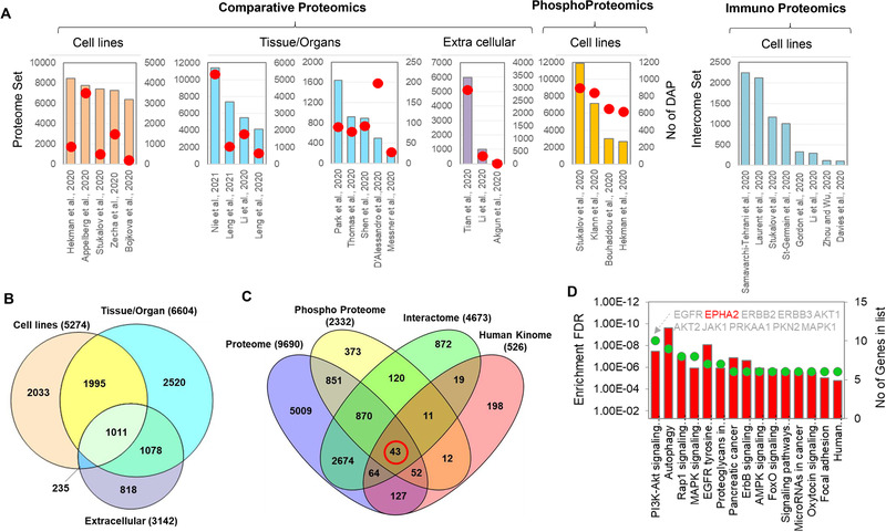
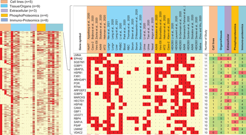
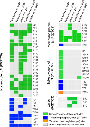
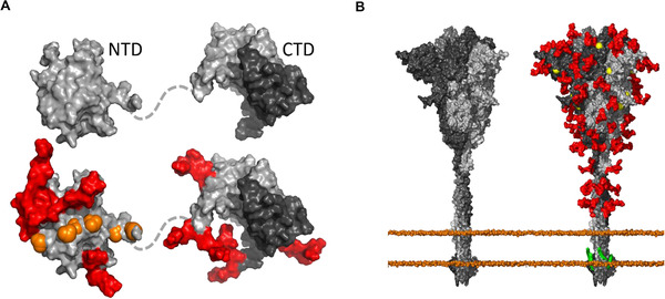
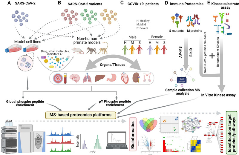
While protein-protein interaction is the first step of the SARS-CoV-2 infection, recent comparative proteomic profiling enabled the identification of over 11,000 protein dynamics, thus providing a comprehensive reflection of the molecular mechanisms underlying the cellular system in response to viral infection. Here we summarize and rationalize the results obtained by various mass spectrometry (MS)-based proteomic approaches applied to the functional characterization of proteins and pathways associated with SARS-CoV-2-mediated infections in humans. Comparative analysis of cell-lines versus tissue samples indicates that our knowledge in proteome profile alternation in response to SARS-CoV-2 infection is still incomplete and the tissue-specific response to SARS-CoV-2 infection can probably not be recapitulated efficiently by in vitro experiments. However, regardless of the viral infection period, sample types, and experimental strategies, a thorough cross-comparison of the recently published proteome, phosphoproteome, and interactome datasets led to the identification of a common set of proteins and kinases associated with PI3K-Akt, EGFR, MAPK, Rap1, and AMPK signaling pathways. Ephrin receptor A2 (EPHA2) was identified by 11 studies including all proteomic platforms, suggesting it as a potential future target for SARS-CoV-2 infection mechanisms and the development of new therapeutic strategies. We further discuss the potentials of future proteomics strategies for identifying prognostic SARS-CoV-2 responsive age-, gender-dependent, tissue-specific protein targets.
Keywords: COVID-19; biomarkers; comparative proteomics; kinase-substrate signaling; post-translational modifications; targeted proteomics; top-down proteomics.
© 2021 Wiley-VCH GmbH.
Conflict of interest statement
The authors declare no conflict of interest.
Figures
Similar articles
-
Analysis of phosphomotifs coupled to phosphoproteome and interactome unveils potential human kinase substrate proteins in SARS-CoV-2.Front Cell Infect Microbiol. 2025 Jul 9;15:1554760. doi: 10.3389/fcimb.2025.1554760. eCollection 2025. Front Cell Infect Microbiol. 2025. PMID: 40703672 Free PMC article.
-
Antibody tests for identification of current and past infection with SARS-CoV-2.Cochrane Database Syst Rev. 2022 Nov 17;11(11):CD013652. doi: 10.1002/14651858.CD013652.pub2. Cochrane Database Syst Rev. 2022. PMID: 36394900 Free PMC article.
-
Rapid, point-of-care antigen tests for diagnosis of SARS-CoV-2 infection.Cochrane Database Syst Rev. 2022 Jul 22;7(7):CD013705. doi: 10.1002/14651858.CD013705.pub3. Cochrane Database Syst Rev. 2022. PMID: 35866452 Free PMC article.
-
Kinetic Multi-omic Analysis of Responses to SARS-CoV-2 Infection in a Model of Severe COVID-19.J Virol. 2021 Sep 27;95(20):e0101021. doi: 10.1128/JVI.01010-21. Epub 2021 Jul 28. J Virol. 2021. PMID: 34319784 Free PMC article.
-
The effect of sample site and collection procedure on identification of SARS-CoV-2 infection.Cochrane Database Syst Rev. 2024 Dec 16;12(12):CD014780. doi: 10.1002/14651858.CD014780. Cochrane Database Syst Rev. 2024. PMID: 39679851 Free PMC article.
Cited by
-
Live and let die: signaling AKTivation and UPRegulation dynamics in SARS-CoVs infection and cancer.Cell Death Dis. 2022 Oct 3;13(10):846. doi: 10.1038/s41419-022-05250-5. Cell Death Dis. 2022. PMID: 36192392 Free PMC article. Review.
-
Aqueous humor proteomics analyzed by bioinformatics and machine learning in PDR cases versus controls.Clin Proteomics. 2024 May 19;21(1):36. doi: 10.1186/s12014-024-09481-w. Clin Proteomics. 2024. PMID: 38764026 Free PMC article.
-
Levels of soluble complement regulators predict severity of COVID-19 symptoms.Front Immunol. 2022 Oct 18;13:1032331. doi: 10.3389/fimmu.2022.1032331. eCollection 2022. Front Immunol. 2022. PMID: 36330526 Free PMC article.
-
Multi-omic profiling of plasma reveals molecular alterations in children with COVID-19.Theranostics. 2021 Jul 6;11(16):8008-8026. doi: 10.7150/thno.61832. eCollection 2021. Theranostics. 2021. PMID: 34335977 Free PMC article.
-
Nimotuzumab for COVID-19: case series.Immunotherapy. 2021 Nov 22;14(3):185-193. doi: 10.2217/imt-2021-0269. Immunotherapy. 2021. PMID: 34806405 Free PMC article.
References
-
- Wadman, M. , Couzin‐Frankel, J. , Kaiser, J. , & Matacic, C. (2020). A Rampage Through the Body. Science, 368, 356–360. - PubMed
-
- Appelberg, S. , Gupta, S. , Akusjärvi, S. S. , Ambikan, A. T. , Mikaeloff, F. , Saccon, E. , Végvári, Á. , Benfeitas, R. , Sperk, M. , Ståhlberg, M. , Krishnan, S. , Singh, K. , Penninger, J. M. , Mirazimi, A. , & Neogi, U. (2020). Dysregulation in Akt/mTOR/HIF‐1 signaling identified by proteo‐transcriptomics of SARS‐CoV‐2 infected cells. Emerg Microbes Infect, 9, 1748–1760. - PMC - PubMed
-
- Grenga, L. , Gallais, F. , Pible, O. , Gaillard, J. C. , Gouveia, D. , Batina, H. , Bazaline, N. , Ruat, S. , Culotta, K. , Miotello, G. , Debroas, S. , Roncato, M. A. , Steinmetz, G. , Foissard, C. , Desplan, A. , Alpha‐Bazin, B. , Almunia, C. , Gas, F. , Bellanger, L. , & Armengaud, J. (2020). Shotgun proteomics analysis of SARS‐CoV‐2‐infected cells and how it can optimize whole viral particle antigen production for vaccines. Emerg Microbes Infect, 9, 1712–1721. - PMC - PubMed
-
- Zecha, J. , Lee, C. Y. , Bayer, F. P. , Meng, C. , Grass, V. , Zerweck, J. , Schnatbaum, K. , Michler, T. , Pichlmair, A. , Ludwig, C. , & Kuster, B. (2020). Data, reagents, assays and merits of proteomics for SARS‐CoV‐2 research and testing. Molecular & Cellular Proteomics, 19, 1503–1522. - PMC - PubMed
Publication types
MeSH terms
Substances
Grants and funding
LinkOut - more resources
Full Text Sources
Other Literature Sources
Medical
Research Materials
Miscellaneous
