. 2022 Feb;58(1):108-117.
doi: 10.23736/S1973-9087.21.06853-2. Epub 2021 Apr 16.

From histo-anatomy to sonography in lymphedema: EURO-MUSCULUS/USPRM approach

Affiliations

From histo-anatomy to sonography in lymphedema: EURO-MUSCULUS/USPRM approach

Vincenzo Ricci et al. Eur J Phys Rehabil Med. 2022 Feb.

Abstract

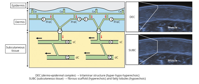
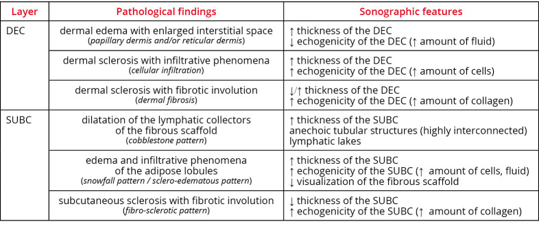
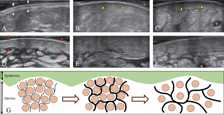
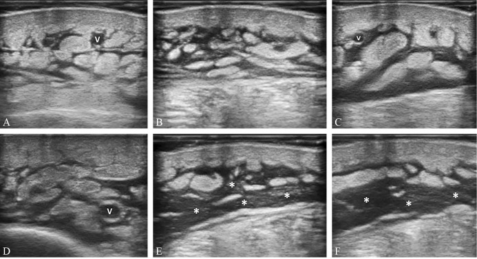
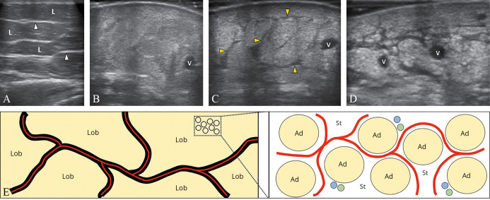
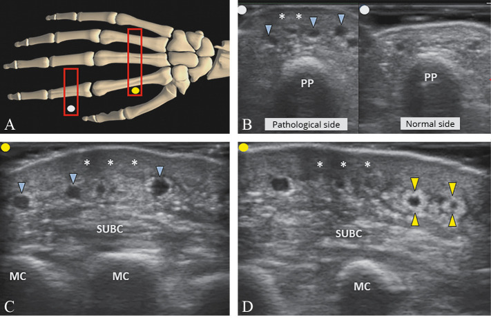
Lymphedema is a disorder characterized by the accumulation of protein-rich lymphatic fluid in the cutaneous and subcutaneous tissue. Based on the underlying causes, it is classified into primary and secondary forms. The use of ultrasound has recently become widespread in the field of lymphedema - especially for its diagnosis and treatment planning. In this study, we briefly reviewed the anatomy and histology of the skin and subcutaneous tissue - to propose a standardized ultrasound assessment of the superficial tissues in patients with upper-/lower-limb lymphedema. We believe that identification of the sono-histological patterns of the dermo-epidermal complex and subcutaneous tissue has place to serve as a simple and reproducible strategy to evaluate their edema diseases that are often subject to an inaccurate diagnosis in daily clinical practice.


Conflict of interest statement

Conflicts of interest.—The authors certify that there is no conflict of interest with any financial organization regarding the material discussed in the manuscript.

Figures

Figure 1
Figure 1
—Schematic drawing shows the lymphatic capillaries in communication with the precollectors in the dermis, and the superficial and deep lymphatic collectors in the subcutaneous tissue. Corresponding sonograms show the trilaminar structure of the normal dermo-epidermal complex and the echotexture of the subcutaneous tissue. Light grey arrow (red in the online version): vertical lymphatic flow; black arrow: horizontal lymphatic flow; black circle: unidirectional valve. Ca: capillaries; PreC: precollectors; sC: superficial; dC: deep; DEC: dermo-epidermal complex; SUBC: subcutaneous tissue.
Figure 2
Figure 2
—Normal papillary and reticular dermis with the superficial lymphovascular plexus (black arrow) made of blood and lymphatic vessels (A) (H&E, Original magnification ×100). Mild inflammatory infiltrate (black arrow) of the papillary dermis with no involvement of the reticular dermis (B) (H&E, Original magnification ×80). Edema and moderate inflammatory infiltrate of the papillary dermis with telangiectasia (black arrow) of the superficial lymphovascular plexus (C) (H&E, Original magnification ×100). Severe inflammatory infiltrate of the papillary and reticular dermis with disorganization of the physiological architecture of the dermo-epidermal complex (D) (H&E, Original magnification ×80). H&E: hematoxylin and eosin; P: papillary; R: reticular.
Figure 3
Figure 3
—Mature adipocites – separated by a thin intercellular matrix – are arranged in fatty lobules sustained by a thicker fibrous scaffold (A, B) (black arrow): A) H&E, Original magnification ×200; B) H&E, Original magnification ×50. Moderate (C) and severe (D) thickening of the fibrous scaffold with rarefaction of the adipocytic lobules: C) H&E, Original magnification ×100; D) H&E, Original magnification ×50. Edema, telangiectasia of the lymphatic and blood vessels, and inflammatory infiltrate (black arrow) (E, F) of the intercellular matrix of adipose lobules: E) H&E, Original magnification ×80; and F) CD3, Original magnification ×100). H&E: hematoxylin and eosin; CD3: cluster of differentiation 3, immunohistochemical stain for T-cell lymphocytes; Ad: adipocites; L: lobule.
Figure 4
Figure 4
—Not-stained large venous vessel (black arrow) and stained small lymphatic vessels in the normal fibrous scaffold: A) D2-40, Original magnification ×50; B) D2-40, Original magnification ×100). Not-stained small capillaries (black arrow) and stained small lymphatic vessels in the intercellular matrix of the fatty lobule (C) (D2-40, Original magnification ×200). Normal papillary dermis with stained small lymphatic vessels forming the superficial lymphovascular plexus just beneath the epidermis (D) (D2-40, Original magnification ×100). D2-40: sialoglycoprotein, immunohistochemical podoplanin stain for the lymphatic endothelium, Ad: adipocyte; P: papillary; E: epidermis.
Figure 5
Figure 5
—Pathological findings and sonographic features to be examined during the basic scanning protocol of the dermo-epidermal complex and the subcutaneous tissue. DEC: dermo-epidermal complex; SUBC: subcutaneous tissue.
Figure 6
Figure 6
—The normal trilaminar structure of the dermo-epidermal complex (white arrowheads) (A). This pattern gradually disappears in dermal edema (arrowheads; yellow in the online version), usually starting from the papillary dermis (B) and progressively/also involving the reticular dermis (C). Dermal sclerosis (arrowheads; red in the online version) which is initially characterized by cellular infiltration and thickening (D), can lead to loss of the dermo-hypodermal interface (E) until the end-stage of dermal fibrosis (F). Schematic drawing shows the different phases of dermal backflow with distension of the subepidermal lymphatic plexus (black lines) and rarefaction of the collagen bundles (light grey dots; brown in the online version) of the interstitial space (G). V: vein.
Figure 7
Figure 7
—Mild (A), moderate (B), and severe (C, D) dilatation of the lymphatic collectors - located inside the fibrous scaffold of the subcutaneous tissue - until the complete disorganization of the subcutis architecture and the development of the lymphatic lakes (white asterisks) (E, F). V: vein.
Figure 8
Figure 8
—The normal subcutaneous tissue shows hypoechoic fatty lobules and hyperechoic fibrous scaffold (white arrowheads) (A). Fluids and cells can infiltrate the intercellular matrix of the adipose lobules generating a “snowfall” pattern with poor visualization of the scaffold (B). Simultaneous dilatation of the lymphatic collectors (black arrowheads; yellow in the online version) can be detected in some patients (C). Fibrotic involution is the end-stage of the disease with a lamellar pattern of the subcutaneous tissue (D). Schematic drawing shows the spatial distribution of the fluids (light grey lines; red in the online version) inside the canalicular system of the fibrous scaffold (black lines) or as widely diffuse in the stromal matrix of the fatty lobules (E). V: vein, Ad: adipocyte, blue dots: blood channels, green dots: lymphatic channels; St: stromal matrix; L/Lob: lobules.
Figure 9
Figure 9
—Positioning the probe (light grey rectangle; red in the online version) over the dorsal surface of the hand/fingers (A). Dermal sclerosis (white asterisks) associated with dilatation of the superficial dorsal veins (light grey arrowheads; blue in the online version) and striped edema of the subcutaneous tissue (SUBC) is clearly visualized (B, C). In the chronic phase, fibrotic tissue (light grey arrowheads; yellow in the online version) wrapping the superficial veins (i.e., perivascular fibrosis) can be identified as well (D). PP: proximal phalanx, MC: metacarpal bone.

References

    1. Lopez M, Roberson ML, Strassle PD, Ogunleye A. Epidemiology of Lymphedema-related admissions in the United States: 2012-2017. Surg Oncol 2020;35:249–53. https://www.ncbi.nlm.nih.gov/entrez/query.fcgi?cmd=Retrieve&db=PubMed&l... 10.1016/j.suronc.2020.09.005 - DOI - PubMed
    1. Mander A, Venosi S, Menegatti E, Byung-Boong L, Neuhardt D, Maietti E, et al. Upper limb secondary lymphedema ultrasound mapping and characterization. Int Angiol 2019;38:334–42. https://www.ncbi.nlm.nih.gov/entrez/query.fcgi?cmd=Retrieve&db=PubMed&l... 10.23736/S0392-9590.19.04176-2 - DOI - PubMed
    1. Lee JH, Shin BW, Jeong HJ, Kim GC, Kim DK, Sim YJ. Ultrasonographic evaluation of therapeutic effects of complex decongestive therapy in breast cancer-related lymphedema. Ann Rehabil Med 2013;37:683–9. https://www.ncbi.nlm.nih.gov/entrez/query.fcgi?cmd=Retrieve&db=PubMed&l... 10.5535/arm.2013.37.5.683 - DOI - PMC - PubMed
    1. Sezgin Ozcan D, Oken O, Dalyan Aras M, Koseoglu BF. Is ultrasonography a useful method to evaluate the effectiveness of complex decongestive therapy in breast cancer-related lymphedema? Lymphology 2017;50:84–94. https://www.ncbi.nlm.nih.gov/entrez/query.fcgi?cmd=Retrieve&db=PubMed&l... - PubMed
    1. Suehiro K, Morikage N, Harada T, Samura M, Nagase T, Mizoguchi T, et al. Regular compression therapy may not be necessary for lymphedema in arms without a subcutaneous echo-free space. Ann Vasc Surg 2020;62:258–62. https://www.ncbi.nlm.nih.gov/entrez/query.fcgi?cmd=Retrieve&db=PubMed&l... 10.1016/j.avsg.2019.04.020 - DOI - PubMed