Review
. 2021 Apr 1;35(7-8):449-469.
doi: 10.1101/gad.346759.120.

Interorganelle communication, aging, and neurodegeneration

Affiliations
Review

Interorganelle communication, aging, and neurodegeneration

Maja Petkovic et al. Genes Dev. .

Abstract

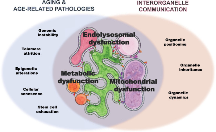
Our cells are comprised of billions of proteins, lipids, and other small molecules packed into their respective subcellular organelles, with the daunting task of maintaining cellular homeostasis over a lifetime. However, it is becoming increasingly evident that organelles do not act as autonomous discrete units but rather as interconnected hubs that engage in extensive communication through membrane contacts. In the last few years, our understanding of how these contacts coordinate organelle function has redefined our view of the cell. This review aims to present novel findings on the cellular interorganelle communication network and how its dysfunction may contribute to aging and neurodegeneration. The consequences of disturbed interorganellar communication are intimately linked with age-related pathologies. Given that both aging and neurodegenerative diseases are characterized by the concomitant failure of multiple cellular pathways, coordination of organelle communication and function could represent an emerging regulatory mechanism critical for long-term cellular homeostasis. We anticipate that defining the relationships between interorganelle communication, aging, and neurodegeneration will open new avenues for therapeutics.

Keywords: aging; cellular homeostasis; communication; contact sites; endolysosomal pathway; interorganelle communication; lipid metabolism; mitochondria; neurodegeneration.


Figures

Figure 1.
Figure 1.
Scheme of cellular communicome highlighting the contact sites and proteins implicated in age and age-related pathologies presented in this review. Please note that we presented only a subset of contacts and contact site proteins. ER-localized proteins are labeled in green. Lysosome proteins are labeled in pink. Cytosolic lipid transfer proteins are labeled in red. Mitochondria proteins are labeled in purple. Lipid droplet localized proteins are labeled in orange. Peroxisome proteins are red, and endosome proteins are fushia. Proteins can be localized to multiple organelles and participate at multiple distinct contact sites.
Figure 2.
Figure 2.
Magnifying glass view of the contact sites between the ER and lysosomes. ER-localized proteins are labeled in green. Lysosome proteins are labeled in pink. Cytosolic lipid transfer proteins are labeled in red. Lipid droplet localized proteins are labeled in orange.
Figure 3.
Figure 3.
Magnifying glass view of the contact sites between the ER, lipid droplet, and peroxisome. ER-localized proteins are labeled in green. Lipid droplet localized proteins are labeled in orange. Peroxisome proteins are red, and endosome proteins are fushia.
Figure 4.
Figure 4.
Magnifying glass view of the contact sites between the ER and mitochondria. ER-localized proteins are labeled in green. Lipid droplet localized proteins are labeled in orange. Lysosome proteins are labeled in pink. Cytosolic lipid transfer proteins are labeled in red. Mitochondria proteins are labeled in purple.
Figure 5.
Figure 5.
Magnifying glass view of the contact sites between the ER and endosome. ER-localized proteins are labeled in green. Lipid droplet localized proteins are labeled in orange. Cytosolic lipid transfer proteins are labeled in red.
Figure 6.
Figure 6.
Venn diagram showing the overlap of processes implicated in age and age-related pathologies as well as supported by interorganelle communication, on which we focused in this review: dysfunction in lipid metabolism, mitochondrial function, and endolysosomal system.

Similar articles

Cited by

References

    1. Allison R, Edgar JR, Pearson G, Rizo T, Newton T, Günther S, Berner F, Hague J, Connell JW, Winkler J, et al. 2017. Defects in ER–endosome contacts impact lysosome function in hereditary spastic paraplegia. J Cell Biol 216: 1337–1355. 10.1083/jcb.201609033 - DOI - PMC - PubMed
    1. Allison R, Edgar JR, Reid E. 2019. Spastin MIT domain disease-associated mutations disrupt lysosomal function. Front Neurosci 13: 1179. 10.3389/fnins.2019.01179 - DOI - PMC - PubMed
    1. Al-Saif A, Al-Mohanna F, Bohlega S. 2011. A mutation in σ1 receptor causes juvenile amyotrophic lateral sclerosis. Ann Neurol 70: 913–919. 10.1002/ana.22534 - DOI - PubMed
    1. Anchisi L, Dessì S, Pani A, Mandas A. 2012. Cholesterol homeostasis: a key to prevent or slow down neurodegeneration. Front Physiol 3: 486. - PMC - PubMed
    1. Area-Gomez E, del Carmen Lara Castillo M, Tambini MD, Guardia-Laguarta C, de Groof AJC, Madra M, Ikenouchi J, Umeda M, Bird TD, Sturley SL, et al. 2012. Upregulated function of mitochondria-associated ER membranes in Alzheimer disease: upregulated function of MAM in AD. EMBO J 31: 4106–4123. 10.1038/emboj.2012.202 - DOI - PMC - PubMed

Publication types