. 2021 Apr 16;15(4):e0009319.
doi: 10.1371/journal.pntd.0009319. eCollection 2021 Apr.

Trichomonas vaginalis infection impairs anion secretion in vaginal epithelium

Affiliations

Trichomonas vaginalis infection impairs anion secretion in vaginal epithelium

Jian-Bang Xu et al. PLoS Negl Trop Dis. .

Abstract

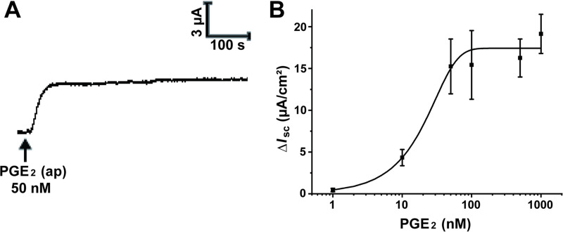
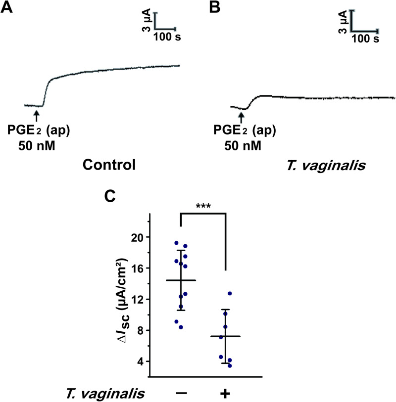
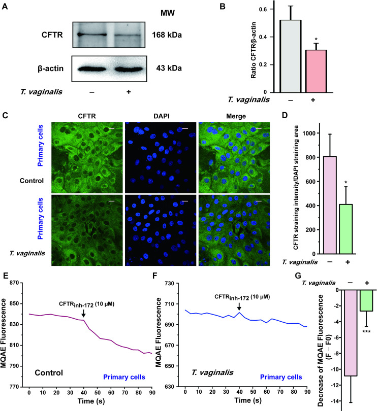
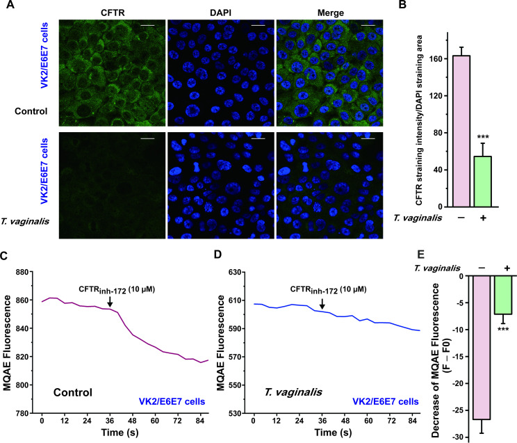
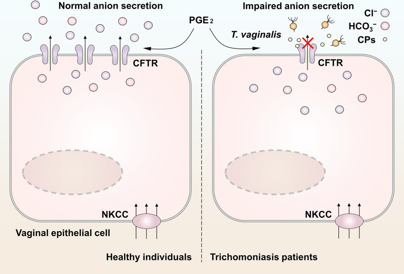
Trichomonas vaginalis is a common protozoan parasite, which causes trichomoniasis associated with severe adverse reproductive outcomes. However, the underlying pathogenesis has not been fully understood. As the first line of defense against invading pathogens, the vaginal epithelial cells are highly responsive to environmental stimuli and contribute to the formation of the optimal luminal fluid microenvironment. The cystic fibrosis transmembrane conductance regulator (CFTR), an anion channel widely distributed at the apical membrane of epithelial cells, plays a crucial role in mediating the secretion of Cl- and HCO3-. In this study, we investigated the effect of T. vaginalis on vaginal epithelial ion transport elicited by prostaglandin E2 (PGE2), a major prostaglandin in the semen. Luminal administration of PGE2 triggered a remarkable and sustained increase of short-circuit current (ISC) in rat vaginal epithelium, which was mainly due to Cl- and HCO3- secretion mediated by the cAMP-activated CFTR. However, T. vaginalis infection significantly abrogated the ISC response evoked by PGE2, indicating impaired transepithelial anion transport via CFTR. Using a primary cell culture system of rat vaginal epithelium and a human vaginal epithelial cell line, we demonstrated that the expression of CFTR was significantly down-regulated after T. vaginalis infection. In addition, defective Cl- transport function of CFTR was observed in T. vaginalis-infected cells by measuring intracellular Cl- signals. Conclusively, T. vaginalis restrained exogenous PGE2-induced anion secretion through down-regulation of CFTR in vaginal epithelium. These results provide novel insights into the intervention of reproductive complications associated with T. vaginalis infection such as infertility and disequilibrium in vaginal fluid microenvironment.


Conflict of interest statement

The authors have declared that no competing interests exist.

Figures

Fig 1
Fig 1. Apical administration of prostaglandin E2 (PGE2) stimulated an increase in short-circuit current (ISC) across rat vaginal epithelium.
(A) Representative trace showing the effect of the apical (ap) application of PGE2 (50 nM) on ISC response in rat vaginal epithelium. (B) The concentration-dependent curve of PGE2 induced ISC responses with a half-maximal effective concentration (EC50) of 24.9 nM. Symbols and bars indicate the means ± S.D. (n = 3–5).
Fig 2
Fig 2. Trichomonas vaginalis infection impaired the anion transport induced by prostaglandin E2 (PGE2) in rat vaginal epithelium.
(A-B) Representative trace showing the short-circuit current (ISC) response induced by apical (ap) PGE2 (50 nM) with (B) or without (A) intravaginal T. vaginalis infection in rats. (C) Statistical analysis showing the effect of T. vaginalis infection on the ISC currents induced by apical PGE2 (50 nM) in rat vaginal epithelium. Symbols and bars indicate the mean ± S.D. (n = 7–11, *** P < 0.001).
Fig 3
Fig 3. Trichomonas vaginalis infection induced down-regulation of cystic fibrosis transmembrane conductance regulator (CFTR) in the primary cultured rat vaginal epithelial cells.
(A) Representative blots showing the expression of CFTR in primary cultured vaginal epithelial cells infected with live 1 × 10⁶ T. vaginalis for 3 h, using β-actin as a loading control. MW, molecular weight. (B) Statistical analysis of Western blot (CFTR/β-actin ratio) showing the effect of T. vaginalis infection on the expression of CFTR. Symbols and bars indicate the mean ± S.D. (n = 3, * P < 0.05 versus the non-infected group). (C) Immunofluorescence images showing the expression of CFTR in primary cultured rat vaginal epithelial cells, in the absence or presence of 1 × 10⁶ T. vaginalis infection, with (D) the corresponding quantification analysis (n = 3, * P < 0.05 versus the non-infected group). Scale bar = 20 μm. (E) Representative trace showing the change of MQAE fluorescence elicited by CFTRinh-172 (10 μM) in primary cultured rat vaginal epithelial cells. (F) Representative trace showing the change of MQAE fluorescence elicited by CFTRinh-172 (10 μM) after 1 × 10⁶ T. vaginalis infection for 3 h. (G) Statistical analysis showing the change of MQAE fluorescence intensity elicited by CFTRinh-172 (10 μM), with or without T. vaginalis infection. Symbols and bars indicate the mean ± S.D. (n = 31–42 cells for each group, *** P < 0.001 versus the non-infected group).
Fig 4
Fig 4. Trichomonas vaginalis infection induced down-regulation of cystic fibrosis transmembrane conductance regulator (CFTR) in human vaginal epithelial VK2/E6E7 cells.
(A) Immunofluorescence images showing the expression of CFTR in VK2/E6E7 cells, in the absence or presence of 2×10⁵ T. vaginalis infection for 3 h, with (B) the corresponding quantification analysis (n = 3, *** P < 0.001 versus the non-infected group). Scale bar = 20 μm. (C) Representative trace showing the change of MQAE fluorescence elicited by CFTRinh-172 (10 μM) in VK2/E6E7 cells. (D) Representative trace showing the change of MQAE fluorescence elicited by CFTRinh-172 (10 μM) after 2×10⁵ T. vaginalis infection for 3 h. (E) Statistical analysis showing the change of MQAE fluorescence intensity elicited by CFTRinh-172 (10 μM), with or without T. vaginalis infection. Symbols and bars indicate the mean ± S.D. (n = 39–41 cells for each group, *** P < 0.001, versus the non-infected group).
Fig 5
Fig 5. Schematic model of the impaired anion secretion triggered by Trichomonas vaginalis infection in vaginal epithelium.
Seminal prostaglandin E2 (PGE2) facilitated anion secretion by activating cystic fibrosis transmembrane conductance regulator (CFTR) in vaginal epithelium. The basolateral Na+-K+-2Cl cotransporter (NKCC) guaranteed the maintenance of steady anion secretion by supplying Cl. After T. vaginalis infection, the CFTR was markedly down-regulated via cysteine proteases secreted by the pathogens, which restrained the exogenous PGE2-induced anion secretion. This may be a cause of the abnormal vaginal fluid microenvironment in patients with trichomoniasis.

Similar articles

Cited by

References

    1. Wira CR, Grant-Tschudy KS, Crane-Godreau MA. Epithelial cells in the female reproductive tract: A central role as sentinels of immune protection. Am J Reprod Immunol. 2005;53(2):65–76. 10.1111/j.1600-0897.2004.00248.x - DOI - PubMed
    1. Chan HC, Chen H, Ruan Y, Sun T. Physiology and pathophysiology of the epithelial barrier of the female reproductive tract: role of ion channels. Adv Exp Med Biol. 2012;763:193–217. 10.1007/978-1-4614-4711-5_10 - DOI - PubMed
    1. Csanady L, Vergani P, Gadsby DC. Structure, Gating, and Regulation of the CFTR Anion Channel. Physiol Rev. 2019;99(1):707–38. Epub 2018/12/06. 10.1152/physrev.00007.2018 - DOI - PubMed
    1. Chan HC, Shi QX, Zhou CX, Wang XF, Xu WM, Chen WY, et al.. Critical role of CFTR in uterine bicarbonate secretion and the fertilizing capacity of sperm. Mol Cell Endocrinol. 2006;250(1–2):106–13. Epub 2006/01/18. 10.1016/j.mce.2005.12.032 - DOI - PubMed
    1. Chan HC, Ruan YC, He Q, Chen MH, Chen H, Xu WM, et al.. The cystic fibrosis transmembrane conductance regulator in reproductive health and disease. J Physiol. 2009;587(Pt 10):2187–95. Epub 2008/11/19. 10.1113/jphysiol.2008.164970 - DOI - PMC - PubMed

Publication types

MeSH terms