Review
. 2021 Apr 16;478(7):1377-1397.
doi: 10.1042/BCJ20200502.

Nuts and bolts of the salt-inducible kinases (SIKs)

Affiliations
Review

Nuts and bolts of the salt-inducible kinases (SIKs)

Nicola J Darling et al. Biochem J. .

Abstract

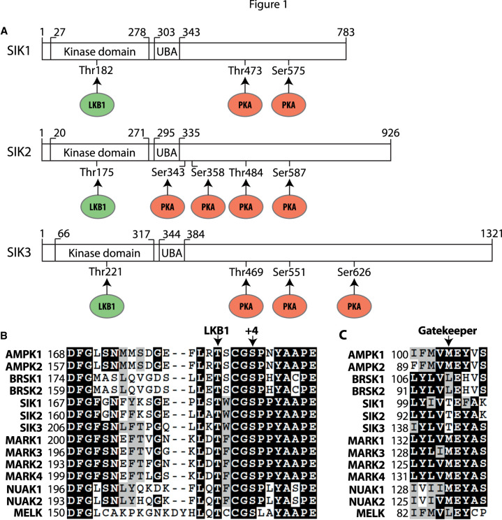
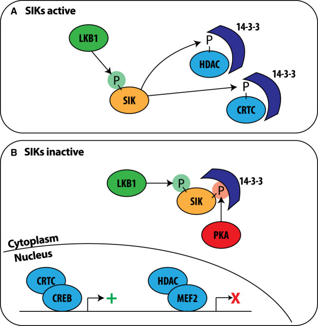
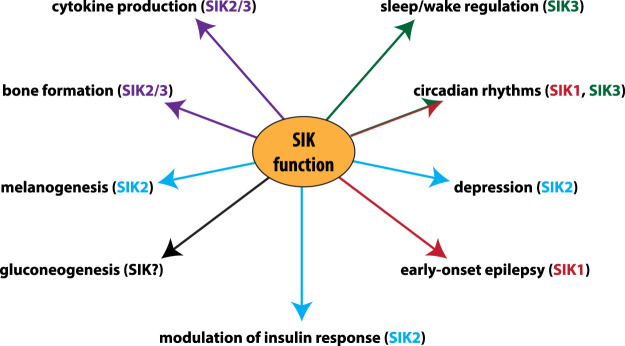
The salt-inducible kinases, SIK1, SIK2 and SIK3, most closely resemble the AMP-activated protein kinase (AMPK) and other AMPK-related kinases, and like these family members they require phosphorylation by LKB1 to be catalytically active. However, unlike other AMPK-related kinases they are phosphorylated by cyclic AMP-dependent protein kinase (PKA), which promotes their binding to 14-3-3 proteins and inactivation. The most well-established substrates of the SIKs are the CREB-regulated transcriptional co-activators (CRTCs), and the Class 2a histone deacetylases (HDAC4/5/7/9). Phosphorylation by SIKs promotes the translocation of CRTCs and Class 2a HDACs to the cytoplasm and their binding to 14-3-3s, preventing them from regulating their nuclear binding partners, the transcription factors CREB and MEF2. This process is reversed by PKA-dependent inactivation of the SIKs leading to dephosphorylation of CRTCs and Class 2a HDACs and their re-entry into the nucleus. Through the reversible regulation of these substrates and others that have not yet been identified, the SIKs regulate many physiological processes ranging from innate immunity, circadian rhythms and bone formation, to skin pigmentation and metabolism. This review summarises current knowledge of the SIKs and the evidence underpinning these findings, and discusses the therapeutic potential of SIK inhibitors for the treatment of disease.

Keywords: AMPK-related kinase; CREB; CREB-regulated transcriptional co-activator (CRTC); histone deacetylase (HDAC); myocyte enhancer factor 2 (MEF2); salt-inducible kinase (SIK).


Conflict of interest statement

The authors declare that there are no competing interests associated with the manuscript.

Figures

Figure 1.
Figure 1.. Domain structure of the SIKs and sites of regulatory phosphorylation.
(A) Schematic of the domain structures of human SIK1 (Uniprot P57059), SIK2 (Q9H0K1) and SIK3 (Q9Y2K2) showing the threonine in the N-terminal kinase domain phosphorylated by LKB1 (green), the ubiquitin-associated (UBA) domain and the C-terminal tail with the sites of phosphorylation by PKA (red). The equivalent phosphorylation sites in murine SIK1 (Q60670) are Thr182, Thr475 and Ser577; the equivalent phosphorylation sites in murine SIK2 (Q8CFH6) are Thr175, Ser343, Ser358, Thr484 and Ser587 and the equivalent phosphorylation sites in murine SIK3 (Q6P4S6) are Thr163, Thr411, Ser493 and Ser616. (B) Alignment of the activation loop sequences of human AMPK-related kinases. Marked are the Thr phosphorylated by LKB1 and the conserved Ser at +4 reported to be a site of autophosphorylation. Identical residues are shaded in black and conserved residues are shaded in grey. (C) As in B except that the gatekeeper Thr residue is marked.
Figure 2.
Figure 2.. Consequences of the activation and inactivation of the SIKs by phosphorylation.
(A) SIKs (orange) are activated by LKB1-dependent phosphorylation (green P) and phosphorylate (P) CRTC family members and Class 2a HDACs (HDAC4/5/7/9). These phosphorylated substrates (light blue) bind to 14-3-3s which retains them in the cytosol. (B) The SIKs are phosphorylated by PKA-dependent phosphorylation (red P) inducing binding to 14-3-3 proteins (dark blue) and inactivation. PKA-dependent phosphorylation may also promote SIK1 translocation from the nucleus to the cytoplasm (not shown). Inactivation of SIKs leads to the dephosphorylation and nuclear translocation of CRTCs and Class 2a HDACs. Within the nucleus CRTCs promote (green +) CREB-dependent gene transcription and Class 2a HDACs inhibit (red X) MEF2-dependent transcription.
Figure 3.
Figure 3.. Chemical structures of compounds inhibiting SIKs.
Chemical structures of compounds inhibiting SIKs at low nM concentrations that have been used to study the functions of SIKs in cells and in vivo. KIN-112, HG-9-91-01, YKL-05-099, YKL-06-061, YKL-06-062, Dasatinib and Bosutinib target the gatekeeper site on SIKs (Figure 1C) whereas MRT67307 and MRT199665 do not. *Pterosin B treatment mimics several effects of SIK3 KO in hepatocytes and chondrocytes but is not thought to be a direct inhibitor of SIK3.
Figure 4.
Figure 4.. Physiological roles of different SIK isoforms identified by genetic analysis.
The contribution of individual SIK isoforms to the regulation of physiological processes identified by the use of knock-out and/or knock-in mice. The major SIK isoform/s responsible for each role is colour coded; SIK1 (red), SIK2 (blue), SIK3 (green), SIK2 and SIK3 (purple). (SIKs) The SIKs have a role in modulating gluconeogenesis in mouse hepatocytes, but which SIK isoform(s) is responsible is unknown. In humans, mutations in SIK1 have been identified in patients with early-onset epilepsy. Consistent with the role in bone formation, a point mutation in SIK3 has been identified in a human patient with skeletal defects.

References

    1. Hardie, D.G., Ross, F.A. and Hawley, S.A. (2012) AMPK: a nutrient and energy sensor that maintains energy homeostasis. Nat. Rev. Mol. Cell Biol. 13, 251–262 10.1038/nrm3311 - DOI - PMC - PubMed
    1. Rena, G., Pearson, E.R. and Sakamoto, K. (2013) Molecular mechanism of action of metformin: old or new insights? Diabetologia 56, 1898–1906 10.1007/s00125-013-2991-0 - DOI - PMC - PubMed
    1. LaMoia, T.E. and Shulman, G.I. (2021) Cellular and molecular mechanisms of metformin action. Endocr. Rev. 42, 77–96 10.1210/endrev/bnaa023 - DOI - PMC - PubMed
    1. Wang, Z., Takemori, H., Halder, S.K., Nonaka, Y. and Okamoto, M. (1999) Cloning of a novel kinase (SIK) of the SNF1/AMPK family from high salt diet-treated rat adrenal. FEBS Lett. 453, 135–139 10.1016/S0014-5793(99)00708-5 - DOI - PubMed
    1. Horike, N., Takemori, H., Katoh, Y., Doi, J., Min, L., Asano, T.et al. (2003) Adipose-specific expression, phosphorylation of Ser794 in insulin receptor substrate-1, and activation in diabetic animals of salt-inducible kinase-2. J. Biol. Chem. 278, 18440–18447 10.1074/jbc.M211770200 - DOI - PubMed

Publication types

MeSH terms

Substances