. 2021 Jun 21;31(12):2507-2519.e4.
doi: 10.1016/j.cub.2021.03.041. Epub 2021 Apr 15.

The nucleolus as a polarized coaxial cable in which the rDNA axis is surrounded by dynamic subunit-specific phases

Affiliations

The nucleolus as a polarized coaxial cable in which the rDNA axis is surrounded by dynamic subunit-specific phases

Alan M Tartakoff et al. Curr Biol. .

Abstract

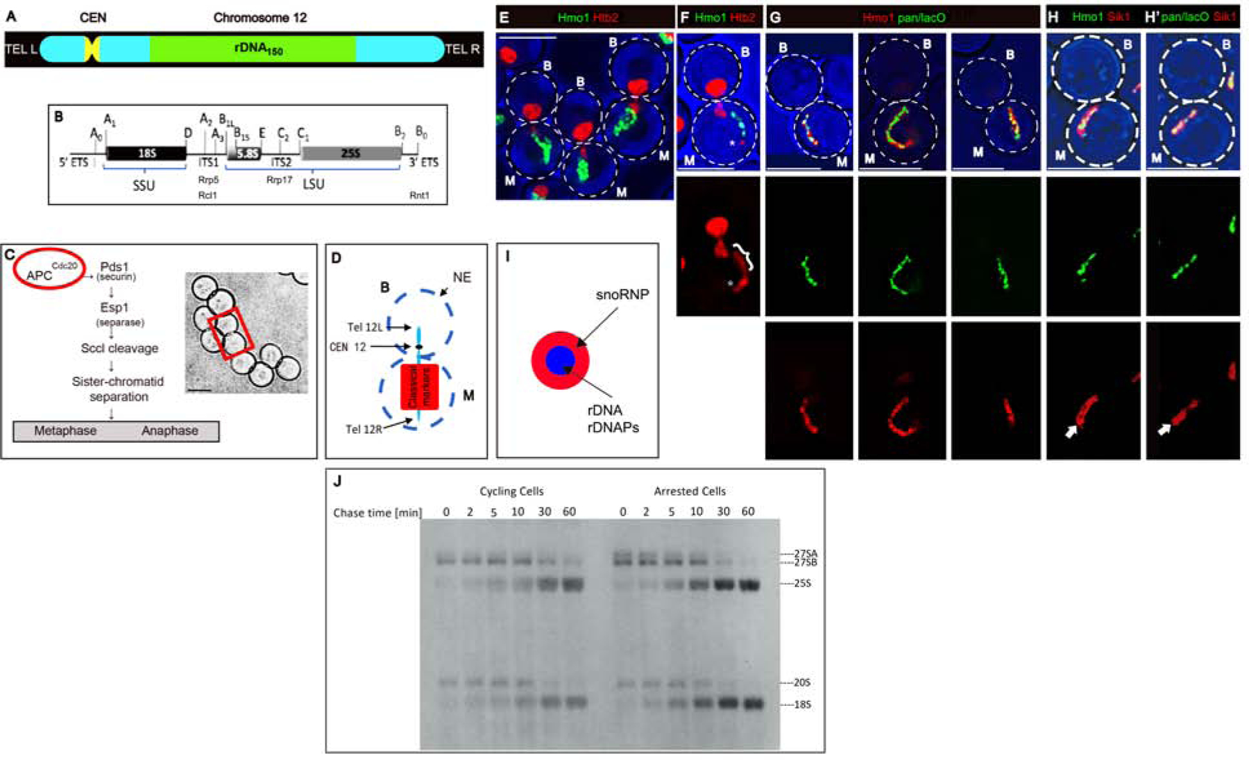
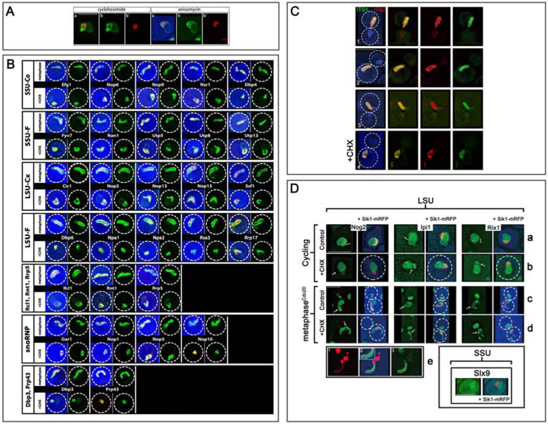
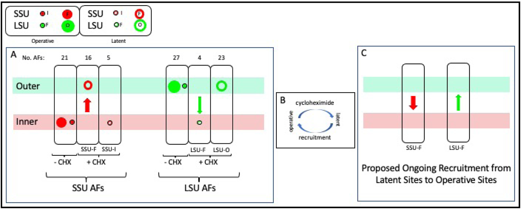
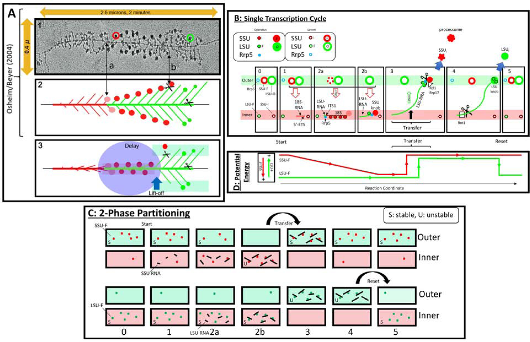
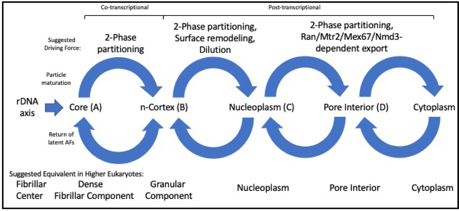
In ribosomal DNA (rDNA) repeats, sequences encoding small-subunit (SSU) rRNA precede those encoding large-subunit (LSU) rRNAs. Processing the composite transcript and subunit assembly requires >100 subunit-specific nucleolar assembly factors (AFs). To investigate the functional organization of the nucleolus, we localized AFs in S. cerevisiae in which the rDNA axis was "linearized" to reduce its dimensionality, thereby revealing its coaxial organization. In this situation, rRNA synthesis and processing continue. The axis is embedded in an inner layer/phase of SSU AFs that is surrounded by an outer layer/phase of LSU AFs. When subunit production is inhibited, subsets of AFs differentially relocate between the inner and outer layers, as expected if there is a cycle of repeated relocation whereby "latent" AFs become "operative" when recruited to nascent subunits. Recognition of AF cycling and localization of segments of rRNA make it possible to infer the existence of assembly intermediates that span between the inner and outer layers and to chart the cotranscriptional assembly of each subunit. AF cycling also can explain how having more than one protein phase in the nucleolus makes possible "vectorial 2-phase partitioning" as a driving force for relocation of nascent rRNPs. Because nucleoplasmic AFs are also present in the outer layer, we propose that critical surface remodeling occurs at this site, thereby partitioning subunit precursors into the nucleoplasm for post-transcriptional maturation. Comparison to observations on higher eukaryotes shows that the coaxial paradigm is likely to be applicable for the many other organisms that have rDNA repeats.

Keywords: 2-phase partitioning; assembly factors; nucleolar domain separation; nucleolar subcompartments; nucleolus; protein phase; rDNA; ribosomal subunit biogenesis; yeast.


Conflict of interest statement

Declaration of interests The authors declare no competing interests.

Figures

Figure 1
Figure 1. In Arrested Cells the rDNA Axis is Colinear with Hmo1 and Processing of pre-rRNA Continues
A) Organization of chromosome XII. B) A single rDNA repeat, indicating segments that code for SSU and LSU rRNAs and the sites of binding (Rrp5) or potential cleavage (Rcl1, Rrp17, Rnt1). C) Left: Overview of the consequences of activation of the anaphase promoting complex. Right: Arrested cells. The rectangle encloses a typical mother/bud pair. D) Diagram of the position of chromosome XII and classical markers in arrested cells. For all panels, M: mother; B: bud. The scale bar in all figures is 5 microns. E) Projected image of arrested cells that express the histone, Htb2-mRFP, and Hmo1-GFP. Strain: ATY10569. F) Single image plane of an arrested cell showing that the elongated rDNA segment has a Htb2-mRFP at the distal extremity (*). The bracket designates the Hmo1-GFP-positive segment. Strain: ATY10569. G) Single image planes of arrested cells that express pan-lacO-tagged rDNA (green) and Hmo1-Apple (red). Strain: ATY10682. H) Comparison of the filamentous Hmo1-Apple (H) and pan-lacO (H’) with the broader Sik1/Nop56-mRFP signal. In each case, the Sik1-mRFP domain includes a non-fluorescent central element (arrows) that approximately coincides with rDNA. Strains: ATY10342, ATY10747. I) Scheme indicating that rDNA and rDNAPs define an axis internal to snoRNP proteins. J) To compare the synthesis and turnover of pre-rRNAs in cycling cells and arrested cells, metaphaseCdc20 cells carrying the URA3 plasmid, pRS316, were pre-grown in medium lacking methionine and uracil. Half of the culture was transferred to synthetic media including methionine for 3 hours. Both samples were pulse-labeled for 10 min with [5.6-3 H]-uridine and chased with an excess of unlabeled uridine for the indicated times. Pre-rRNAs and mature rRNA species are labeled. Due to long pulse, the earliest pre-rRNA processing intermediates (35S, 32S) had already been processed to intermediates. Strain: ATY10402 [pRS316]. Related to Figure S1.
Figure 2
Figure 2. Nucleolar Assembly Factors Localize to Coaxial Layers
A) Distributions of AFs in Cycling Cells. In panels (1–5), Sik1-mRFP is included to define the crescent. Panels (1–2): pan-lacO dots are throughout the crescent. Panels (3–5) illustrate AFs that localize either to the crescent with a weak signal along the NE and a weak signal throughout the nucleoplasm (Mak11), to the nucleoplasm (Rea1), or exclusively to the crescent (Rrp9). In (3), the small boxes illustrate the single colors. Panel (6) illustrates Nmd3, that localizes largely to the cytoplasm, co-expressed with a marker of the NE/ER: mRFP-HDEL. Strains: ATY10659, ATY8300, ATY7833, ATY7838, ATY8119. B) In arrested cells, the snoRNP, GFP-Nop1, colocalizes with Sik1-mRFP. The panels show the two colors separately or combined. Strain: ATY10412. C) In arrested cells, the SSU AF, Efg1-GFP, colocalizes with Sik1-mRFP. Strain: ATY10383. D) In arrested cells, the LSU AF, Mak11-GFP, surrounds Sik1-mRFP. Strain: ATY8297. E) Comparison of the LSU AF, Ytm1-Apple, to the SSU AF, Utp30-GFP. (E’) comparison of Mak11-GFP to Utp30-CFP, all in arrested cells. Strains: ATY10589, ATY10400. F) Comparison of LSU AFs, Ytm1-Apple and Mak11-GFP, to markers of the NE/ER: GFP-HDEL and mRFP-HDEL, all in arrested cells. Strains: ATY10717, ATY10722. G) Scheme of the coaxial organization of the nucleolar cable. H/I) Schematic of Distributions of AFs in Cells that are Making Subunits. Left) This two-phase diagram (T-diagram) depicts the inner and outer layers/phases as two separated horizontal bands (pink, green). Inner layer AFs are snoRNP proteins, Rpa subunits, Dbp3, Prp43, SSU AFs, Rrp5, and Rnt1. Among outer layer AFs are Rcl1 and many LSU AFs, including Rrp17. SSU AFs are red and LSU AFs are green. Proteins that contribute to both subunits have both colors. Right) This related diagram includes the nucleoplasmic compartment in blue and the axis in yellow. As is described later in this text, multiple AFs that are conspicuous in the nucleoplasm (dark blue) are also detected in the outer layer for arrested cells. Htb2 is found both along the axis and throughout the nucleoplasm. By contrast, Hmo1 (green) is associated with the axis but is not detected in the nucleoplasm. Related to Figure S2.
Figure 3
Figure 3. Localizations of Nucleolar Assembly Factors and rRNA
A) Cycling cells expressing Mak11-GFP and Sik1-mRFP were treated with cycloheximide or anisomycin for 30 min. Note the conspicuous domain separation of the two colors, with GFP surrounding the red signal. Before treatment, the two colors were extensively intermixed. Note that a faint GFP signal extends around the nucleus. Strain: ATY8112. B) Cells expressing the indicated AF-GFP fusions, as well as Sik1-mRFP, were either arrested (metaphase) or, when cycling, were treated with cycloheximide (+ CHX). In each case, we include both a 3-color image and an image of the GFP-tagged protein by itself. For the arrested cells, only the mother domain is included. The dotted circle indicates the cell perimeter in all figures. 16 further examples of SSU-F and LSU-Ou AFs are in Figure S2. C) To localize different portions of rRNA in arrested cells, we used fluorescent in situ hybridization. As illustrated for three representative cells (1–3), sequences upstream of cleavage site A2 in ITS1 emphasize the inner layer (green signal), while sequences within ITS2 have more external localization (red signal). The blue signal is DAPI. The final row of images (4) illustrates the impact of cycloheximide (30 min) before fixation. Note that the two signals have become coincident. Strain: ATY10402. D) Nucleoplasmic AFs fill the outer layer but chromatin is absent. Rows (a-d): The distributions of three LSU AFnpls (Nog2, Ipi1, Rix1) are illustrated +/− Sik1-mRFP. Each protein is abundant in the nucleoplasm and can also be detected (weakly) in the nucleolar crescent (bracket) where the interface with the nucleoplasm is often highlighted (row (a)). The SSU AF, Slx9, that is also present in the cytoplasm, is included in the separate panel in the lower right. Upon arrest (row c), Nog2, Ipi1 and Rix1 continue to be visible throughout the nucleoplasm (asterisks) and along the outer layer (arrows). They do not coincide with Sik1-mRFP. When cycling cells are treated with cycloheximide (row (b) vs row (a)) these LSU AFnpls still fill the nucleoplasm. It is unclear whether they become depleted from the region occupied by Sik1-mRFP (arrows). In arrested cells, the nucleoplasmic signal persists, as does highlighting of the outer layer (arrows). It is not obvious that the intensity of this highlighting is accentuated by comparison to controls (row d vs row c). There is little or no overlap with the condensed Sik1-mRFP. Strains: ATY8105, ATY8126, ATY8280, ATY10770, ATY10774, ATY10785. Row (e). Cells expressing Mak11-GFP and Htb2-mRFP were arrested and imaged. Note the elongated domain (in the mother) in which the Htb2-mRFP-labeled axis is flanked by the GFP signal. Strain: ATY10157. Related to Figures S2 and S5.
Figure 4
Figure 4. Differential Relocation of Subunit-Specific Assembly Factors
A) Three ovals at the left: In cells that are making subunits, both SSU-In and SSU-F AFs localize to the inner layer (first oval). When assembly is inhibited, SSU-F AFs relocate (red arrow) to the outer layer while SSU-In AFs remain in the inner layer. Second group of three ovals: LSU-Ou and LSU-F AFs concentrate along the outer layer when subunits are being produced. The LSU-F subset relocates (green arrow) to the inner layer when assembly is inhibited, while the LSU-Ou subset remains along the outer layer. The size of the symbols reflects the relative number of AFs in each group. B) We hypothesize that each AF repeatedly cycles between an operative state and a latent state and that cycloheximide stops recruitment to the operative state. C) We propose that relocation of SSU-F AFs from the inner layer to the outer layer upon addition of cycloheximide (and inverse relocation of LSU-F AFs) signifies that these AFs normally transit in the opposite directions during each cycle of transcription. Related to Figure S3 and Figure S2B.
Figure 5
Figure 5. Transcript Maturation, the Polarity of rDNA and Energy Relations
A) Transcript elongation. Panels (1–2) are modified from. The rDNA and rRNA segments are color-coded: red for 5’ETS and SSU rRNA/DNA, green for LSU rRNA/DNA. Panel 1: EM of a spread of yeast rDNA during transcription. Note the horizontal rDNA axis and the lateral emergence of transcripts. A SSU terminal knob is circled in red, and a putative LSU knob is circled in green. ITS1 is indicated as (a). The removal of SSU knobs occurs at (b). Panel 2: Once transcription reaches ITS1, SSU pre-knobs (pink) and knobs (red) appear. They persist until transcription has reached into LSU sequences. Panel 3: Since cleavage at ITS1 is delayed well beyond the point at which the 3’ extremity of the SSU rRNA coding sequences has been reached, we propose that removal requires transfer to the outer layer. This could allow them to undergo further maturation and, likely, to be cleaved by Rcl1 or Utp24. Further elongation, processing, and formation of particulate intermediates would also occur along the outer layer. Final cleavage occurs at site Bo near the 3’ extremity. B) Suggested sequential processing of rRNA. See the text for a detailed description of these T-Diagrams. In frames (2a) and (3), the interruption of the perimeter of the large red circles designates progressive release of AFs. AFs that are required for both subunits are not included. The solid circular symbols imply that the indicated AFs are associated with maturing subunits. When not associated, the symbols have a white center. C) Vectorial 2-Phase Partitioning. Schematic of the relocation of SSU-F AFs and LSU-F AFs. The upper two rows pertain to SSU maturation and the lower two rows pertain to LSU maturation. We consider the localization of these AFs after cycloheximide treatment to be an indication of the phase in which they are most stable (S). By contrast, once they become associated with nascent transcripts, they localize to phases in which they are relatively unstable (U). D) Energy Relations. During a single cycle, the present observations suggest that 5’-ETS AFs and SSU-F AFs begin in the outer layer and relocate to the inner layer as they load onto nascent rRNA. Phase transfer then allows them to return to the outer layer. This is energetically downhill. Reciprocally, the LSU-F AFs (green line) begin in the inner layer, but shift to the outer layer, perhaps in conjunction with transfer of SSU-Fs. At the end of the cycle, their downhill return to the inner layer resets the system for repeated use. The vertical arrows at the left designate the energy relations. Coupling of inner-to-outer flux of both SSU precursors and LSU-F AFs (states 2b/3) could make their reciprocal flux energetically neutral. Related to Figure S6
Figure 6
Figure 6. Proposed Steps of Translocation of Immature Subunits and Assembly Factors
Cycle I can be attributed to vectorial 2-phase partitioning. It is bidirectional since latent SSU-F and latent LSU-F AFs relocate in opposite directions between inner and outer layers. Only during this first cycle do the immature subunits include nascent rRNAs. Cycle II relies on surface AFs that make it possible for subunit precursors to intermix with chromatin. Cycle III involves binding to export factors (exportins, Mex67/Mtr2, Nmd3) that confer compatibility with the interior of the nuclear pore. Cycle IV corresponds to release into the cytoplasm and return of export factors. Related to Figure S2 and S6.

Similar articles

Cited by

References

    1. Nerurkar P, Altvater M, Gerhardy S, Schutz S, Fischer U, Weirich C, et al. Eukaryotic Ribosome Assembly and Nuclear Export. Int Rev Cell Mol Biol. 2015;319:107–40. - PubMed
    1. Klinge S, Woolford JL Jr. Ribosome assembly coming into focus. Nature reviews Molecular cell biology. 2019;20(2):116–31. - PMC - PubMed
    1. Pena C, Hurt E, Panse VG. Eukaryotic ribosome assembly, transport and quality control. Nature structural & molecular biology. 2017;24(9):689–99. - PubMed
    1. Bassler J, Hurt E. Eukaryotic Ribosome Assembly. Annu Rev Biochem. 2019;88:281–306. - PubMed
    1. Osheim YN, French SL, Keck KM, Champion EA, Spasov K, Dragon F, et al. Pre-18S ribosomal RNA is structurally compacted into the SSU processome prior to being cleaved from nascent transcripts in Saccharomyces cerevisiae. Molecular cell. 2004;16(6):943–54. - PubMed

Publication types

MeSH terms

LinkOut - more resources