. 2021 Jan-Jun:296:100661.
doi: 10.1016/j.jbc.2021.100661. Epub 2021 Apr 14.

The position of single-base deletions in the VNTR sequence of the carboxyl ester lipase (CEL) gene determines proteotoxicity

Affiliations

The position of single-base deletions in the VNTR sequence of the carboxyl ester lipase (CEL) gene determines proteotoxicity

Anny Gravdal et al. J Biol Chem. 2021 Jan-Jun.

Abstract

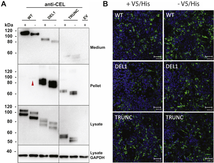
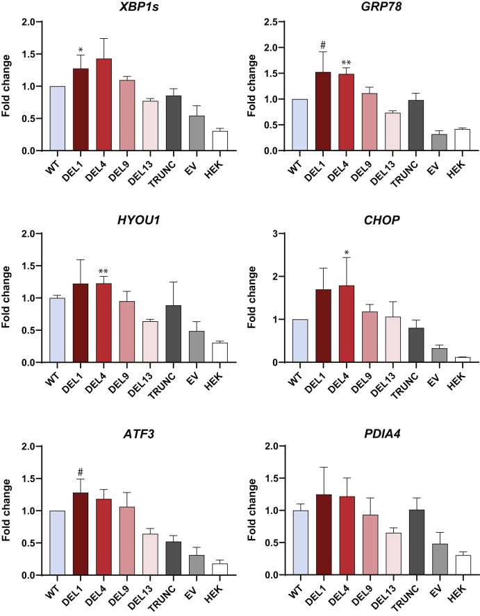
Variable number of tandem repeat (VNTR) sequences in the genome can have functional consequences that contribute to human disease. This is the case for the CEL gene, which is specifically expressed in pancreatic acinar cells and encodes the digestive enzyme carboxyl ester lipase. Rare single-base deletions (DELs) within the first (DEL1) or fourth (DEL4) VNTR segment of CEL cause maturity-onset diabetes of the young, type 8 (MODY8), an inherited disorder characterized by exocrine pancreatic dysfunction and diabetes. Studies on the DEL1 variant have suggested that MODY8 is initiated by CEL protein misfolding and aggregation. However, it is unclear how the position of single-base deletions within the CEL VNTR affects pathogenic properties of the protein. Here, we investigated four naturally occurring CEL variants, arising from single-base deletions in different VNTR segments (DEL1, DEL4, DEL9, and DEL13). When the four variants were expressed in human embryonic kidney 293 cells, only DEL1 and DEL4 led to significantly reduced secretion, increased intracellular aggregation, and increased endoplasmic reticulum stress compared with normal CEL protein. The level of O-glycosylation was affected in all DEL variants. Moreover, all variants had enzymatic activity comparable with that of normal CEL. We conclude that the longest aberrant protein tails, resulting from single-base deletions in the proximal VNTR segments, have highest pathogenic potential, explaining why DEL1 and DEL4 but not DEL9 and DEL13 have been observed in patients with MODY8. These findings further support the view that CEL mutations cause pancreatic disease through protein misfolding and proteotoxicity, leading to endoplasmic reticulum stress and activation of the unfolded protein response.

Keywords: CEL; MODY8; O-glycosylation; endoplasmic reticulum stress; protein misfolding; single-base deletions; unfolded protein response.


Conflict of interest statement

Conflict of interest The authors declare that they have no conflicts of interest with the contents of this article.

Figures

Figure 1
Figure 1
Schematic overview of the different CEL protein variants encoded by constructs used in the present study.A, the normal CEL protein (CEL-WT) with 16 repeated segments, each consisting of 11 amino acids and encoded by the VNTR. This is the most common CEL variant in the general population. The yellow box symbolizes the N-terminal signal peptide, whereas light blue boxes represent the normal repeats. The unique C-terminal sequence of CEL-WT is symbolized by a gray box. Also indicated are residues required for bile salt binding, N- and O-glycosylation, phosphorylation, and catalysis. B, overview of the different CEL protein variants investigated. Predicted molecular mass (kDa) and the isoelectric point (pI) of each variant are shown on the right side. All DEL constructs were based on a 16-repeat backbone. Light blue boxes represent normal repeat sequences, whereas red boxes indicate aberrant repeats resulting from the frameshifts introduced by single–base deletions in the CEL VNTR. CEL-TRUNC is an artificial variant lacking the VNTR region and was included as control construct. Elements of A and B are not drawn to scale. DEL, deletion; VNTR, variable number of tandem repeat.
Figure 2
Figure 2
Secretion and intracellular distribution of tagged versus untagged CEL variants in HEK293 cells. HEK293 cells were transiently transfected with plasmids encoding CEL-WT, CEL-DEL1, or CEL-TRUNC with or without the V5/His tag (+/−). The expressed CEL proteins were examined by Western blotting (A) and immunocytochemistry (B). Representative images from n = 3 independent experiments are shown. A, secretion was evaluated as CEL levels detected in the conditioned medium. Intracellular distribution was assessed by analyzing the soluble (lysate) and insoluble (pellet) cellular fractions. Cells transfected with the empty vector (EV) were used as the negative control, whereas GAPDH expression was monitored for control of loading. The red arrow points at a very weak band for untagged CEL-WT protein in the pellet fraction. The stippled line indicates an unrelated lane removed from the images. B, transfected cells were subjected to immunostaining and confocal microscopy. CEL protein is stained green and cell nuclei are stained blue. Scale bars represent 50 μm. DEL, deletion; HEK293, human embryonic kidney 293.
Figure 3
Figure 3
Secretion and cellular distribution of different CEL protein variants.A, HEK293 cells were transiently transfected with plasmids encoding the CEL variants shown in Figure 1 and subjected to cellular fractionation and Western blotting. Cells transfected with the empty vector (EV) were used as negative control, whereas GAPDH expression was monitored for control of loading. Secretion was assessed as CEL levels detected in the conditioned medium. Intracellular distribution was evaluated by analyzing the soluble (lysate) and insoluble (pellet) fractions after lysis of the cells. The stippled line indicates an unrelated lane removed from the images. Representative images from n = 3 independent experiments are shown. B, quantification of Western blot band intensities in the three experiments, adjusted to the GAPDH levels and normalized to the CEL-WT lane. Error bars are SD. Statistical significance is indicated as follows for band intensities different from that of CEL-WT: ∗p ≤ 0.05; ∗∗p ≤ 0.01; ∗∗∗p ≤ 0.001. HEK293, human embryonic kidney 293.
Figure 4
Figure 4
Expression of CEL variants in an O-glycosylation-deficient cellular model. HEK293 cells (A) and HEK293 cells with Cosmc KO (SimpleCells) (B) were transiently transfected with plasmids encoding the CEL variants shown in Figure 1 and analyzed by Western blotting. Cells transfected with the empty vector (EV) were used as negative control. Tubulin expression was monitored for control of loading. C-terminal O-glycosylation was assessed as shifts in protein migration in the SimpleCells when compared with expression in nonaltered HEK293 cells. The positions of the heaviest protein bands in each lane (except TRUNC), present in unmodified HEK293 cells but missing in the SimpleCells, are indicated in the lower image by red arrowheads. This reflects the difference in CEL O-glycosylation level between the two cell lines. Secretion was evaluated based on loading equal amounts of protein from the conditioned media. The band observed at ∼60 kDa in all lanes is an unspecific band occasionally observed in the lysate fraction with the antibody used. Representative images from n = 3 independent experiments are shown. HEK293, human embryonic kidney 293.
Figure 5
Figure 5
Effect of CEL variants on ER stress markers at the mRNA level. HEK293 cells were transiently transfected with plasmids encoding the CEL variants shown in Figure 1, and mRNA levels were analyzed by real-time quantitative PCR (n = 3). Cells transfected with the empty vector (EV), and untransfected cells (HEK) were used as negative controls. For each marker, the mRNA expression level was corrected to the geometric mean of three reference genes (OAZ1, GAPDH, and ACTB) and then normalized to the marker's expression in cells transfected with CEL-WT. Error bars are SD. Statistical significance is indicated as follows for the mRNA level different from that of the CEL-WT experiment: ∗p ≤ 0.05; ∗∗p ≤ 0.01. Borderline significance (#) for two markers was observed for the DEL1 variant (GRP78, p = 0.055; ATF3, p = 0.059). DEL, deletion; ER, endoplasmic reticulum; HEK293, human embryonic kidney 293.
Figure 6
Figure 6
Effect of CEL variants on ER stress markers at the protein level.A, HEK293 cells were transiently transfected with plasmids encoding the CEL variants shown in Figure 1, and expression of ER stress markers was assessed by Western blotting of the soluble (lysate) and insoluble (pellet) fractions. Cells transfected with the empty vector (EV) were used as negative control, whereas GAPDH expression was monitored for control of loading. For each of the ER stress markers GRP78, calnexin, and PERK, representative images from n = 3 independent experiments are shown. Stippled lines for two of the markers indicate unrelated lanes removed from the images. B, quantification of marker band intensities after adjustment to the GAPDH levels and normalization to the expression in CEL-WT–transfected cells. Error bars are SD. The asterisk denotes statistical significance (p ≤ 0.05) for band intensities different from that of the CEL-WT experiment. ER, endoplasmic reticulum; HEK293, human embryonic kidney 293.
Figure 7
Figure 7
Enzymatic activity of the studied CEL variants. HEK293T cells were transiently transfected with plasmids encoding the CEL variants shown in Figure 1. A, SDS-PAGE of CEL variants purified from the conditioned medium and stained with Gel-Code Blue. The same amount of protein was loaded in each lane. The stippled line indicates an unrelated lane removed from the image. A representative image from n = 3 independent experiments is shown. B, CEL lipolytic activity in the medium measured with trioctanoate as the substrate. The activity was adjusted for band density (as in panel A) and molecular weight of each variant. Error bars are SD. HEK293, human embryonic kidney 293.

References

    1. Brookes K.J. The VNTR in complex disorders: The forgotten polymorphisms? A functional way forward? Genomics. 2013;101:273–281. - PubMed
    1. Jeffreys A.J., Wilson V., Thein S.L. Individual-specific ‘fingerprints’ of human DNA. Nature. 1985;316:76–79. - PubMed
    1. Cai C.Q., Zhang T., Breslin M.B., Giraud M., Lan M.S. Both polymorphic variable number of tandem repeats and autoimmune regulator modulate differential expression of insulin in human thymic epithelial cells. Diabetes. 2011;60:336–344. - PMC - PubMed
    1. Simpson J., Vetuz G., Wilson M., Brookes K.J., Kent L. The DRD4 receptor Exon 3 VNTR and 5' SNP variants and mRNA expression in human post-mortem brain tissue. Am. J. Med. Genet. B Neuropsychiatr. Genet. 2010;153:1228–1233. - PubMed
    1. Johansson B.B., Fjeld K., El Jellas K., Gravdal A., Dalva M., Tjora E., Ræder H., Kulkarni R.N., Johansson S., Njølstad P.R., Molven A. The role of the carboxyl ester lipase (CEL) gene in pancreatic disease. Pancreatology. 2018;18:12–19. - PubMed

Publication types

LinkOut - more resources