

'Visual' cortices of congenitally blind adults are sensitive to response selection demands in a go/no-go task
- PMID: 33862241
- PMCID: PMC8249356
- DOI: 10.1016/j.neuroimage.2021.118023
'Visual' cortices of congenitally blind adults are sensitive to response selection demands in a go/no-go task
Abstract
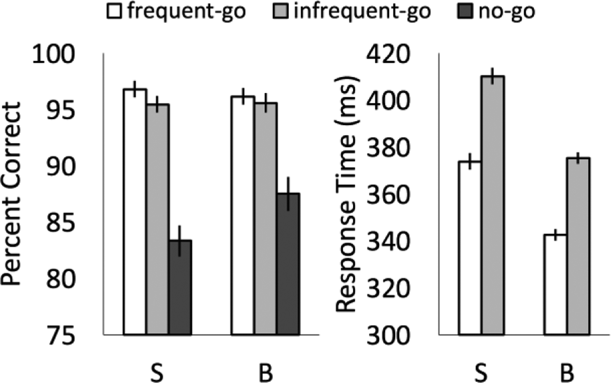
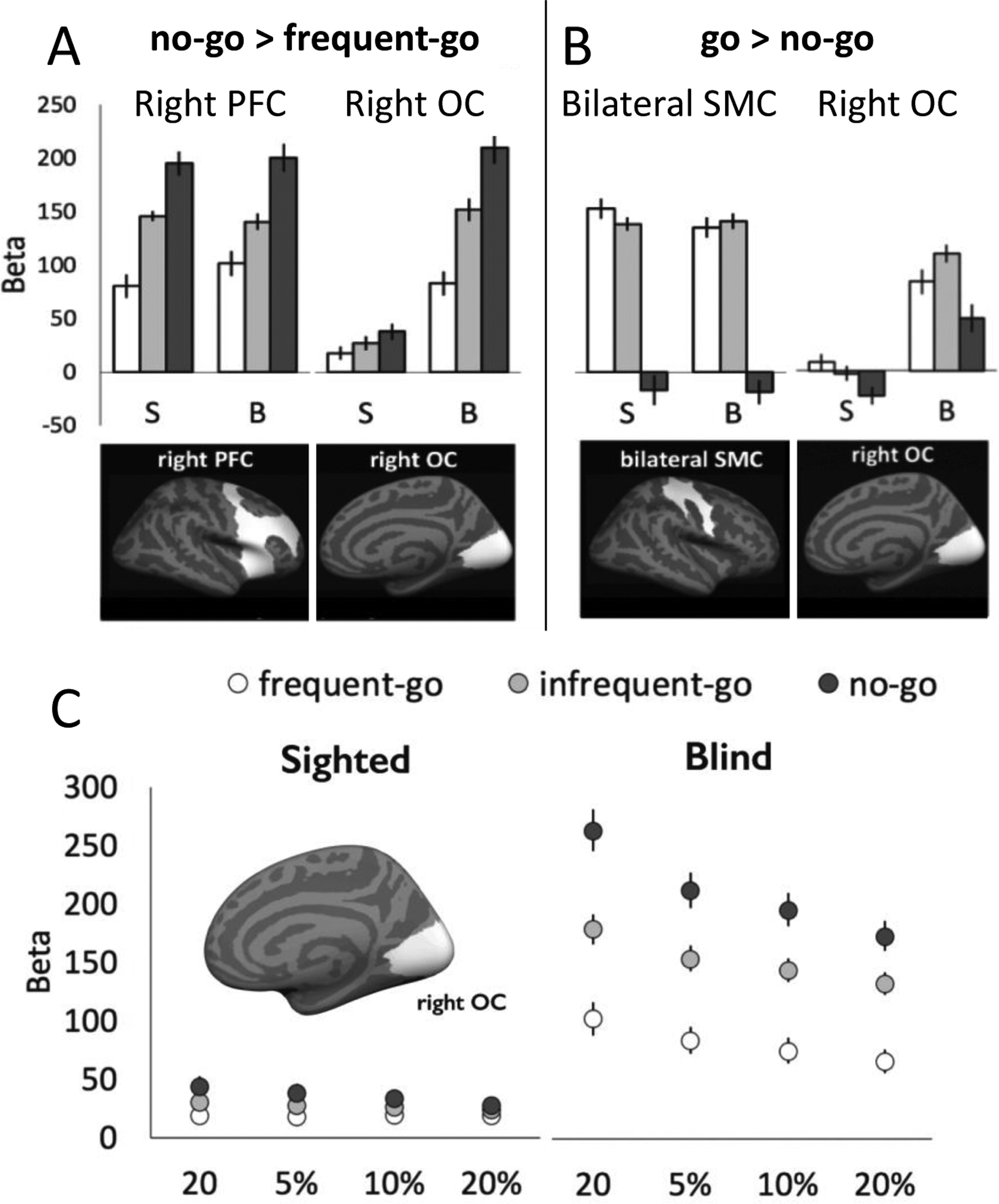
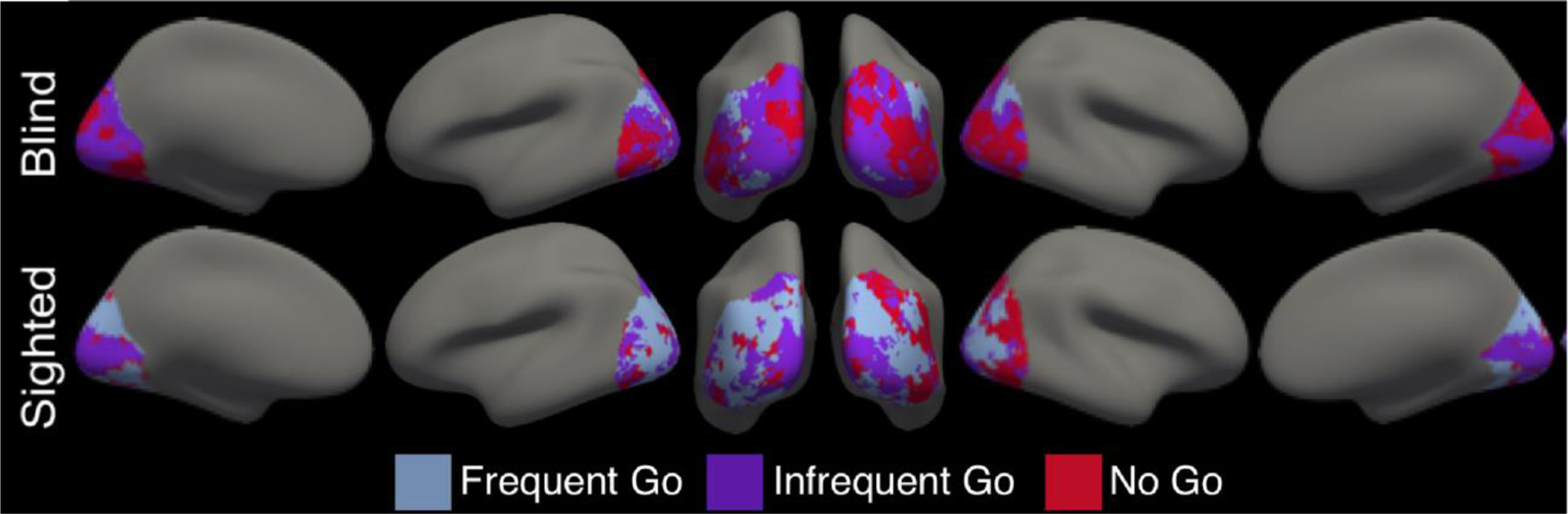
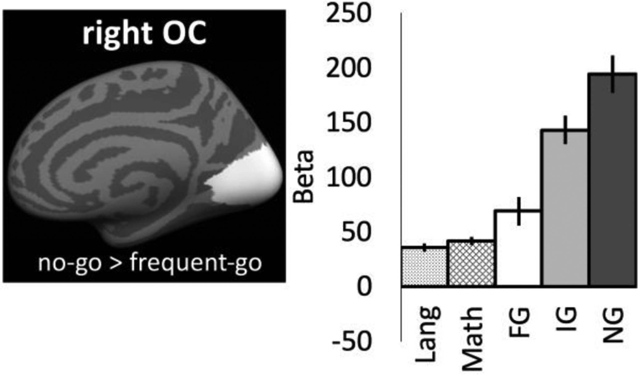
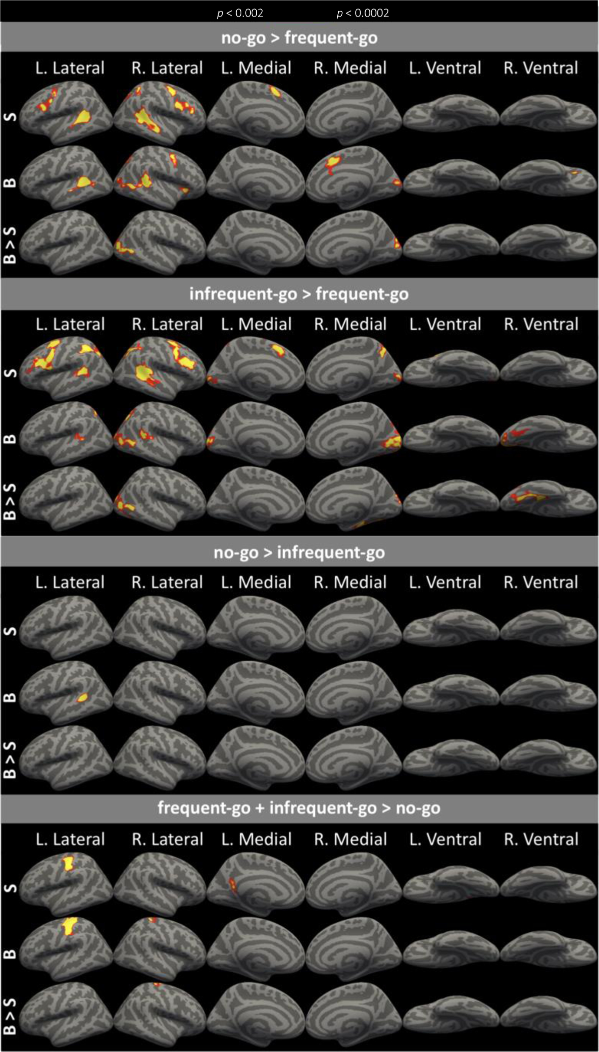
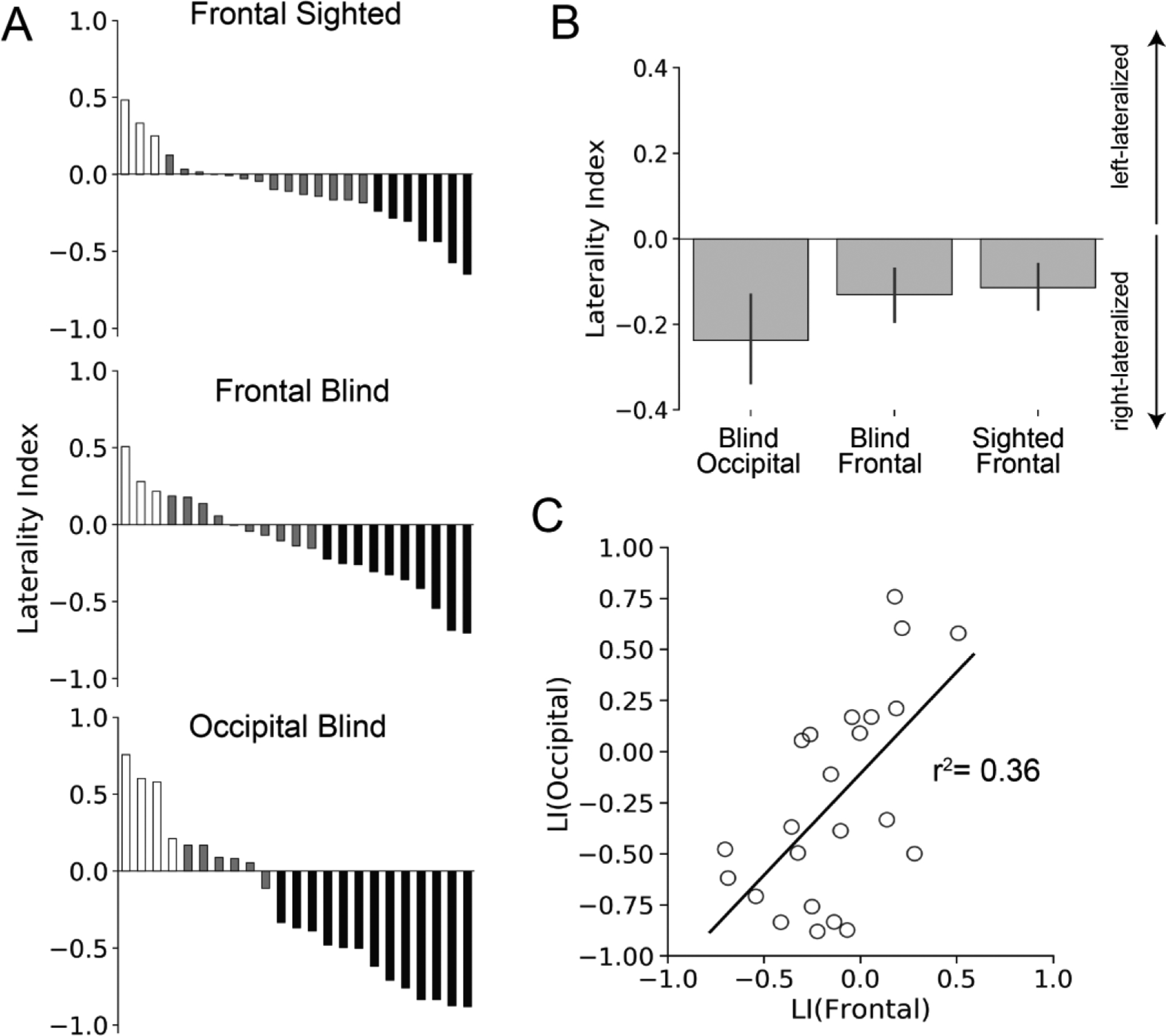
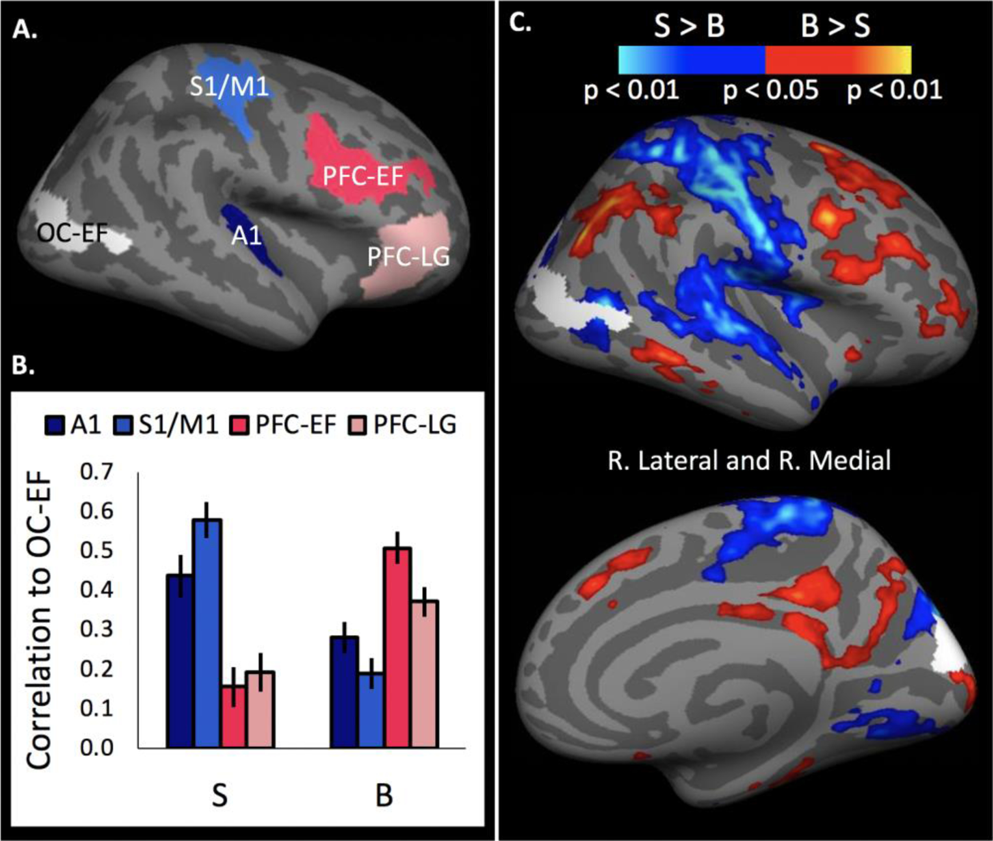
Studies of occipital cortex plasticity in blindness provide insight into how intrinsic constraints interact with experience to determine cortical specialization. We tested the cognitive nature and anatomical origins of occipital responses during non-verbal, non-spatial auditory tasks. In a go/no-go task, congenitally blind (N=23) and sighted (N=24) individuals heard rapidly occurring (<1/s) non-verbal sounds and made one of two button presses (frequent-go 50%, infrequent-go 25%) or withheld a response (no-go, 25%). Rapid and frequent button presses heighten response selection/inhibition demands on the no-go trials: In sighted and blind adults a right-lateralized prefrontal (PFC) network responded most to no-go trials, followed by infrequent-go and finally frequent-go trials. In the blind group only, a right-lateralized occipital network showed the same response profile and the laterality of occipital and PFC responses was correlated across blind individuals. A second experiment with spoken sentences and equations (N=16) found that no-go responses in occipital cortex are distinct from previously identified occipital responses to spoken language. Finally, in resting-state data (N=30 blind, N=31 blindfolded sighted), no-go responsive 'visual' cortex of blind relative to sighted participants was more synchronized with PFC and less synchronized with primary auditory and sensory-motor cortices. No-go responsive occipital cortex showed higher resting-state correlations with no-go responsive PFC than language responsive inferior frontal cortex. We conclude that in blindness, a right-lateralized occipital network responds to non-verbal executive processes, including response selection. These results suggest that connectivity with fronto-parietal executive networks is a key mechanism for plasticity in blindness.
Keywords: Blindness; Executive demand; Executive function; Plasticity; Pluripotency; Response selection.
Copyright © 2021. Published by Elsevier Inc.
Conflict of interest statement
Declaration of Competing Interest The authors declare no competing interests.
Figures
Similar articles
-
Occipital cortex of blind individuals is functionally coupled with executive control areas of frontal cortex.J Cogn Neurosci. 2015 Aug;27(8):1633-47. doi: 10.1162/jocn_a_00807. Epub 2015 Mar 24. J Cogn Neurosci. 2015. PMID: 25803598
-
Functional specialization for auditory-spatial processing in the occipital cortex of congenitally blind humans.Proc Natl Acad Sci U S A. 2011 Mar 15;108(11):4435-40. doi: 10.1073/pnas.1013928108. Epub 2011 Feb 28. Proc Natl Acad Sci U S A. 2011. PMID: 21368198 Free PMC article.
-
"Visual" Cortex of Congenitally Blind Adults Responds to Syntactic Movement.J Neurosci. 2015 Sep 16;35(37):12859-68. doi: 10.1523/JNEUROSCI.1256-15.2015. J Neurosci. 2015. PMID: 26377472 Free PMC article.
-
Plasticity of multisensory dorsal stream functions: evidence from congenitally blind and sighted adults.Restor Neurol Neurosci. 2010;28(2):193-205. doi: 10.3233/RNN-2010-0500. Restor Neurol Neurosci. 2010. PMID: 20404408 Review.
-
Do blind people hear better?Trends Cogn Sci. 2022 Nov;26(11):999-1012. doi: 10.1016/j.tics.2022.08.016. Epub 2022 Oct 5. Trends Cogn Sci. 2022. PMID: 36207258 Review.
Cited by
-
Beyond the Visual Word Form Area - a cognitive characterization of the left ventral occipitotemporal cortex.Front Hum Neurosci. 2023 Jul 28;17:1199366. doi: 10.3389/fnhum.2023.1199366. eCollection 2023. Front Hum Neurosci. 2023. PMID: 37576470 Free PMC article. Review.
-
"Touching" the brain: braille reading mitigates the SC-FC decoupling of brain networks in congenital blindness.Brain Struct Funct. 2025 Jul 7;230(6):114. doi: 10.1007/s00429-025-02975-9. Brain Struct Funct. 2025. PMID: 40622597
-
Anatomical and Functional Impacts of Congenital Bilateral Visual Deprivation on the Visual Pathway-A Comprehensive Review.J Clin Med. 2024 Mar 20;13(6):1775. doi: 10.3390/jcm13061775. J Clin Med. 2024. PMID: 38541998 Free PMC article. Review.
-
Sensory modality and spoken language shape reading network in blind readers of Braille.Cereb Cortex. 2023 Mar 10;33(6):2426-2440. doi: 10.1093/cercor/bhac216. Cereb Cortex. 2023. PMID: 35671478 Free PMC article.
-
The visual cortex in the blind but not the auditory cortex in the deaf becomes multiple-demand regions.Brain. 2024 Oct 3;147(10):3624-3637. doi: 10.1093/brain/awae187. Brain. 2024. PMID: 38864500 Free PMC article.
References
-
- Abboud S, Cohen L (2019) Distinctive Interaction Between Cognitive Networks and the Visual Cortex in Early Blind Individuals. Cerebral Cortex, 1–18. - PubMed
-
- Amedi A, Floel A, Knecht S, Zohary E, Cohen LG (2004) Transcranial magnetic stimulation of the occipital pole interferes with verbal processing in blind subjects. Nature Neuroscience 7(11), 1266–1270. - PubMed
-
- Amedi A, Raz N, Pianka P, Malach R, Zohary E (2003) Early “visual” cortex activation correlates with superior verbal memory performance in the blind. Nature Neuroscience 6(7), 758–766. - PubMed
-
- Aron AR, Robbins TW, Poldrack RA (2004) Inhibition and the right inferior frontal cortex. Trends in Cognitive Sciences 8(4), 170–177. - PubMed
Publication types
MeSH terms
Grants and funding
LinkOut - more resources
Full Text Sources
Other Literature Sources
Miscellaneous
