Review
. 2021 Jul 13:23:407-432.
doi: 10.1146/annurev-bioeng-091520-025358. Epub 2021 Apr 16.

Vascularized Microfluidics and Their Untapped Potential for Discovery in Diseases of the Microvasculature

Affiliations
Review

Vascularized Microfluidics and Their Untapped Potential for Discovery in Diseases of the Microvasculature

David R Myers et al. Annu Rev Biomed Eng. .

Abstract

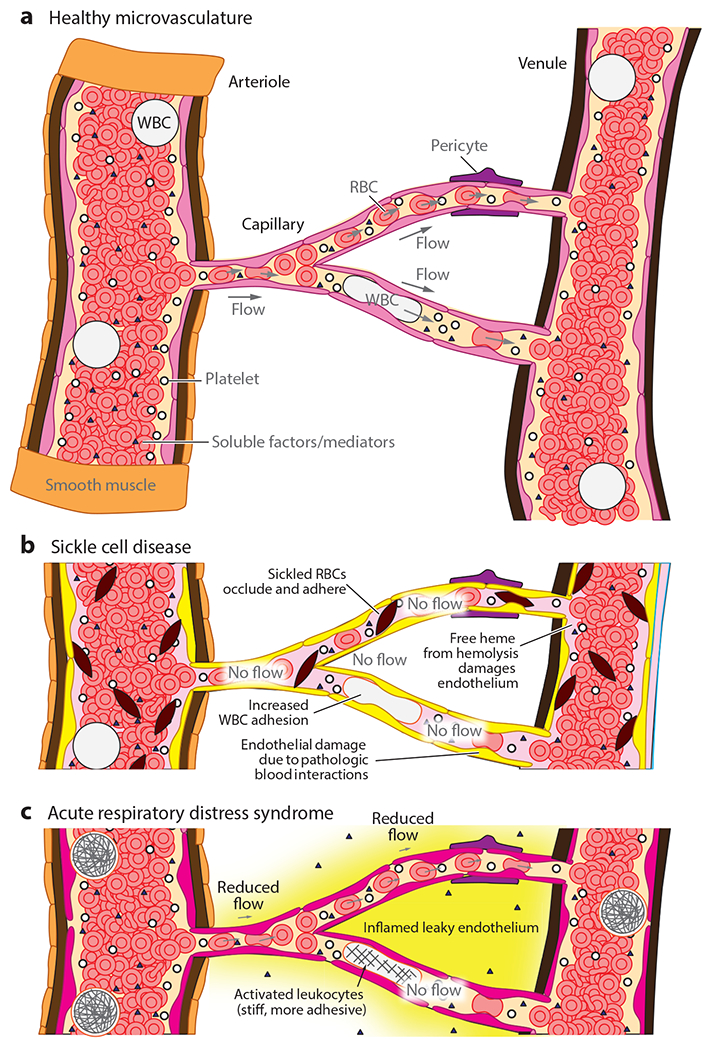
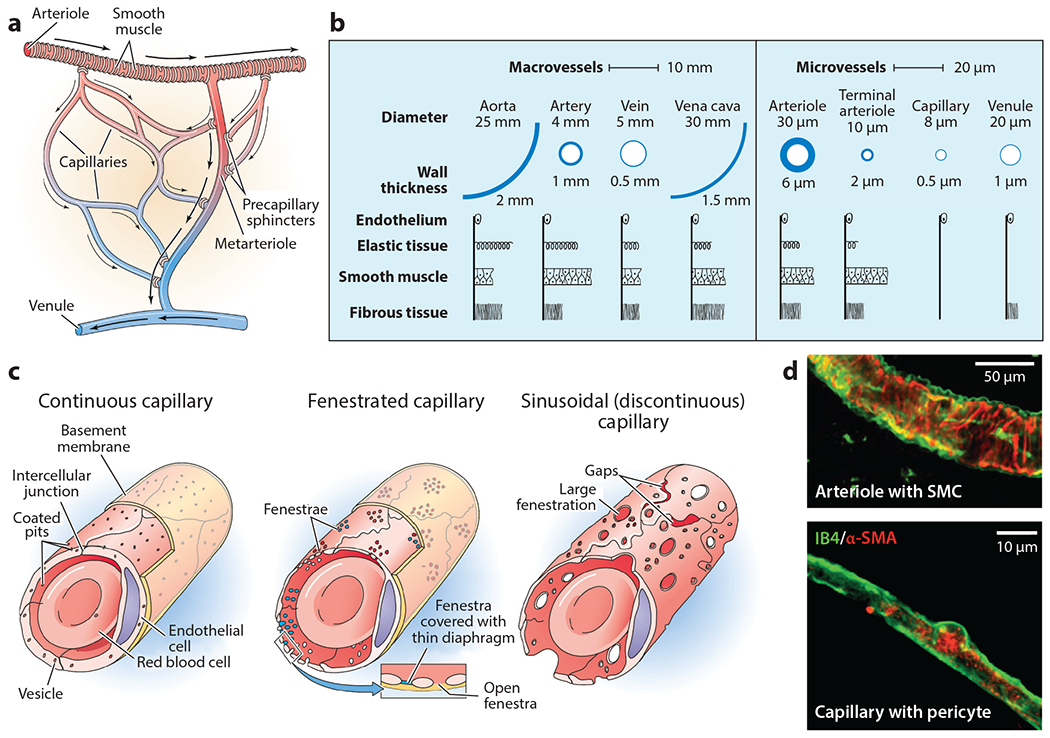
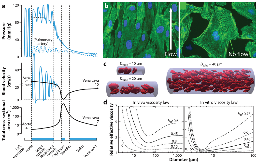
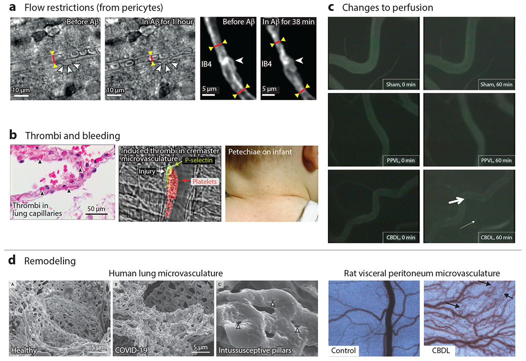
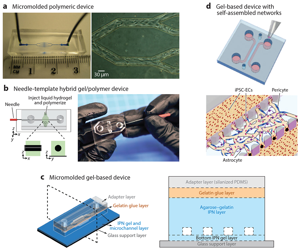
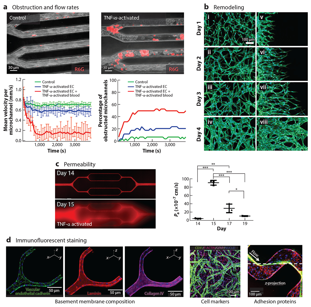
Microengineering advances have enabled the development of perfusable, endothelialized models of the microvasculature that recapitulate the unique biological and biophysical conditions of the microcirculation in vivo. Indeed, at that size scale (<100 μm)-where blood no longer behaves as a simple continuum fluid; blood cells approximate the size of the vessels themselves; and complex interactions among blood cells, plasma molecules, and the endothelium constantly ensue-vascularized microfluidics are ideal tools to investigate these microvascular phenomena. Moreover, perfusable, endothelialized microfluidics offer unique opportunities for investigating microvascular diseases by enabling systematic dissection of both the blood and vascular components of the pathophysiology at hand. We review (a) the state of the art in microvascular devices and (b) the myriad of microvascular diseases and pressing challenges. The engineering community has unique opportunities to innovate with new microvascular devices and to partner with biomedical researchers to usher in a new era of understanding and discovery of microvascular diseases.

Keywords: blood; endothelial; microfluidics; microvasculature; pathology.


Figures

Figure 1
Figure 1
Diseases of the microvasculature can be caused by changes to the endothelium, blood, or their ensuing pathological interactions. (a) The microvasculature is composed of arterioles, capillaries, and venules. Blood cells must nominally deform to pass through the microvasculature, and changes to the adhesiveness or stiffness significantly influence transport. Soluble factors in blood can modulate endothelial cell, smooth muscle cell, and pericyte activity. (b) In sickle cell disease, sickled red blood cells (RBCs) are stiffer and occlude endothelium, white blood cells (WBCs) are more adhesive, and free heme damages the endothelium. (c) Acute respiratory distress syndrome involves an inflamed, leaky endothelium and activated leukocytes, which are stiff and more adhesive.
Figure 2
Figure 2
Anatomy and structure of the microvasculature. (a) The microvasculature includes valves that are present as precapillary sphincters and valves in the venules. Panel a adapted from Reference . (b) The microvascular vessels are defined as less than 100 μm thick and are much simpler in structure than arteries and veins. Panel b adapted from References and . (c) Endothelial cells are highly heterogeneous, especially with regard to the anatomical source, which should be considered when designing an in vitro system. Panel c adapted from Reference . (d) Arterioles and capillaries are relatively simple structures with smooth muscle cells and pericytes, respectively Panel d adapted from Reference . Abbreviations: α-SMA, alpha smooth muscle actin; IB4, isolectin B4; SMC, smooth muscle cell.
Figure 3
Figure 3
Biofluid mechanics of the microvasculature. (a) As blood transitions from the arteries to the arterioles, capillaries, and venules, there is a significant drop in the applied pressure and blood velocity due to a significant increase in the cross-sectional area of the vessels. Panel a adapted from Reference . (b) Endothelial cells strongly respond to shear stress, changing morphologically and with altered signaling pathways related to growth factors, coagulation, inflammation, extracellular matrix degradation, cell division, differentiation, migration, apoptosis, and permeability. Panel b adapted from Reference . (c) A low hematocrity (0.15) simulation illustrates the Fåhræus-Lindqvist effect and unique noncontinuum flow physics governing the microvasculature. Panel c adapted from Reference . (d) Mathematical models corrected with experimental data provide an estimate of the expected relative viscosity as a function of bulk hematocrit (HD). Notably, there can be up to a fourfold increase in smaller vessels in vivo, informed by rat mesentery microvascular measurements, as compared to in vitro, informed by glass capillary experiments. Empirically derived equations for both are available from Reference .
Figure 4
Figure 4
Microvascular dysfunction in vivo occurs from flow restrictions, thrombi, bleeding, microvascular remodeling, and changes to perfusion. (a) Bright-field and two-photon fluorescence of rat cortical slices show constriction near pericytes. Less conspicuous, albeit significant, systemic 25% changes in diameter can cause reductions in blood flow on the order of 50% in the brain (39). (b) Microthrombi, especially when systemically distributed, can cause organ damage and death. Here, microthrombi are shown from the alveolar capillaries of a patient who died from COVID-19 (left) or induced by laser injury in murine cremaster microvasculature (middle). Petechiae are from capillary bleeding, seen here on an infant with viral illness (right). Panel b adapted from References (left subpanel), (middle subpanel), and (right subpanel). (c) Common bile duct ligation causes changes to the vascular permeability as seen by the increased fluorescent signal in the interstitium. Panel c adapted from Reference . (d) Microvasculature can also remodel in response to pathological conditions, shown here for a COVID-19 patient. In particular, intussusceptive angiogenesis appears to be occurring as evidenced by the intussusceptive pillars. Changes to the microvascular density in the visceral peritoneum of rats can be seen with common bile duct ligation. Panel d adapted from Reference . Abbreviations: Aβ, amyloid beta; CBDL, common bile duct ligation; IB4, isolectin B4; PPVL, partial portal vein ligation.
Figure 5
Figure 5
Approaches to creating in vitro microvasculature can be categorized as using polymeric or very soft gel materials with template-based, self-assembled, or directed networks of endothelial cells. (a) Polymeric-based micromolded structures offer high control over spatial dimensions and typically have a fast time to confluency. Panel a adapted from Reference . (b) Needle-templated hybrid structures offer key advantages of convenience and ease of fabrication, while using soft gels for perfusion studies, but typically are larger than most microvasculature (>100 μm). Panel b adapted from Reference . (c) Stiffer gels cleverly bonded with adapter layers led to the creation of the smallest in vitro vessels (~20 μm) that were still in a soft gel material for perfusion studies (35). (d) Pillars separating three fluidic channels enable gels laden with endothelial cells, pericytes, and astrocytes to be cast in the center gel and have perfusion in the adjacent channels to support culture and subsequent seeding after vascularization. Panel d adapted from Reference . Abbreviations: IPN, interpenetrating polymer network; iPSC-EC, induced pluripotent stem cell–derived endothelial cell; PDMS, polydimethylsiloxane.
Figure 6
Figure 6
Measurements of in vitro microvasculature can measure all modes of dysfunction and help provide mechanistic data. As in vitro microvasculature is much easier to visualize than corresponding in vivo structures, it can provide a rich amount of quantitative information. (a) In vitro postcapillary venule obstruction and flow rates can be quantitated, here shown in response to TNF-α. Panel a adapted from Reference . (b) In vitro changes to microvasculature morphology and shape. Panel b adapted from Reference . (c) Exceptional permeability measurements can be performed and quantitated, especially ones lasting multiple days, to examine recovery of the microvasculature. Shown here is the permeability of the endothelium to BSA-AF549 tracer before and after TNF-α perfusion between days 14 and 15. P values were calculated using one-way analysis of variance with Bonferroni’s post hoc test (*P < 0.05, **P < 0.01, ***P < 0.001). Panel c adapted from Reference . (d) Immunofluorescent staining can be used to identify basement membrane composition, cell markers, and adhesion proteins, such as the von Willebrand factor, an adhesion that mediates platelet adhesion at high shear rates. Panel d adapted from References , , and , respectively. Abbreviations: EC, endothelial cell; GFAP, glial fibrillary acidic protein; TNF-α, tumor necrosis factor alpha.

References

    1. Pappano AJ, Wier WG. 2019. Cardiovascular Physiology. Philadelphia, PA: Elsevier
    1. Aird WC. 2005. Spatial and temporal dynamics of the endothelium. J. Thromb. Haemost 3:1392–406 - PubMed
    1. Feletou M. 2011. The Endothelium. San Rafael, CA: Morgan & Claypool Life Sciences - PubMed
    1. Jackson SP, Darbousset R, Schoenwaelder SM. 2019. Thromboinflammation: challenges of therapeutically targeting coagulation and other host defense mechanisms. Blood 133:906–18 - PubMed
    1. Koeppen BM, Stanton BA. 2018. Overview of circulation. In Berne & Levy Physiology, pp. 301–3. Philadelphia, PA: Elsevier

Publication types

LinkOut - more resources