. 2021 Apr 16;23(1):117.
doi: 10.1186/s13075-021-02504-z.

Toll-like receptor 2 induced senescence in intervertebral disc cells of patients with back pain can be attenuated by o-vanillin

Affiliations

Toll-like receptor 2 induced senescence in intervertebral disc cells of patients with back pain can be attenuated by o-vanillin

Matthew Mannarino et al. Arthritis Res Ther. .

Abstract

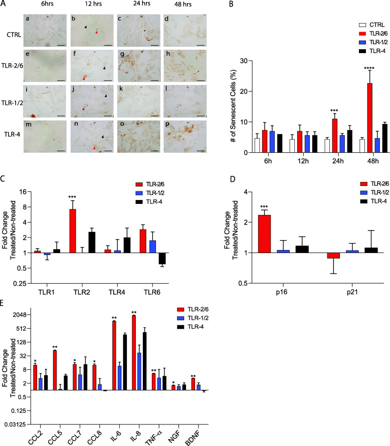
Background: There is an increased level of senescent cells and toll-like teceptor-1, -2, -4, and -6 (TLR) expression in degenerating intervertebral discs (IVDs) from back pain patients. However, it is currently not known if the increase in expression of TLRs is related to the senescent cells or if it is a more general increase on all cells. It is also not known if TLR activation in IVD cells will induce cell senescence.

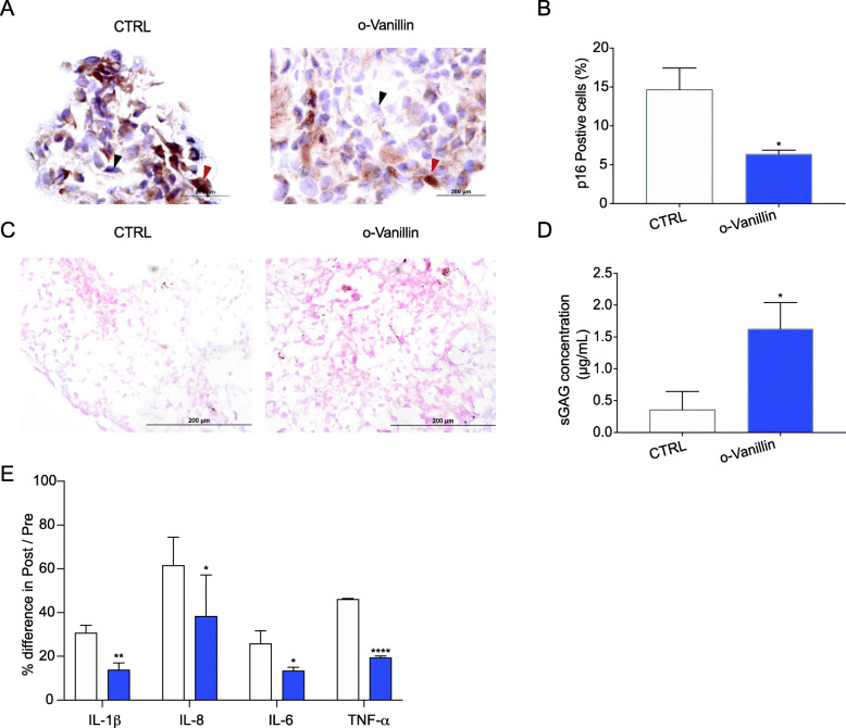
Methods: Cells from non-degenerate human IVD were obtained from spine donors and cells from degenerate IVDs came from patients undergoing surgery for low back pain. Gene expression of TLR-1,2,4,6, senescence and senescence-associated secretory phenotype (SASP) markers was evaluated by RT-qPCR in isolated cells. Matrix synthesis was verified with safranin-O staining and Dimethyl-Methylene Blue Assay (DMMB) confirmed proteoglycan content. Protein expression of p16INK4a, SASP factors, and TLR-2 was evaluated by immunocytochemistry (ICC) and/or by enzyme-linked immunosorbent assay (ELISA).

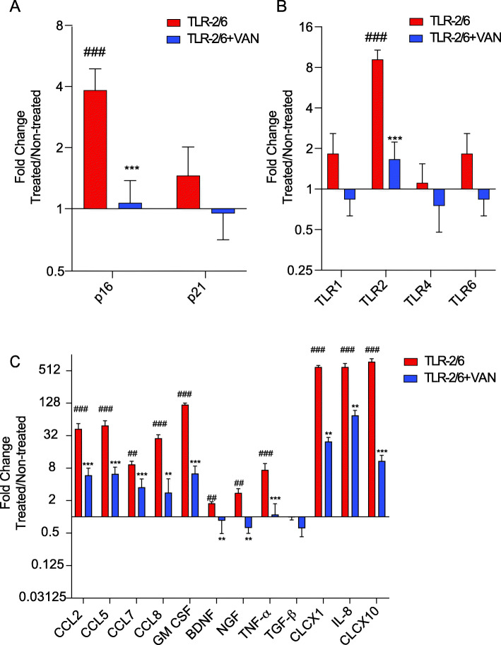
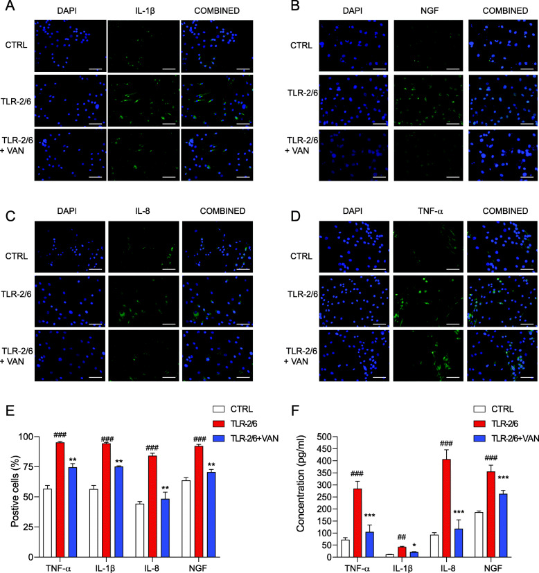
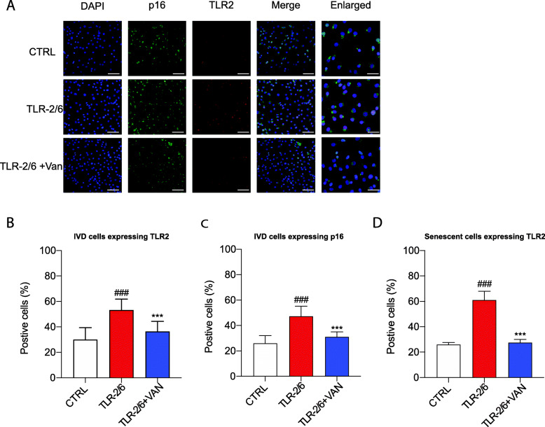
Results: An increase in senescent cells was found following 48-h induction with a TLR-2/6 agonist in cells from both non-degenerate and degenerating human IVDs. Higher levels of SASP factors, TLR-2 gene expression, and protein expression were found following 48-h induction with TLR-2/6 agonist. Treatment with o-vanillin reduced the number of senescent cells, and increased matrix synthesis in IVD cells from back pain patients. Treatment with o-vanillin after induction with TLR-2/6 agonist reduced gene and protein expression of SASP factors and TLR-2. Co-localized staining of p16INK4a and TLR-2 demonstrated that senescent cells have a high TLR-2 expression.

Conclusions: Taken together our data demonstrate that activation of TLR-2/6 induce senescence and increase TLR-2 and SASP expression in cells from non-degenerate IVDs of organ donors without degeneration and back pain and in cells from degenerating human IVD of patients with disc degeneration and back pain. The senescent cells showed high TLR-2 expression suggesting a link between TLR activation and cell senescence in human IVD cells. The reduction in senescence, SASP, and TLR-2 expression suggest o-vanillin as a potential disease-modifying drug for patients with disc degeneration and back pain.

Keywords: Back pain; Degeneration; Inflammation; Intervertebral disc; Senescence; Senolytics; Toll-like receptor 2; o-Vanillin.


Conflict of interest statement

The authors declare that they have no competing interests.

Figures

Fig. 1
Fig. 1
Cell activation with a TLR-2/6 agonist, caused an increase in the number of senescent cells and SASP factor release in cells from non-degenerate human IVDs. A p16ink4a immunostaining images of untreated cells (ad) from healthy donor IVDs or treated with either TLR-2/6 agonist (eh), TLR-1/2 agonist (il), or TLR-4 agonist (mp) cultured for 6, 12, 24, or 48 h. Scale bars = 25 μm. B Quantification of the percentage of p16ink4a positive cells in the control and treated cells for the four time points. Examples of positive cell are indicated by the red arrow and for negative cells by the black arrow. CE qPCR was performed using cells from healthy donor IVDs cultured for 48 h with TLR-2/6 agonist, TLR-1/2 agonist, or TLR-4 agonist. Gene expression for C TLR-1, -2, -4, and -6; D senescence markers p16ink4a and p21; and E SASP factors (CCL2, CCL5, CCL7, CCL8, IL-6, IL-8, TNF-α, NGF. and BDNF). Fold changes were normalized relative to the non-treated control (CTRL). BE Two-way ANOVA with Tukey’s multiple comparisons test, n = 5; significance was evaluated between treated groups compared to control group. Data shown as mean ± SD, *p < 0.05, **p < 0.01, ***p < 0.001, ****p < 0.0001
Fig. 2
Fig. 2
o-Vanillin reduced the number of senescent cells and enhanced proteoglycan production in cell pellet cultures from degenerate IVDs. All experiments were performed on day 21. a Representative photomicrographs of p16ink4a immunohistochemistry staining in pellet cultures from degenerate IVD cells either treated with o-vanillin or not (CTRL). Examples of positive cell are indicated by the red arrow and for negative cells by the black arrow. b Quantification of the percentage of p16ink4a positive cells in the pellet cultures; n = 3. c Images for Safranin O staining for proteoglycan content in CTRL and o-vanillin cell pellet cultures from degenerate IVD. d sGAG concentration measured using DMMB assay in the culture media of CTRL and o-vanillin cell pellets; n = 5. e Percentage of change in concentration of IL-1β, IL-8, IL-6, and TNF-α in pellet culture media from untreated and o-vanillin treated degenerate IVD cells measured by ELISAs. Percent difference was evaluated by normalizing the post-treated media to the pre-treated media; n = 5. *p < 0.05, **p < 0.01, ****p < 0.0001. b, d, e Mean ± SD, Statistical analysis was done using paired t-test
Fig. 3
Fig. 3
o-Vanillin reduced gene expression of p16, TLR-2 and SASP factors following TLR-2 activation in IVD cells from patients with back pain and IVD degeneration. ac Gene expression of a senescence markers p16ink4a and p21; b TLR-1, -2, -4, and -6; and c SASP factors (CCL2, CCL5, CCL7, CCL8, GM CSF, BDNF, NGF, TNF-α, TGF-β, CLCX1, CLCX8, CLCX10) of disc cells from degenerate IVDs cultured for 48 h with TLR-2/6 agonist with o-vanillin (TLR-2/6 + VAN) or without o-vanillin (TLR-2/6) treatment for 6 h or no induction with TLR-2/6. Fold changes were normalized relative to non-induced control. #p < 0.05, ##p < 0.01, ###p < 0.001 indicate significant difference between the TLR-2/6 treated to the non-induced control and *p < 0.05, **p < 0.01, ***p < 0.001 indicate significant difference between groups TLR-2/6 + VAN and TLR-2/6. ac Mean ± SD, measured by two-way ANOVA with Sidak’s multiple comparisons test. Values are expressed in average fold change for n = 5
Fig. 4
Fig. 4
o-Vanillin reduced the protein expression of SASP factors (IL-1β, NGF, IL-8, and TNF-α) following TLR-2/6 activation of IVD cells from patients with back pain and IVD degeneration. Disc cells from degenerate IVD were cultured for 48 h with TLR-2/6 with o-vanillin (TLR-2/6 + VAN) or without o-vanillin (TLR-2/6) treatment for 6 h or no induction with TLR-2/6 (CTRL). ad Using Immunocytochemistry, untreated and treated IVD cells were stained for DAPI and either a IL-1β, b NGF, c IL-8, and d TNF-α, respectively. Scale Bars = 25 μm. e Quantification of the percentage of the cells that stained positive for IL-1β, NGF, IL-8, and TNF-α when non-induced, treated with TLR-2/6 agonist or treated with TLR-2/6 agonist and o-vanillin; n = 5. f ELISAs were performed to measure the concentration of TNF-α, IL-1β, and IL-8 in cell media and NGF from cell lysate in non-induced, TLR-2/6 agonist treated or the combined treatment TLR-2/6 agonist and o-vanillin treated samples. Percentage of positive cells in e were the average for n = 5, . #p < 0.05, ##p < 0.01, and ###p < 0.001 indicate significant difference between the TLR-2/6 agonist treated to the non-induced control and *p < 0.05, **p < 0.01, and ***p < 0.001 indicate significant difference between the tested groups. e, f Mean ± SD, statistical analysis was done using paired t-test
Fig. 5
Fig. 5
o-Vanillin reduced the number of cells co-expressing TLR-2 and p16ink4a in cells exposed to TLR-2/6 agonist. Disc cells from degenerate IVDs were induced with TLR-2/6 agonist for 48 h with (TLR-2/6 + VAN) or without (TLR-2/6) o-vanillin treatment for 6 h or no induction with TLR-2/6 agonist (CTRL). a Photomicrographs of IVD cells stained for DAPI (blue) and either p16ink4a (green), TLR-2 (red), or the merge (p16ink4a and TLR-2) as revealed by Immunocytochemistry. DAPI, p16, TLR2, and merge images scale bars = 25 μm. Enlarged images scale bar: 10 μm. bd Quantification of the percentage of IVD cells that stained positive for b TLR-2, c p16ink4a, or d co-localized cell for TLR-2 and p16ink4a. Percentage of positive cells in e were the average for n = 5. #p < 0.05, ##p < 0.01, and ###p < 0.001 indicate significant difference between the TLR-2/6 agonist treated to the non-induced control and *p < 0.05, **p < 0.01, and ***p < 0.001 indicate significant difference between the TLR-2/6 agonist with o-vanillin treated to the TLR-2/6 agonist treated. bd Mean ± SD, statistical analysis was done using paired t-test

Similar articles

Cited by

References

    1. Ahmed S, Anuntiyo J, Malemud CJ, Haqqi TM. Biological basis for the use of botanicals in osteoarthritis and rheumatoid arthritis: a review. Evid Based Complement Alternat Med. 2005;2(3):301–308. doi: 10.1093/ecam/neh117. - DOI - PMC - PubMed
    1. Elmali N, Baysal O, Harma A, Esenkaya I, Mizrak B. Effects of resveratrol in inflammatory arthritis. Inflammation. 2007;30(1–2):1–6. doi: 10.1007/s10753-006-9012-0. - DOI - PubMed
    1. Feng H, Danfelter M, Strömqvist B, Heinegård D. Extracellular matrix in disc degeneration. J Bone Joint Surg Am. 2006;88(Suppl 2):25–29. - PubMed
    1. Geurts JW, Willems PC, Kallewaard JW, van Kleef M, Dirksen C. The impact of chronic discogenic low back pain: costs and patients’ burden. Pain Res Manag. 2018;2018:4696180. doi: 10.1155/2018/4696180. - DOI - PMC - PubMed
    1. Phillips CJ. The cost and burden of chronic pain. Rev Pain. 2009;3(1):2–5. doi: 10.1177/204946370900300102. - DOI - PMC - PubMed

Publication types