Case Reports
. 2021 Apr 16;21(1):35.
doi: 10.1186/s40644-021-00404-8.

Intra-patient comparison of physiologic 68Ga-PSMA-11 and 18F-DCFPyL PET/CT uptake in ganglia in prostate cancer patients: a pictorial essay

Affiliations
Case Reports

Intra-patient comparison of physiologic 68Ga-PSMA-11 and 18F-DCFPyL PET/CT uptake in ganglia in prostate cancer patients: a pictorial essay

Medhat M Osman et al. Cancer Imaging. .

Abstract

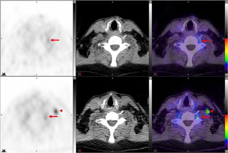
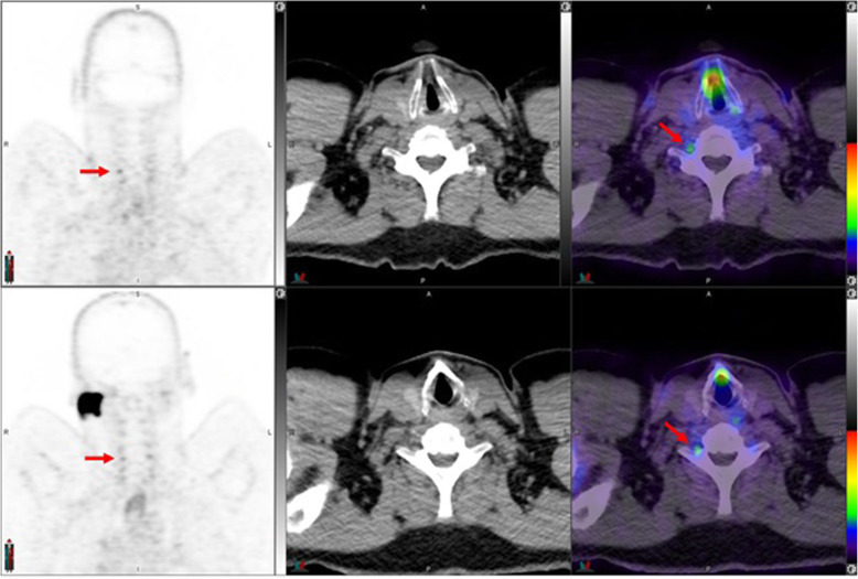
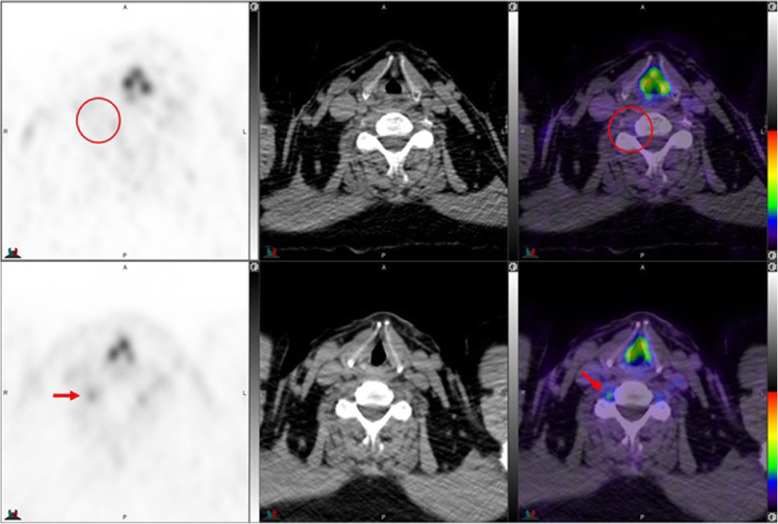
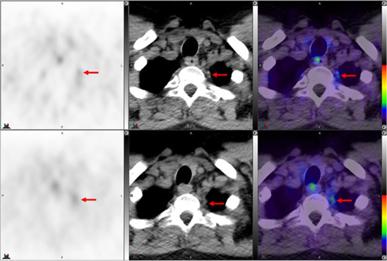
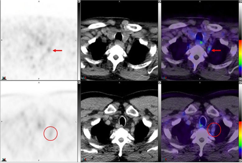
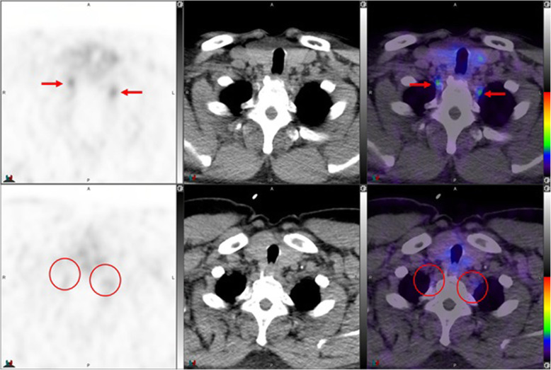
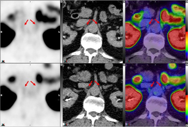
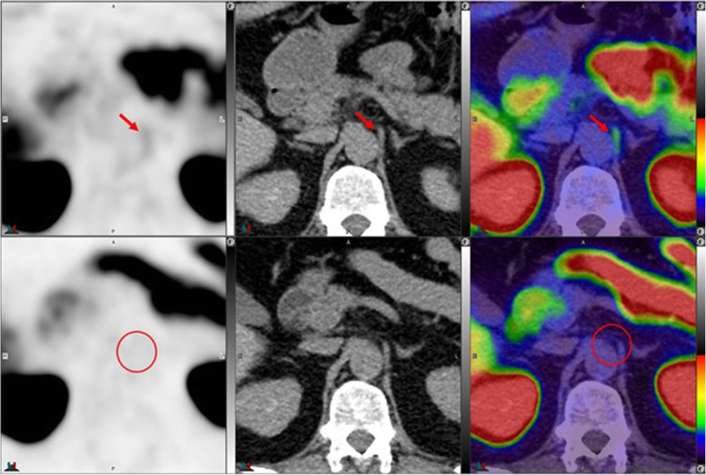
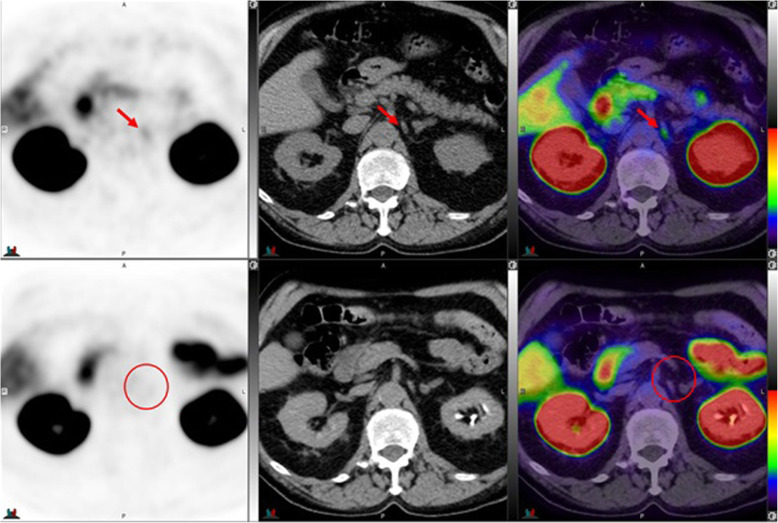
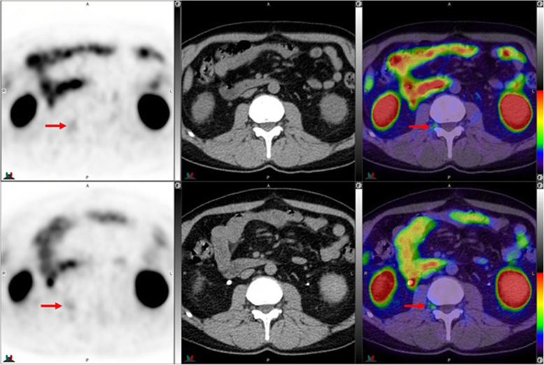
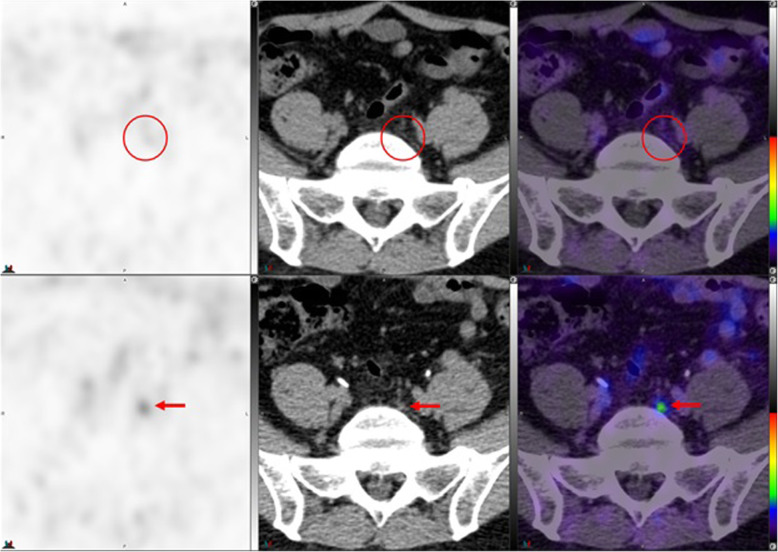
Background: Recent studies reported metabolic uptake in at least one of the evaluated ganglia in 98.5% of patients undergoing 68Ga -PSMA-11 and in 96.9% of patients undergoing 18F-DCFPyL PET/CT examination. We have observed different patterns of ganglion visualization with 18F-DCFPyL compared to 68Ga-PSMA-11. This includes more frequent visualization of cervical and sacral ganglia, which may be attributable to better imaging characteristics with 18F PET imaging.

Case presentation: This pictorial essay is to illustrate and compare, in the same patient, various representative cases of 68Ga-PSMA-11 and 18F-DCFPyL PET/CT uptake in ganglia at different anatomic locations, with different patterns and distribution of metabolic activity.

Conclusion: Reading physicians should be aware of the frequently encountered and occasionally different physiologic uptake of 68Ga-PSMA-11 and 18F DCFPyL in different ganglia.

Keywords: 18F-DCFPyL PET/CT; 68Ga-PSMA11 PET/CT; Ganglia; PSMA.


Conflict of interest statement

The authors declare that they have no competing interests.

Figures

Fig. 1
Fig. 1
Top panel 68Ga-PSMA-11: PET, CT and PET/CT. Left cervical ganglion (arrow) maximum Standard Uptake Value (SUVmax) = 2.2. Lower panel 18F –DCFPyL: PET, CT and PET/CT. Left cervical ganglion (arrow) SUVmax = 2.6, Right cervical ganglion SUVmax = 2.3. Of note, 18F –DCFPyL shows new left supraclavicular metastases (arrow head). The color intensity has been slightly adjusted to highlight the uptake in the ganglia. There is higher and bilateral lower cervical ganglia uptake with 18F –DCFPyL. By CT, there is typically no anatomic structure detectable in the region of cervical ganglia; however, location, bilaterality and multiplicity would provide clues
Fig. 2
Fig. 2
Top panel 68Ga-PSMA-11: coronal PET, axial CT and axial fused PET/CT. Right cervical ganglion (arrow) SUVmax = 2.4. Lower panel 18F –DCFPyL: PET, CT and PET/CT. Right cervical ganglion (arrow) SUVmax = 2.5. The color intensity has been slightly adjusted to highlight the uptake in the ganglia. Higher and bilateral uptake in lower cervical ganglia noted with 18F –DCFPyL
Fig. 3
Fig. 3
Top panel 68Ga-PSMA-11: axial PET, axial CT and axial fused PET/CT. Region of the Right lower cervical ganglion (circle). Lower panel 18F –DCFPyL: PET, CT and PET/CT. Right lower cervical ganglion uptake (SUVmax = 2.5) noted with 18F –DCFPyL (arrow)
Fig. 4
Fig. 4
Top panel 68Ga-PSMA-11: axial PET, axial CT and axial fused PET/CT. Lower panel 18F –DCFPyL: axial PET, axial CT and axial fused PET/CT. The color intensity has been slightly adjusted to highlight the uptake in the ganglia (arrows). Left stellate ganglion with 18F –DCFPyL (SUVmax = 2.5) > 68Ga-PSMA-11 (SUVmax = 2.1). Metabolic activity is more commonly noted in the left side. By CT, stellate ganglia are typically located anterior to first rib and have curvilinear or nodular shape
Fig. 5
Fig. 5
Top panel 68Ga-PSMA-11: axial PET, axial CT and axial fused PET/CT. Lower panel 18F –DCFPyL: axial PET, axial CT and axial used PET/CT. The color intensity has been slightly adjusted to highlight the uptake in the ganglia (arrows in the top panel and circles in the lower panel). Left stellate ganglion uptake with 68Ga-PSMA-11 only (SUVmax = 1.8)
Fig. 6
Fig. 6
Top panel 68Ga-PSMA-11: axial PET, axial CT and axial fused PET/CT. Lower panel 18F –DCFPyL: axial PET, axial CT and axial fused PET/CT. The color intensity has been slightly adjusted to highlight the uptake in the ganglia (arrows in the top panel and circles in the lower panel). Bilateral stellate ganglia uptake with 68Ga-PSMA-11 (left and right stellate ganglia SUVmax = 2.7 and 3.0, respectively) but not with 18F –DCFPyL
Fig. 7
Fig. 7
Top panel 68Ga-PSMA-11: axial PET, axial CT and axial fused PET/CT. Lower panel 18F –DCFPyL: axial PET, axial CT and axial fused PET/CT. Left coeliac ganglion uptake with 68Ga-PSMA-11 (SUVmax 2.4) > 18F –DCFPyL (SUVmax 2.2) (left arrows). Right coeliac ganglion shows similar uptake with both tracers (SUVmax = 2.1) (right arrows). By CT, coeliac ganglia are typically located adjacent to coeliac axis and have discoid, band or teardrop shape
Fig. 8
Fig. 8
Top panel 68Ga-PSMA-11: axial PET, axial CT and axial fused PET/CT. Lower panel 18F –DCFPyL: axial PET, axial CT and axial fused PET/CT. The color intensity has been slightly adjusted to highlight the uptake in the ganglia. Left coeliac ganglion uptake with 68Ga-PSMA-11 (SUVmax = 2.7) (arrows) but not with 18F –DCFPyL (circles)
Fig. 9
Fig. 9
Top panel 68Ga-PSMA-11: axial PET, axial CT and axial fused PET/CT. Lower panel 18F –DCFPyL: axial PET, axial CT and axial fused PET/CT. Left coeliac ganglion uptake with 68Ga-PSMA (SUVmax = 2.4) (arrows) but not with 18F –DCFPyL (circles). By CT, the left coeliac ganglion is typically located between the coeliac trunk and left adrenal gland
Fig. 10
Fig. 10
Top panel 68Ga-PSMA-11: axial PET, axial CT and axial fused PET/CT. Lower panel 18F –DCFPyL: axial PET, axial CT and axial fused PET/CT. The color intensity has been slightly adjusted to highlight the uptake in the ganglia. Right upper lumbar ganglion (arrows) uptake with 18F –DCFPyL (SUVmax=2.4) > with 68Ga-PSMA-11 (SUVmax=2.3). By CT, similar to cervical ganglia, there are typically no anatomic structures detectable in the region of lumbar ganglia; however, paravertebral location, bilaterality and multiplicity would provide clues
Fig. 11
Fig. 11
Top panel 68Ga-PSMA-11: axial PET, axial CT and axial fused PET/CT. Lower panel 18F –DCFPyL: axial PET, axial CT and axial fused PET/CT. Left sacral ganglion uptake with 18F –DCFPyL (SUVmax = 3.1) (arrows) but not with 68Ga-PSMA-11 (circles). By CT, similar to lumbar ganglia, there are typically no anatomic structures detectable in the region of paravertebral ganglia; however, location, bilaterality and multiplicity would provide clues. Prevertebral sacral ganglion, however, may appear band like, linear or curvilinear, and given the proximity to primary sites, it is probably the most challenging ganglia to differentiate from nodal metastasis. This is particularly true if uptake in the ganglia is similar to that of metastatic lymph nodes [25]

Similar articles

Cited by

References

    1. Hofman MS, Lawrentschuk N, Francis RJ, Tang C, Vela I, Thomas P, Rutherford N, Martin JM, Frydenberg M, Shakher R, Wong LM, Taubman K, Ting Lee S, Hsiao E, Roach P, Nottage M, Kirkwood I, Hayne D, Link E, Marusic P, Matera A, Herschtal A, Iravani A, Hicks RJ, Williams S, Murphy DG. Prostate-specific membrane antigen PET-CT in patients with high-risk prostate cancer before curative-intent surgery or radiotherapy (proPSMA): a prospective, randomised, multicentre study. Lancet. 2020;395(10231):1208–1216. doi: 10.1016/S0140-6736(20)30314-7. - DOI - PubMed
    1. Eiber M, Maurer T, Souvatzoglou M, Beer AJ, Ruffani A, Haller B, Graner FP, Kubler H, Haberhorn U, Eisenhut M, Wester HJ, Gschwend JE, Schwaiger M. Evaluation of hybrid (6)(8) Ga-PSMA ligand PET/CT in 248 patients with biochemical recurrence after radical prostatectomy. J Nucl Med. 2015;56(5):668–674. doi: 10.2967/jnumed.115.154153. - DOI - PubMed
    1. Afshar-Oromieh A, Avtzi E, Giesel FL, Holland-Letz T, Linhart HG, Eder M, Eisenhut M, Boxler S, Hadaschik BA, Kratochwil C, Weichert W, Kopka K, Debus J, Haberkorn U. The diagnostic value of PET/CT imaging with the (68) Ga-labelled PSMA ligand HBED-CC in the diagnosis of recurrent prostate cancer. Eur J Nucl Med Mol Imaging. 2015;42(2):197–120. doi: 10.1007/s00259-014-2949-6. - DOI - PMC - PubMed
    1. Demirci E, Sahin OE, Ocak M, Akovali B, Nematyazar J, Kabasakal L. Normal distribution pattern and physiological variants of 68Ga-PSMA-11 PET/CT imaging. Nucl Med Commun. 2016;37(11):1169–1179. doi: 10.1097/MNM.0000000000000566. - DOI - PubMed
    1. Osman MM, Iravani A, Hicks RJ, Hofman MS. Detection of Synchronous Primary Malignancies with 68Ga-Labeled Prostate-Specific Membrane Antigen PET/CT in Patients with Prostate Cancer: Frequency in 764 Patients. J Nucl Med. 2017;58(12):1938–1942. doi: 10.2967/jnumed.117.190215. - DOI - PubMed

Publication types

MeSH terms