Review
. 2021 Jun;42(6):461-474.
doi: 10.1016/j.tips.2021.03.004. Epub 2021 Apr 13.

Delivering AAV to the Central Nervous and Sensory Systems

Affiliations
Review

Delivering AAV to the Central Nervous and Sensory Systems

Cole W Peters et al. Trends Pharmacol Sci. 2021 Jun.

Abstract

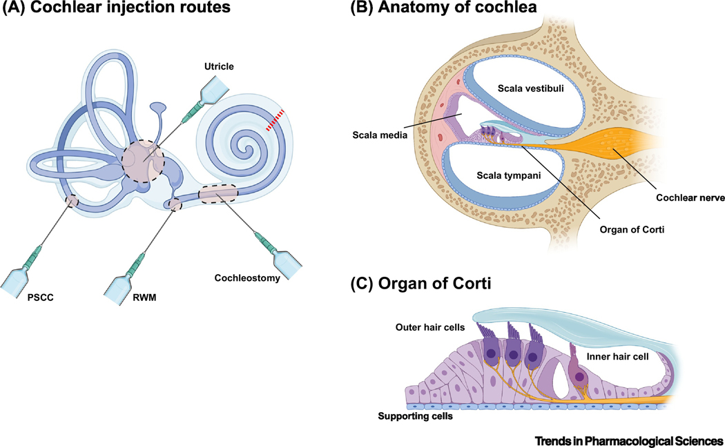
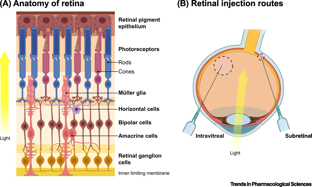
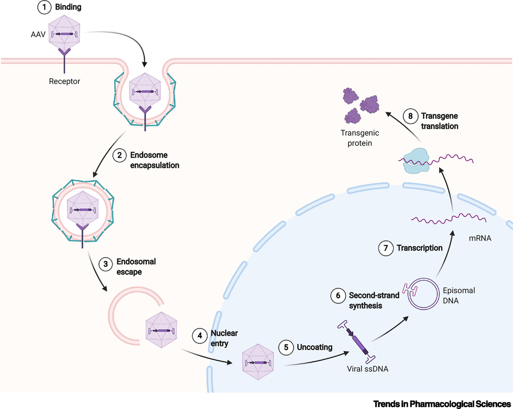
As gene therapy enters mainstream medicine, it is more important than ever to have a grasp of exactly how to leverage it for maximum benefit. The development of new targeting strategies and tools makes treating patients with genetic diseases possible. Many Mendelian disorders are amenable to gene replacement or correction. These often affect post-mitotic tissues, meaning that a single stably expressing therapy can be applied. Recent years have seen the development of a large number of novel viral vectors for delivering specific therapies. These new vectors - predominately recombinant adeno-associated virus (AAV) variants - target nervous tissues with differing efficiencies. This review gives an overview of current gene therapies in the brain, ear, and eye, and describes the optimal approaches, depending on cell type and transgene. Overall, this work aims to serve as a primer for gene therapy in the central nervous and sensory systems.

Keywords: AAV; brain; gene therapy; inner ear; retina.


Figures

Figure 1.
Figure 1.. Cochlear Architecture.
a. Routes of injection employed in the cochlea. The round window membrane (RWM) is the most commonly used approach for neonatal mice, as well as NHPs. Cochleostomy involves penetrating the lateral wall and is used after the cochlear bone calcifies in adult subjects. Targeting the cochlea via the vestibular system is also an option. The posterior semicircular canal (PSCC) is also used for treating adult mice. More recently, injecting via the utricle has been shown to deliver high level expression in the cochlea with a number of serotypes. However, more invasive procedures like the PSCC and cochleostomy can carry a greater risk of damaging hair cells, obviating any therapeutic benefit[39,40]. b. A cross-section of the cochlea (red line in a) is shown. The scalae vestibuli and tympani are perilymph-filled vessels surrounding the organ of Corti, where the sensory apparatus resides. This perilymph must remain separate from the endolymph of the scala media; as such, any injection directed towards the organ of Corti must be minimal to limit the puncture size. c. Focus on the organ of Corti. Inner and outer hair cells are tightly nestled among a number of classes of supporting cells called Dieter, Claudius, and Hensen cells.
Figure 2.
Figure 2.. Retinal architecture.
a. Anatomy of the retina. The retina is made up of seven main types (and over sixty subtypes) of cells. Rods and cones within the retina receive light input; this signal is transferred to retinal ganglion cells (RGCs) via bipolar cells. The signal is decomposed and processed by inhibitory horizontal and amacrine cells, before undergoing further processing by RGCs and transmittance to the brain via the optic nerve. Müller glia support retinal neurons through homeostatic maintenance and cycling of neurotransmitter levels[118]. Glia also help form the inner limiting membrane (ILM), a barrier that separates the retina from the vitreous. b. Routes of administration to the retina. Intravitreal (IVit) and subretinal (SR) injections are the primary routes for drugs being delivered to the retina. IVit involves direct delivery to the vitreous humor of the eye – a well tolerated procedure millions of patients receive monthly for diseases such as age-related macular degeneration. While safe and routine to perform, the ILM prevents passage of most AAV serotypes beyond RGCs[119]. Subretinal injection delivers to the space between the retinal pigment epithelium (RPE) and photoreceptors. It is the most effective method for transducing these cell types. However, this more invasive surgical method involves isolated detachment of the retina through blebbing, which – particularly in vulnerable, diseased retinas – can lead to damage. Further, AAV injections into the subretinal space of larger animals result in only a small area of the retina immediately surrounding the bleb being transduced.
Box 1 Figure I:
Box 1 Figure I:
Schematic of AAV transduction.

References

    1. Keeler AM and Flotte TR (2019) Recombinant Adeno-Associated Virus Gene Therapy in Light of Luxturna (and Zolgensma and Glybera): Where Are We, and How Did We Get Here? Annu. Rev. Virol. 6, 601–621 - PMC - PubMed
    1. Sonntag F et al. (2010) A viral assembly factor promotes AAV2 capsid formation in the nucleolus. Proc. Natl. Acad. Sci. U. S. A. 107, 10220–10225 - PMC - PubMed
    1. Ogden PJ et al. (2019) Comprehensive AAV capsid fitness landscape reveals a viral gene and enables machine-guided design. Science (80-. ). 366, 1139–1143 - PMC - PubMed
    1. Meier AF et al. The interplay between adeno-associated virus and its helper viruses., Viruses. (2020), 12(6), 662 - PMC - PubMed
    1. Hsu H-L et al. (2020) Structural characterization of a novel human adeno-associated virus capsid with neurotropic properties. Nat. Commun. 11, 3279. - PMC - PubMed