. 2021 Nov;26(6):e13042.
doi: 10.1111/adb.13042. Epub 2021 Apr 16.

Cocaine experience induces functional adaptations in astrocytes: Implications for synaptic plasticity in the nucleus accumbens shell

Affiliations

Cocaine experience induces functional adaptations in astrocytes: Implications for synaptic plasticity in the nucleus accumbens shell

Bernadette O'Donovan et al. Addict Biol. 2021 Nov.

Abstract

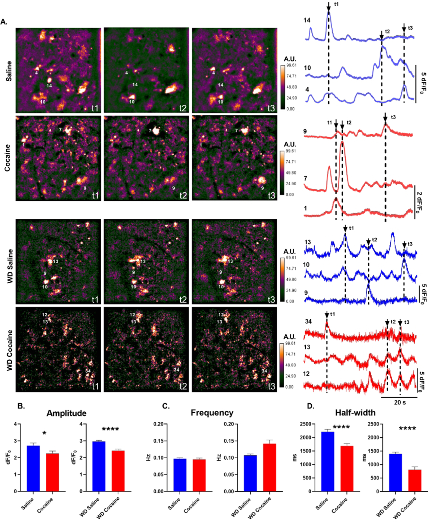
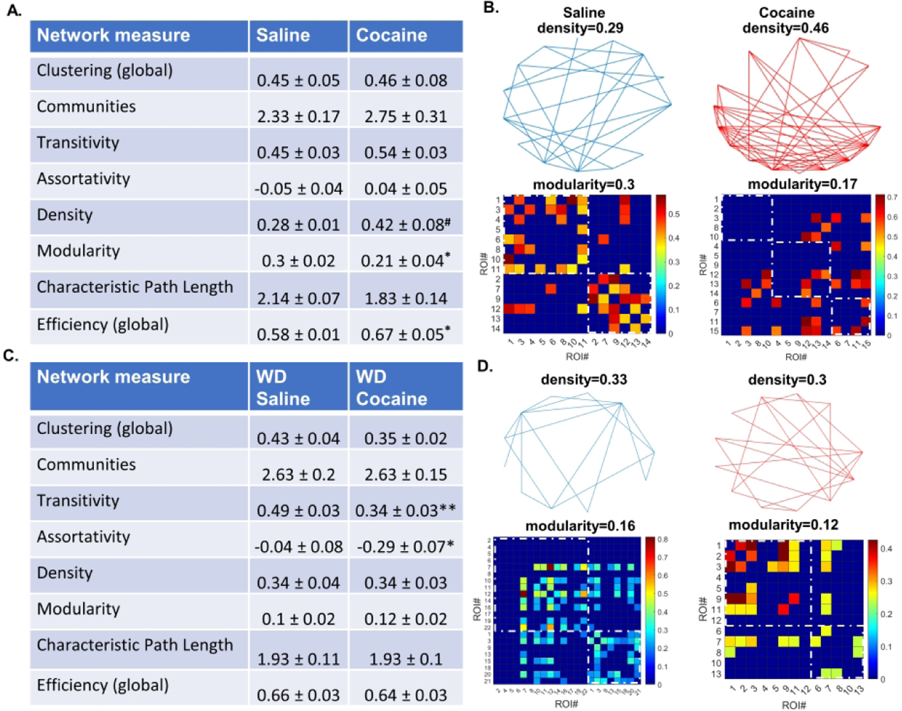
Astrocytes have become established as an important regulator of neuronal activity in the brain. Accumulating literature demonstrates that cocaine self-administration in rodent models induces structural changes within astrocytes that may influence their interaction with the surrounding neurons. Here, we provide evidence that cocaine impacts astrocytes at the functional level and alters neuronal sensitivity to astrocyte-derived glutamate. We report that a 14-day period of short access to cocaine (2 h/day) decreases spontaneous astrocytic Ca2+ transients and precipitates changes in astrocyte network activity in the nucleus accumbens shell. This is accompanied by increased prevalence of slow inward currents, a physiological marker of neuronal activation by astrocytic glutamate, in a subset of medium spiny neurons. Within, but not outside, of this subset, we observe an increase in duration and frequency of N-methyl-D-aspartate (NMDA) receptor-mediated synaptic events. Additionally, we find that the link between synaptic NMDA receptor plasticity and neuron sensitivity to astrocytic glutamate is maintained independent of drug exposure and is observed in both cocaine and saline control animals. Imaging analyses of neuronal Ca2+ activity show no effect of cocaine self-administration on individual cells or on neuronal network activity in brain slices. Therefore, our data indicate that cocaine self-administration promotes astrocyte-specific functional changes that can be linked to increased glutamate-mediated coupling with principal neurons in the nucleus accumbens. Such coupling may be spatially restricted as it does not result in a broad impact on network structure of local neuronal circuits.

Keywords: astrocyte; cocaine self-administration; synaptic plasticity.


Figures

Figure 1.
Figure 1.. Astrocyte Ca2+ transients after cocaine self-administration.
A) Left: False-color GFAP-GCamp6f Ca2+ fluorescence frames from NAc shell astrocyte videos after 24 hours (saline, cocaine) and 14 days (wd saline, wd cocaine) withdrawal from cocaine self-administration. The color scale is in arbitrary units (A.U.) expressed as percent of maximal fluorescence intensity. Right: Representative dF/F0 traces taken from numbered ROIs. Arrows indicate time points (t1–t3) represented by image frames to the left. B-D) Histograms of amplitude, frequency, duration (half-width) of Ca2+ events in NAc shell astrocytes (n=182 saline cells, 145 cocaine cells, 354 wd saline cells, 133 wd cocaine cells *, p<0.05; ****, p<0.0001; Mann-Whitney U tests).
Figure 2.
Figure 2.. Astrocyte network structure after cocaine self-administration.
A) A table of network measures for astrocytes in the NAc shell of cocaine and yoked saline rats 24 hours after the last operant session. Cocaine self-administration is associated with increased connection density, decreased modularity, and increased network efficiency (# K-S test, p<0.01, Mann-Whitney U, p=0.055. * Unpaired t-test or Mann-Whitney U, as appropriate, p<0.05). B) Graphical representations of astrocytic networks in a single slice from a yoked saline or a cocaine-experienced rat at 24 hours of withdrawal. Top: Vertices represent individual ROIs, lines represent connections between ROIs. Density is the ratio of existing connections to all possible connections. Bottom: ROI × ROI grids representing connections sorted into communities. Color represents connection strength (phi-coefficient). Two communities are shown in the saline condition and three communities in the cocaine condition (dashed squares). Intra-community connections are inside the dashed squares and all colored slots outside of dashed squares indicate inter-community connections. C) A table of network measures for astrocyte in the NAc shell of cocaine and yoked saline rats 14 days after the last operant session. Cocaine self-administration and withdrawal is associated with decreased transitivity and disassortativity (* p < 0.05, ** p < 0.01 Mann-Whitney U-test). D) Graphical representations of astrocytic networks in a single slice from a yoked saline or a cocaine experienced rat at 14 days of withdrawal.
Figure 3.
Figure 3.. Cocaine self-administration does not alter rectification of astroglial potassium currents.
A) Representative traces and B) current-voltage relationship of currents attributed to Kir channels in NAc shell astrocytes from saline control and cocaine-experienced rats. Raw data are presented on the left (main effect of cocaine: p=0.69, two-way RM ANOVA; n=31 saline cells from 7 animals; n=29 cocaine cells from 6 animals). Normalized currents are presented on the right (scale bars are 4 nA and 10 ms; main effect of cocaine: p=0.96, two-way RM ANOVA. C) Representative traces and D) current-voltage relationship of currents attributed to Kdr channels in NAc shell astrocytes from saline control and cocaine-experienced rats. Raw data are presented on the left (scale bars are 4 nA and 200 ms; main effect of cocaine: p=0.21, two-way RM ANOVA). Currents normalized to maximal Kdr are presented on the right (main effect of cocaine: p=0.71, two-way RM ANOVA).
Figure 4.
Figure 4.. Cocaine self-administration increases prevalence of SICs in NAc shell MSNs.
A) Top, Examples of SICs (arrows) in an MSN from a cocaine-experienced animal. SICs, but not AMPA receptor-mediated synaptic currents, are blocked by an NMDA receptor antagonist, DL-AP5 (50 mM). Bottom, Representative SICs from a cocaine-naïve (saline) and a cocaine-experienced animal. B) Cocaine experience increases the fraction of cells with SICs (* p<0.05, Fisher’s). C-D) SIC frequency and amplitude are not affected by cocaine self-administration. Circles indicate individual MSNs. Black horizontal bars indicate distribution means and standard errors, respectively.
Figure 5:
Figure 5:. Neuroglial coupling does not affect synaptic plasticity of mixed AMPA/NMDA receptor currents.
A) Representative traces of sEPSC averages from SIC-positive and SIC-negative MSNs in saline control and cocaine-experienced animals. B-D) Summary histograms of sEPSC amplitude, sEPSC duration, and sEPSC frequency in SIC-positive and SIC-negative cells (n=6–12 cells; main effect of SICs: amplitude, p=0.89; frequency, p=0.27; decay, p=0.37).
Figure 6:
Figure 6:. Neuroglial coupling reports plasticity of NMDA receptor-mediated sEPSCs independent of cocaine experience.
A) Representative traces of pharmacologically isolated NMDA receptor-mediated sEPSC averages from SIC-positive and SIC-negative MSN in saline control and cocaine-experienced animals. B-D) Summary histograms of sEPSC amplitude, sEPSC duration, and sEPSC frequency in SIC-positive and SIC-negative cells. (**, p<0.01, 2-way ANOVAs).
Figure 7.
Figure 7.. Spontaneous Ca2+ transients in neurons after cocaine self-administration.
A) Left: False color hSyn-GCamp6f Ca2+ fluorescence images taken from NAc shell neurons of one rat trained to self-administer cocaine and one saline-yoke control. The color scale is in arbitrary units (A.U.) expressed as percent of maximal fluorescence intensity. Right: Representative dF/F0 traces taken from numbered ROIs. Arrows indicate time points (t1–t3) represented by image frames to the left. B-D) Histograms of amplitude, frequency, and half-width of GCamp6f-reported Ca2+ events in NAC shell MSNs.

References

    1. Parpura V, Heneka MT, Montana V, et al. Glial cells in (patho)physiology. Journal of Neurochemistry. 2012;121(1):4–27. - PMC - PubMed
    1. Ortinski PI, Dong J, Mungenast A, et al. Selective induction of astrocytic gliosis generates deficits in neuronal inhibition. Nat Neurosci. 2010;13(5):584–591. - PMC - PubMed
    1. Kaczor P, Rakus D, Mozrzymas JW. Neuron-astrocyte interaction enhance GABAergic synaptic transmission in a manner dependent on key metabolic enzymes. Front Cell Neurosci. 2015;9:120. - PMC - PubMed
    1. Belanger M, Allaman I, Magistretti PJ. Brain energy metabolism: focus on astrocyte-neuron metabolic cooperation. Cell Metab. 2011;14(6):724–738. - PubMed
    1. Amzica F, Massimini M, Manfridi A. Spatial buffering during slow and paroxysmal sleep oscillations in cortical networks of glial cells in vivo. J Neurosci. 2002;22(3):1042–1053. - PMC - PubMed

Publication types