. 2021 Jun;78(11):4973-4992.
doi: 10.1007/s00018-021-03835-5. Epub 2021 Apr 17.

Aβ1-16 controls synaptic vesicle pools at excitatory synapses via cholinergic modulation of synapsin phosphorylation

Affiliations

Aβ1-16 controls synaptic vesicle pools at excitatory synapses via cholinergic modulation of synapsin phosphorylation

Daniela Anni et al. Cell Mol Life Sci. 2021 Jun.

Abstract

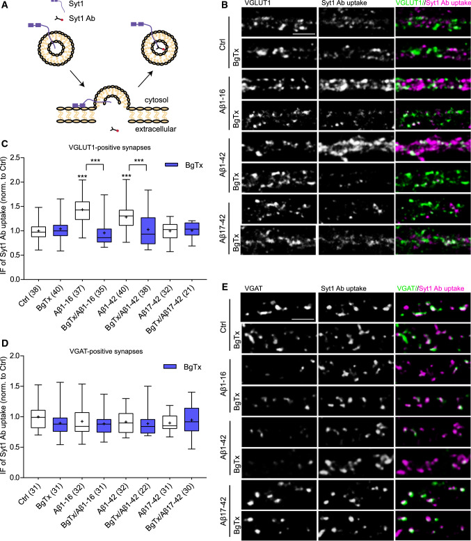
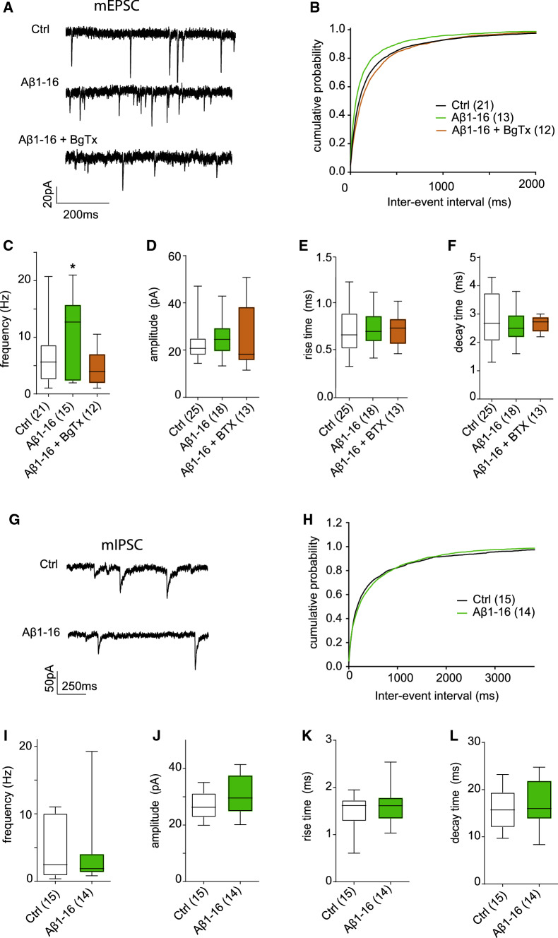
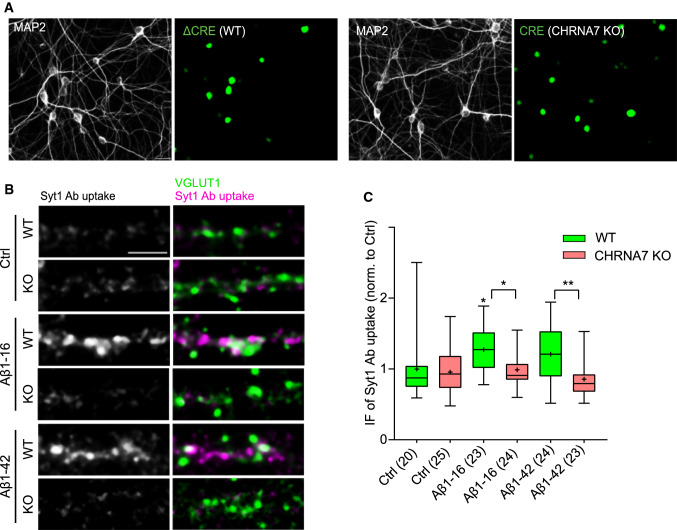
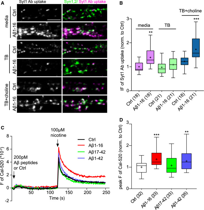
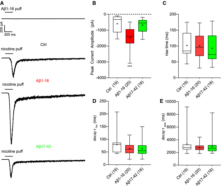
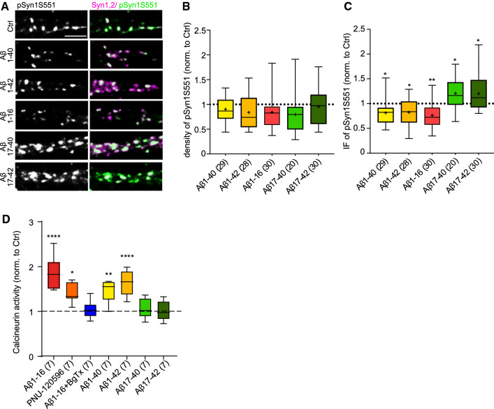
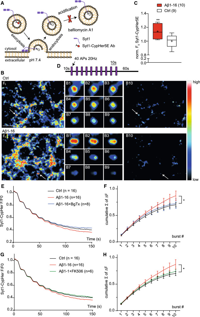
Amyloid beta (Aβ) is linked to the pathology of Alzheimer's disease (AD). At physiological concentrations, Aβ was proposed to enhance neuroplasticity and memory formation by increasing the neurotransmitter release from presynapse. However, the exact mechanisms underlying this presynaptic effect as well as specific contribution of endogenously occurring Aβ isoforms remain unclear. Here, we demonstrate that Aβ1-42 and Aβ1-16, but not Aβ17-42, increased size of the recycling pool of synaptic vesicles (SV). This presynaptic effect was driven by enhancement of endogenous cholinergic signalling via α7 nicotinic acetylcholine receptors, which led to activation of calcineurin, dephosphorylation of synapsin 1 and consequently resulted in reorganization of functional pools of SV increasing their availability for sustained neurotransmission. Our results identify synapsin 1 as a molecular target of Aβ and reveal an effect of physiological concentrations of Aβ on cholinergic modulation of glutamatergic neurotransmission. These findings provide new mechanistic insights in cholinergic dysfunction observed in AD.

Keywords: Alpha7 nicotinic acetylcholine receptor; Amyloid beta; Synapsin 1; Synaptic vesicle dynamics.


Conflict of interest statement

The authors have no competitive interests to declare.

Figures

Fig. 1
Fig. 1
The N-terminal domain of Aβ increases the TRP of SVs via α7nAChRs at excitatory but not inhibitory synapses. a Schematic representation of labelling of SV recycling using Syt1 Ab. Upon depolarization, releasable SVs fuse to the plasma membrane. The lumenal domain of integral SV protein Syt1 becomes exposed to the media and available for Syt1 Ab binding. Compensatory endocytosis drives the uptake of Syt1 Ab inside of retrieved SVs. b, e Representative images of depolarization-induced Syt1 Ab uptake (magenta) in primary cortical neurons (20–21 DIV) treated with vehicle (Ctrl), Aβ1-16, Aβ1-42, or Aβ17-42 (200 pM, 1 h) in the presence or absence of BgTx (50 nM, 90 min). Antibody against VGLUT1 and VGAT (green) was used to mark excitatory and inhibitory synapses, respectively. Scale bar is 5 μm. c, d Quantifications of Syt1 Ab uptake from (B,E). Values in brackets show the number of analysed cells obtained from two to three independent preparations. In the plots, boxes indicate the interquartile range and median, whiskers minimum and maximum values, and + depicts the mean. One-way ANOVA with Dunnett´s post hoc test was used to evaluate statistical significance. Significance of comparison to control are shown about the boxes, the BgTx effects are shown above the brackets; ***p < 0.001
Fig. 2
Fig. 2
Effect of Aβ1-16 on mEPSCs and mIPSC in hippocampal neurons. a Examples of mEPSC traces recorded in neurons under control conditions and after incubation with the Aβ1-16 alone or in presence of BTX (b). Cumulative distributions of the inter-event intervals of mEPSC from the experiment in a. Fragment Aβ1-16 significantly decreased mEPSC inter-event intervals, which is reflected in leftward shift of the cumulative distribution compared to control or Aβ + BTX (p < 0.001, Kolmogorov–Smirnov test). Aβ1-16 increases mean frequency (c), without effect on amplitude (d), rise time (e) and decay time (f) of mEPSC. Treatment with BgTx counteracts this effect. g Examples of mIPSC traces recorded in control neurons and after incubation with the Aβ1-16. h Cumulative distributions of the inter-event intervals of mIPSC in control neurons and after application of Aβ1-16. Aβ1-16 fragment has no effect on the mean frequency (i) amplitude (j), rise (k) and decay time (l) of mIPSC. Values in brackets in B-F and in H–L are number of analysed cells obtained from three independent preparations. Data are displayed as means (b, h) or as means ± SEM (cl), *p < 0.05 compared to control, t test
Fig. 3
Fig. 3
Aβ 1–16-mediated increase in SV recycling requires normal expression of α7nAChRs. a Representative images of primary mouse neurons (16 DIV) infected at 4 DIV either with ΔCRE (WT) or CRE (CHRNA7 KO) and stained with MAP2 to assess neuronal transduction efficiency. Scale bar 20 μm. b Images show the Syt1 Ab-labelled TRP in cortical mouse cultures (16–17 DIV) pretreated with vehicle (Ctrl), Aβ1-16, or Aβ1-42 on the left (magenta) at excitatory presynapses marked with VGLUT1 (green). Scale bar 5 μm. c Quantification of the IF intensity of Syt1 Ab uptake shown in b. Values in brackets indicate the number of analysed cells obtained from two independent experiments. Boxes indicate the interquartile range and median, whiskers minimum and maximum values, and + shows the mean. One-way ANOVA followed by Tukey´s post hoc test was used to assess statistical significance. The comparisons to vehicle-treated WT neurons are above each box, the symbols above brackets indicate the effect of CHRNA7-depletion, *p < 0.05, **p < 0.01
Fig. 4
Fig. 4
N-terminal portion of Aβ requires choline and nicotine to increase TRP and Ca2+ influx via α7nAChRs. a Images show TRP labelled by Syt1 Ab loading (magenta) in rat cortical neurons treated either with vehicle or 200 pMAβ1-16 (1 h, 37 °C) in the neuronal growth media, in Tyrode´s buffer or in Tyrode´s buffer supplemented with 28 μM choline. Synapses were identified by co-staining for Syn1,2 (green). Scale bar 5 μm. b Quantification of the experiment described in a. c Ca2+ imaging was done using Cal-520 AM indicator in HEK293T expressing α7nAChRs. Traces show the time course of the experiment. Arrows depict the sequential applications of human Aβ1-16, human/rat Aβ17-42, human Aβ1-42 (200 pM, 115 μl/s) or vehicle solution and nicotine (100 μM, 115 μl/s). d Quantification of nicotine-induced Cal-520 fluorescence in experiment shown in graph (C). Values in brackets refer to the number of analysed cells (b) or recorded wells (d) obtained from two (b) or four (D) independent preparations. Boxes indicate the interquartile range with median, whiskers minimum and maximum values, and + shows the mean. Significance was assessed by one-way ANOVA followed by Dunnett´s post hoc test; marks (***p < 0.001, **p < 0.01) above boxes indicate comparisons to the Ctrl
Fig. 5
Fig. 5
Aβ1-16, but not Aβ17-42, modulates nicotine-induced inward current of α7nAChRs. a Representative whole-cell current traces of α7nAChRs transiently expressed in HEK293T cells evoked by 300 ms puffs of 200 pM human Aβ1-16 or by 100 μM nicotine in the presence of vehicle (Ctrl), 200 pM human Aβ1-16 or human/rat Aβ17-42. Cells were held at -70 mV. Quantification of nicotine-evoked peak current amplitude (b) and rise time (c). Time constant (τ) of the fast (d) and slow (e) component of current decay in response to nicotine puff application in all conditions. τ was estimated by fitting the fast and slow phase of current decay with a bi-exponential function. Values in brackets refer to the number of recorded cells. Boxes indicate the interquartile range with median, whiskers minimum and maximum values, and + shows the mean. One-way ANOVA with Dunnett´s multiple comparisons test (c) or Kruskal–Wallis test with Dunn’s post hoc test (b, d, e) were used for comparisons to Ctrl; ***p < 0.001.
Fig. 6
Fig. 6
Aβ-derived fragments differently affect phosphorylation of Syn1 at serine 551 and calcineurin activity. a Representative images of mature cortical neurons (17–22 DIV) treated with vehicle (Ctrl), rat Aβ1-40, rat Aβ1-42, human Aβ1-16, human/rat Aβ17-40, human/rat Aβ17-42 for 1 h at 37 °C and stained with antibodies recognizing Syn1 phosphorylation at serine 551 (pSyn1S551, green). Syn1,2 (magenta) was used as a synaptic marker. Scale bar 5 μm. b Quantification of the density and c IF intensity of synaptic puncta positive for pSyn1S551. (D) Quantification of calcineurin activity in cortical neurons (21DIV) treated with Aβ peptides, BgTx and PNU-120596. Data were normalized to the Ctrl. In plots, the boxes show the interquartile range and median, whiskers minimum and maximum values, and + indicates the mean. Values in brackets are the in b and c number of analysed cells obtained from three independent experiments and in d numbers of independent experiments. One-way ANOVA followed by Dunnett´s post hoc test was applied to assess statistical significance of comparison to Ctrl; *p < 0.05, **p < 0.01, ****p < 0.0001
Fig. 7
Fig. 7
The N-terminal Aβ increases availability of SVs during repetitive stimulation dependently on α7nAChR and calcineurin activation. a Schematic representation of the Syt1-CypHer5E imaging. At rest, the fluorescence (F) of CypHer5E is completely unquenched in the low intra-vesicular pH (~ 5.5) and gets quenched upon SV exocytosis in the neutral extracellular media (~ 7.4). Following endocytosis and vesicular reacidification, the CypHer5E F increases again. Blockade of the v-ATPase with bafilomycin A1 prevents vesicular reacidification trapping SVs in the alkaline state. b Representative image F0 shows typical Syt1-CypHer5E loading in hippocampal neurons (17–20 DIV) incubated with either vehicle (Ctrl) or human Aβ1-16 (200 pM) for 1 h at 37 °C in Tyrode´s buffer with choline before Syt1-CypHer5E loading (1 h, 37 °C). c Quantification of the Syt1-CypHer5E loading (F0). Images B1–B9 show the course of destaining of an individual synapse (marked by arrow in the F0 overview image) upon application of stimulation protocol shown in d in the presence of bafilomycin A (1 μM). B10 shows the whole region upon application of the final stimulus. Scale bar is 5 μm in overview and 1 μm in close-up image. d Schematic representation of applied stimulation protocol. After 10 s of baseline, 10 bursts (B1–B10) of 40AP at 20 Hz spaced by 10 s pause were applied via electrical field stimulations. Imaging was continued for additional 60 s. e,g Traces showing average time-course of Syt1-CypHer5E F in response to the stimulation protocol in control and cells treated with Aβ1-16 alone or in presence of either BgTx (E) or FK506 (G). All values were normalized to F0. f, h Cumulative amplitude of Syt1-CypHer5E F calculated from curves shown in e and g. Data are obtained from at least four independent experiments and are expressed as a mean (e, g) or mean ± SEM (f, h) or as boxes depicting the interquartile range and median, with whiskers showing minimum and maximum values, and mean showed as + (c). Values in brackets show the number of analysed coverslips from four independent experiments. Unpaired t test was used to estimate statistical significance; *p < 0.05, **p < 0.01

References

    1. Abramov E, Dolev I, Fogel H, Ciccotosto GD, Ruff E, Slutsky I. Amyloid-beta as a positive endogenous regulator of release probability at hippocampal synapses. Nat Neurosci. 2009;12:1567–1576. doi: 10.1038/nn.2433. - DOI - PubMed
    1. Alkondon M, Pereira EF, Cortes WS, Maelicke A, Albuquerque EX. Choline is a selective agonist of alpha7 nicotinic acetylcholine receptors in the rat brain neurons. Eur J Neurosci. 1997;9:2734–2742. doi: 10.1111/j.1460-9568.1997.tb01702.x. - DOI - PubMed
    1. Brody DL, Magnoni S, Schwetye KE, Spinner ML, Esparza TJ, Stocchetti N, Zipfel GJ, Holtzman DM. Amyloid-beta dynamics correlate with neurological status in the injured human brain. Science. 2008;321:1221–1224. doi: 10.1126/science.1161591. - DOI - PMC - PubMed
    1. Burrone J, Li ZY, Murthy VN. Studying vesicle cycling in presynaptic terminals using the genetically encoded probe synaptopHluorin. Nat Protoc. 2006;1:2970–2978. doi: 10.1038/nprot.2006.449. - DOI - PubMed
    1. Castro NG, Albuquerque EX. Alpha-bungarotoxin-sensitive hippocampal nicotinic receptor-channel has a high-calcium permeability. Biophys J. 1995;68:516–524. doi: 10.1016/S0006-3495(95)80213-4. - DOI - PMC - PubMed

MeSH terms

LinkOut - more resources