

The triangular fibrocartilage complex on high-resolution 3 T MRI in healthy adolescents: the thin line between asymptomatic findings and pathology
- PMID: 33864484
- PMCID: PMC8449761
- DOI: 10.1007/s00256-021-03779-8
The triangular fibrocartilage complex on high-resolution 3 T MRI in healthy adolescents: the thin line between asymptomatic findings and pathology
Abstract
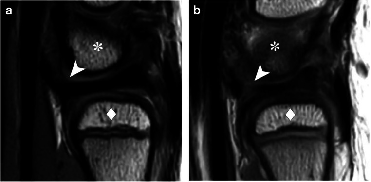
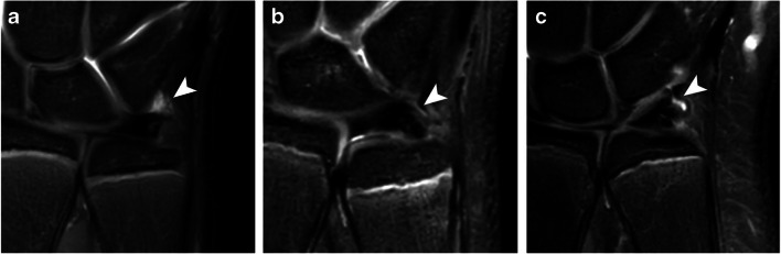
Objective: The objective of the study is to provide a reference for morphology, homogeneity, and signal intensity of triangular fibrocartilage complex (TFCC) and TFCC-related MRI features in adolescents.
Materials and methods: Prospectively collected data on asymptomatic participants aged 12-18 years, between June 2015 and November 2017, were retrospectively analyzed. A radiograph was performed in all participants to determine skeletal age and ulnar variance. A 3-T MRI followed to assess TFCC components and TFCC-related features. A standardized scoring form, based on MRI definitions used in literature on adults, was used for individual assessment of all participants by four observers. Results per item were expressed as frequencies (percentages) of observations by all observers for all participants combined (n = 92). Inter-observer agreement was determined by the unweighted Fleiss' kappa with 95% confidence intervals (95% CI).
Results: The cohort consisted of 23 asymptomatic adolescents (12 girls and 11 boys). Median age was 13.5 years (range 12.0-17.0). Median ulnar variance was -0.7 mm (range - 2.7-1.4). Median triangular fibrocartilage (TFC) thickness was 1.4 mm (range 0.1-2.9). Diffuse increased TFC signal intensity not reaching the articular surface was observed in 30 (33%) observations and a vertical linear increased signal intensity with TFC discontinuation in 19 (20%) observations. Discontinuation between the volar radioulnar ligament and the TFC in the sagittal plane was seen in 23 (25%) observations. The extensor carpi ulnaris was completely dislocated in 10 (11%) observations, more frequent in supinated wrists (p = 0.031). Inter-observer agreement ranged from poor to fair for scoring items on the individual TFCC components.
Conclusion: MRI findings, whether normal variation or asymptomatic abnormality, can be observed in TFCC and TFCC-related features of asymptomatic adolescents. The rather low inter-observer agreement underscores the challenges in interpreting these small structures on MRI. This should be taken into consideration when interpreting clinical MRIs and deciding upon arthroscopy.
Keywords: Adolescent; Magnetic resonance imaging; Triangular fibrocartilage; Wrist injuries.
© 2021. The Author(s).
Conflict of interest statement
The authors declare that they have no conflict of interest.
Figures


References
MeSH terms
LinkOut - more resources
Full Text Sources
Other Literature Sources
Medical
