. 2021 Oct;141(10):2436-2448.
doi: 10.1016/j.jid.2021.03.019. Epub 2021 Apr 15.

IRAK2 Has a Critical Role in Promoting Feed-Forward Amplification of Epidermal Inflammatory Responses

Affiliations

IRAK2 Has a Critical Role in Promoting Feed-Forward Amplification of Epidermal Inflammatory Responses

Shuai Shao et al. J Invest Dermatol. 2021 Oct.

Abstract

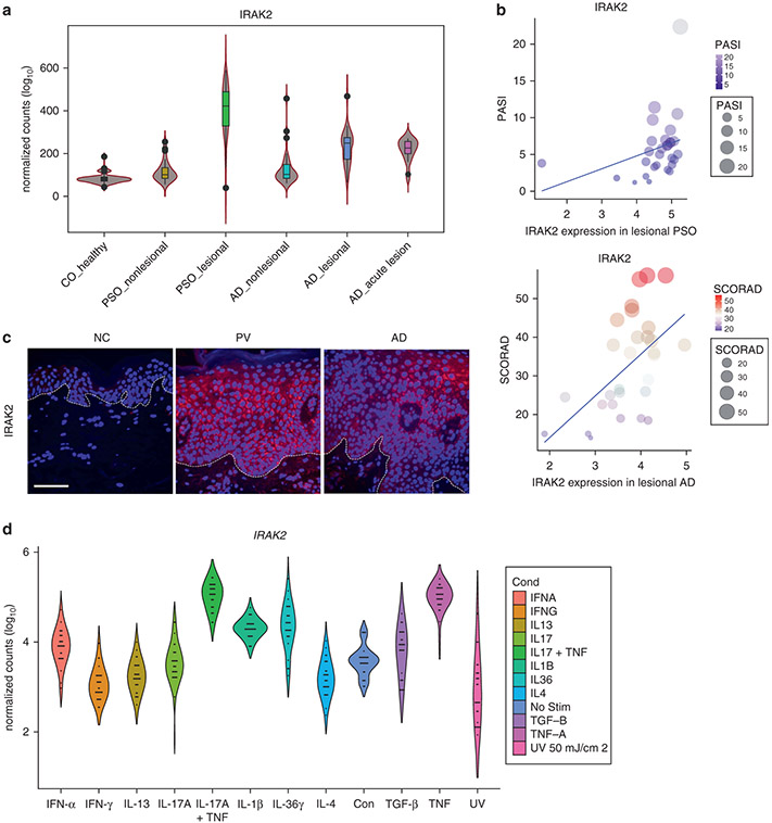
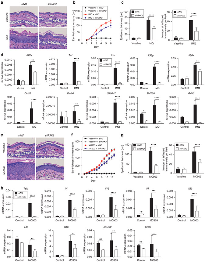
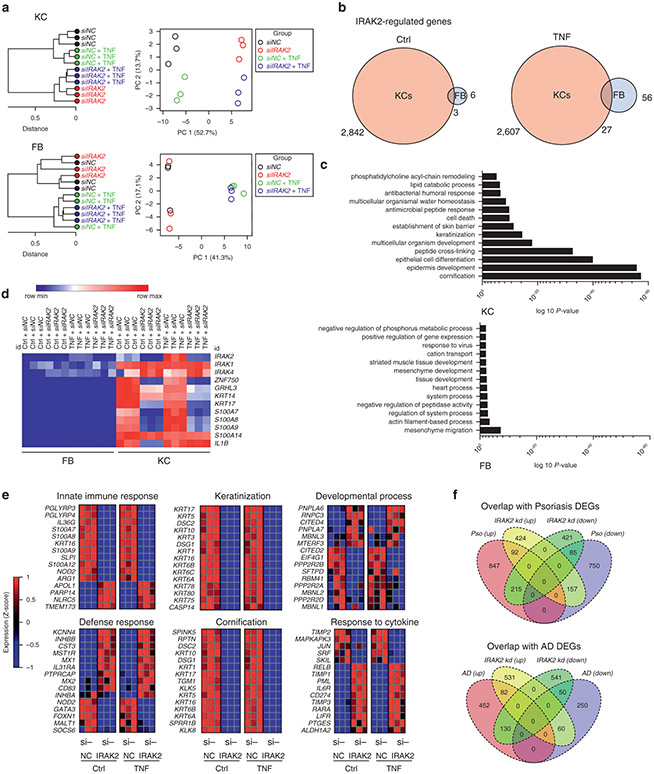
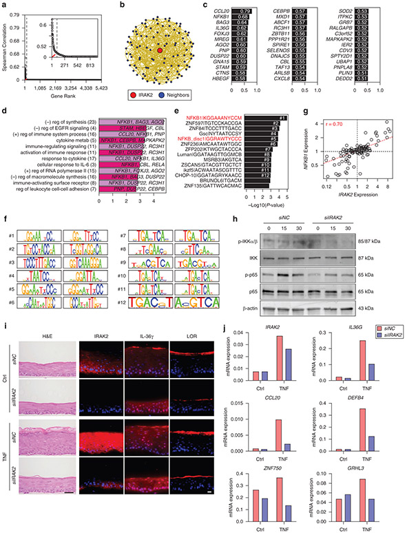
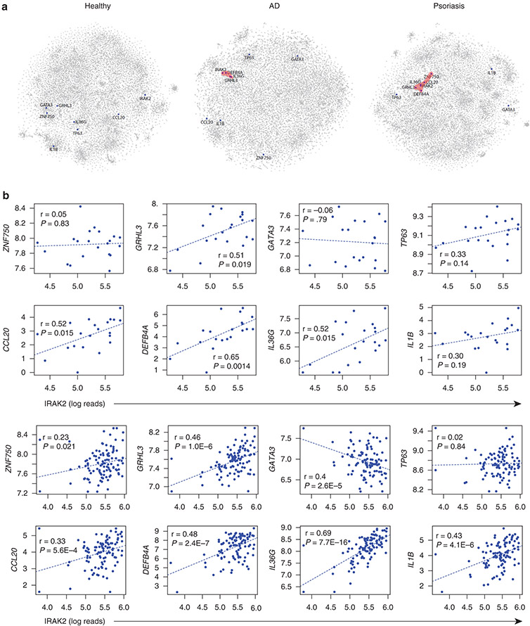
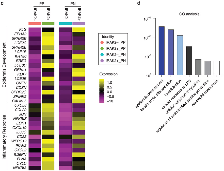
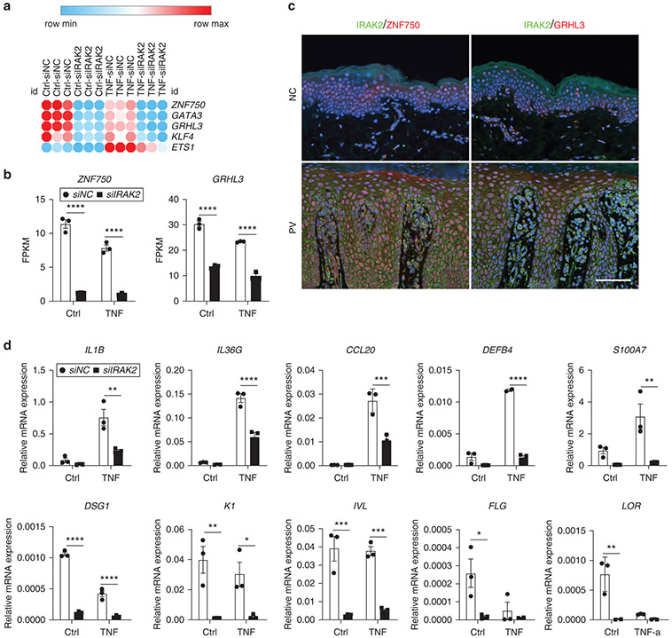
Many inflammatory skin diseases are characterized by altered epidermal differentiation. Whether this altered differentiation promotes inflammatory responses has been unknown. Here, we show that IRAK2, a member of the signaling complex downstream of IL-1 and IL-36, correlates positively with disease severity in both atopic dermatitis and psoriasis. Inhibition of epidermal IRAK2 normalizes differentiation and inflammation in two mouse models of psoriasis- and atopic dermatitis-like inflammation. Specifically, we demonstrate that IRAK2 ties together proinflammatory and differentiation-dependent responses and show that this function of IRAK2 is specific to keratinocytes and acts through the differentiation-associated transcription factor ZNF750. Taken together, our findings suggest that IRAK2 has a critical role in promoting feed-forward amplification of inflammatory responses in skin through modulation of differentiation pathways and inflammatory responses.


Conflict of interest statement

CONFLICT OF INTEREST

This work received support through BIOMAP (Biomarkers in Atopic Dermatitis and Psoriasis), a project funded by the Innovative Medicines Initiative 2 Joint Undertaking under Grant Agreement No. 821511. SW is coprincipal investigator of the German Atopic Eczema Registry TREATgermany; has received institutional research grants from Sanofi Deutschland GmbH, LEO Pharma, and La Roche Posay; has performed consultancies for Sanofi-Genzyme, Regeneron Pharmaceuticals, LEO Pharma, AbbVie, Pfizer, Eli Lilly, Kymab, and Novartis; has lectured at educational events sponsored by Sanofi-Genzyme, Regeneron Pharmaceuticals, LEO Pharma, AbbVie, Novartis, and Galderma; and is involved in performing clinical trials with many pharmaceutical industries that manufacture drugs used for the treatment of psoriasis and atopic eczema. JMK has served on ad boards for AstraZeneca, Provention Bio, Aurinia Pharmaceuticals, Bristol Myers Squibb, Boehringer Ingelheim, Eli Lilly, Viela Bio, and Ventus Therapeutics and has grants from Bristol Myers Squibb and Q32 Bio. JEG has received honoraria from AnaptysBio, Bristol-Myers Squibb Celgene, Sanofi, AstraZeneca, Eli Lilly, Novartis, and Almirall and research grants from Eli Lilly, Kyowa Kirin, Almirall, and Bristol-Myers Squibb Celgene. The other authors state no conflict of interest.

Figures

Figure 1.
Figure 1.. IRAK2 is highly expressed in skin lesions of patients with AD and PSO.
(a) Violin plot showing the expression level of IRAK2 as detected by RNA-seq analysis in lesional skin of patients with PSO (n = 28, FC = 4.9-fold, P = 2 × 10⁻²⁵), nonlesional skin of patients with PSO (n = 27), chronic (n = 28, FC = 2.2-fold, P = 2 × 10⁻¹¹) and acute lesional (n = 11, FC = 1.8-fold, P = 2 × 10⁻¹⁰) skin of patients with AD, nonlesional skin of patients with AD (n = 27), and healthy CO (n = 38). (b) Positive correlation of IRAK2 expression level in psoriatic lesions with PASI (upper panel) and in AD lesions with SCORAD (lower panel). (c) Representative immunofluorescent staining of IRAK2 (red) in lesional AD and PSO skin. White dashed line indicates the epidermal and dermal junction (n = 3, biological replicates). Bar = 100 μm. (d) Violin plots highlighting the expression level of IRAK2 mRNA in primary keratinocytes (n = 38) stimulated with various cytokines, such as TNF (FC = 1.8-fold, FDR = 8.7 × 10⁻¹⁶), combination of TNF + IL-17A (FC = 1.7-fold, FDR = 7.6 × 10⁻¹⁸), or IL-36γ (FC = 1.5-fold, FDR = 2.3 × 10⁻²³) for 24 hours. One-way ANOVA. Error bars represent SEM. AD, atopic dermatitis; CO, control; FC, fold change; FDR, false discovery rate; NC, normal control; PSO, psoriasis; PV, psoriasis vulgaris; RNA-seq, RNA-sequencing.
Figure 2.
Figure 2.. IRAK2 knockdown improves inflammatory responses in both psoriasis and AD mouse models.
siRNA targeting IRAK2 was topically applied to IMQ-induced psoriasis-like mouse or MC903-induced AD-like mouse ears to silence IRAK2 expression. (a) H&E staining of ear sections from IMQ-induced mice at day 6. Bar = 50 μm. (b) Dynamic changes in ear thickness at the indicated time points. (c) Epidermal thickness and inflammatory infiltrates were evaluated based on (a). (d) Quantitative real-time PCR results showing mRNA expression of various cytokines in the ears of IMQ mice on day 6. (e) H&E staining of ear sections from MC903-induced mice at day 15. Bar = 50 μm. (f) Dynamic changes in ear thickness at the indicated time points. (g) Epidermal thickness and inflammatory infiltrates were evaluated based on (e). (h) Quantitative real-time PCR results showing mRNA expression of various cytokines in the ears of MC903 mice on day 15. Two-way ANOVA. Data are presented as mean ± SEM. *P < 0.05, **P < 0.01, ***P < 0.001, ****P < 0.0001. AD, atopic dermatitis; IMQ, imiquimod; ns, nonsignificant; K, keratin; NC, normal control; siIRAK2, IRAK2-silenced; si-NC, NC-silenced; siRNA, small interfering RNA.
Figure 3.
Figure 3.. IRAK2′s role is primarily in KCs instead of FBs.
(a) KCs and FBs were transfected with IRAK2 siRNA or ctrl siRNA and stimulated with TNF (10 ng/ml) for 24 hours (n = 3 samples per group). Cluster analysis of four experimental groups in KCs (upper panel) and FBs (lower panel) are shown based on the expression changes detected by RNA-seq. (b) Venn diagrams showing the number of DEGs similarly altered by IRAK2 siRNA in KCs and FBs with or without TNF stimulation (10 ng/ml). (c) Functional categories enriched in IRAK2-regulated genes in KCs and FBs. (d) Heatmap showing expression level of selected IRAK2-regulated genes enriched in differentiation and innate immune responses in FBs and KCs, respectively. (e) Heatmap showing expression levels of IRAK2-regulated genes in different GO categories with or without TNF stimulation. (f) Overlap between IRAK2-regulated genes (upregulated and downregulated) and DEGs in AD and psoriasis skin lesions. AD, atopic dermatitis; ctrl, control; DEG, differentially expressed gene; FB, fibroblast; GO, Gene Ontology; KC, keratinocyte; max, maximum; min, minimum; NC, normal control; PC, principal component; RNA-seq, RNA-sequencing; siIRAK2, IRAK2-silenced; siNC, NC-silenced; siRNA, small interfering RNA.
Figure 4.
Figure 4.. IRAK2 regulatory network and effect on epidermal differentiation during inflammatory state.
(a) Correlation coefficients relative to the ranked list of 5,423 genes detected by RNA-seq. The vertical axis is exponentially scaled (r7) to emphasize higher correlations. The red line represents the minimal distance between the lower-left origin and correlation curve, which defines a set of 108 genes having IRAK2-correlated expression (rs ≥ 0.45). (b) IRAK2-correlated genes are plotted in a spherical network with IRAK2 at center. The top three intranetwork correlations were used for each gene to draw connections. (c) Network genes most strongly correlated with IRAK2. (d) GO biological process terms most strongly enriched with respect to the 108 IRAK2-correlated genes. The number of genes associated with each term are shown (parentheses, left margin) and exemplar genes are listed within the figure. (e, f) DNA motifs (e) most heavily enriched in 5-kb regions upstream of IRAK2-correlated genes, with (f) the 12 most significant motifs shown (see corresponding names in e). (g) The correlation of IRAK2 with NFKB1 expression. (h) Representative western blot showing the activation of IKK and NF-κB p65 signaling in siIRAK2 versus Ctrl keratinocytes in response to TNF stimulation. (i) Representative H&E staining (left panel) of HSEs showing the effect of IRAK2 silencing with or without TNF stimulation, and immunofluorescent staining (right panel) of IRAK2, IL-36γ, and LOR (all red) at day 6. The experiment was repeated twice. Bar = 100 μm. (j) Representative quantitative real-time PCR analysis of proinflammatory and differentiation genes in siIRAK2-treated 3D HSE with or without TNF stimulation. 3D, three-dimensional; Ctrl, control; GO, Gene Ontology; HSE, human skin equivalent; IKK, IκB kinase; kb, kilo base; NC, normal control; p-IKKα/β, phosphorylated IκB kinaseα/β; p-P65, phosphorylated p65; reg, region; RNA-seq, RNA-sequencing; siIRAK2, IRAK2-silenced; siNC, NC-silenced.
Figure 5.
Figure 5.. IRAK2-associated genes in keratinocytes involve inflammatory response genes and markers of epidermal development.
(a) 2DCoexpression networks of gene expression (RNA-seq) of healthy skin, psoriatic, and AD skin lesions with labeled IRAK2 and key epidermal differentiation genes showing grouping and tight clustering of these genes in psoriatic skin but not normal skin. (b) The correlation analysis of IRAK2 with selected inflammatory and epidermal differentiated genes in skin lesions of AD (upper panel) and psoriasis (lower panel) based on the RNA-seq data. (c) Heatmap and (d) GO categories of epidermis development and inflammatory response of IRAK2+ keratinocytes based on single-cell RNA-seq of psoriasis skin lesions. 2D, two-dimensional; AD, atopic dermatitis; GO, Gene Ontology; LPS, lipopolysaccharide; NC, normal control; PN, psoriatic non-lesional skin; PP, psoriatic lesional skin; RNA-seq, RNA-sequencing.
Figure 6.
Figure 6.. IRAK2 affects ZNF750 expression to modulate epidermal differentiation and proinflammatory responses.
(a) Heatmap analysis showing the critical differentiation genes affected by siIRAK2 in keratinocytes. (b) RNA-seq validation of ZNF750 and GRHL3 mRNA expression by siIRAK2 in keratinocytes (n = 3). (c) Immunofluorescent costaining of IRAK2 (green) against ZNF750 and GRHL3 (all red) in normal and lesional psoriatic skin. Bar = 100 μm. (d) Quantitative real-time PCR for mRNA expression of differentiation and proinflammatory genes in ZNF750-silenced keratinocytes with or without TNF stimulation (n = 3). Two-way ANOVA. Data are presented as mean ± SEM. *P < 0.05, **P < 0.01, ***P < 0.001, ****P < 0.0001. Ctrl, control; DSG, desmoglein; FPKM, Fragments Per Kilobase of transcript per Million mapped reads; id, identification; K, keratin; max, maximum; min, minimum; NC, normal control; ns, no significance; PV, psoriasis vulgaris; RNA-seq, RNA-sequencing; siIRAK2, IRAK2-silenced; siNC, NC-silenced.

Similar articles

Cited by

References

    1. Arnette C, Koetsier JL, Hoover P, Getsios S, Green KJ. In vitro model of the epidermis: connecting protein function to 3D structure. Methods Enzymol 2016;569:287–308. - PMC - PubMed
    1. Birnbaum RY, Hayashi G, Cohen I, Poon A, Chen H, Lam ET, et al. Association analysis identifies ZNF750 regulatory variants in psoriasis. BMC Med Genet 2011;12:167. - PMC - PubMed
    1. Boehncke WH, Schön MP. Psoriasis. Lancet 2015;386:983–94. - PubMed
    1. Cataisson C, Salcedo R, Hakim S, Moffitt BA, Wright L, Yi M, et al. IL-1R-MyD88 signaling in keratinocyte transformation and carcinogenesis. J Exp Med 2012;209:1689–702. - PMC - PubMed
    1. Conner JR, Smirnova II, Poltorak A. A mutation in Irak2c identifies IRAK-2 as a central component of the TLR regulatory network of wild-derived mice. J Exp Med 2009;206:1615–31. - PMC - PubMed

Publication types