

Antiviral activities of four marine sulfated glycans against adenovirus and human cytomegalovirus
- PMID: 33864843
- PMCID: PMC8212419
- DOI: 10.1016/j.antiviral.2021.105077
Antiviral activities of four marine sulfated glycans against adenovirus and human cytomegalovirus
Abstract
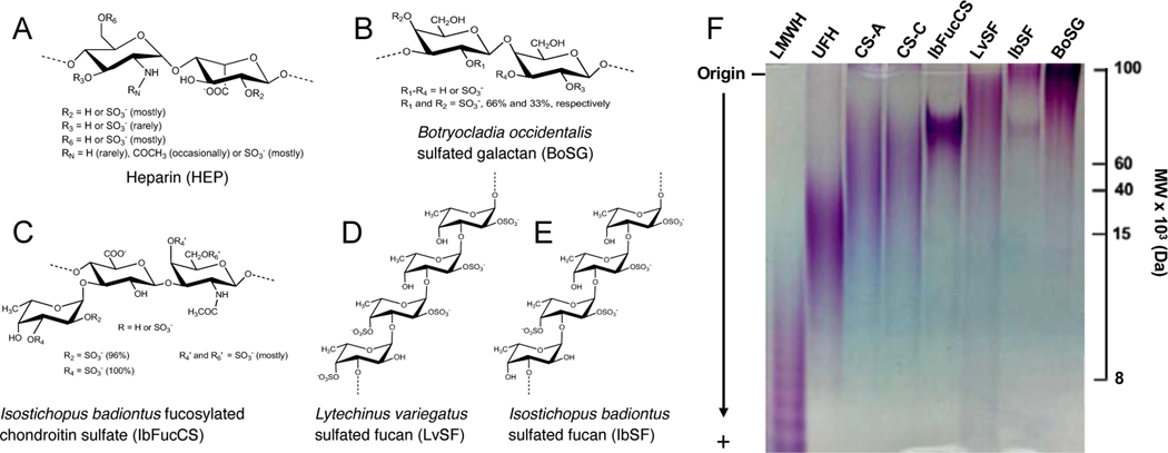
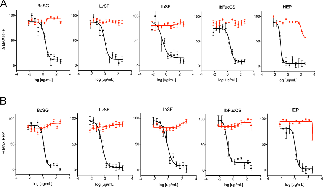
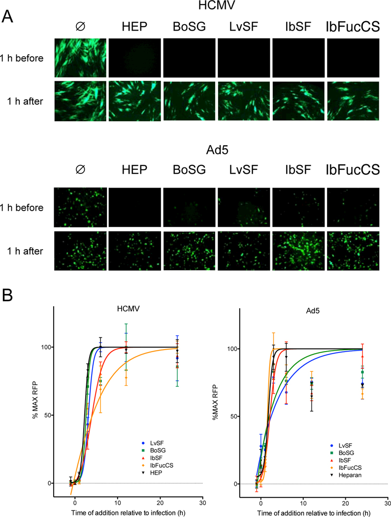
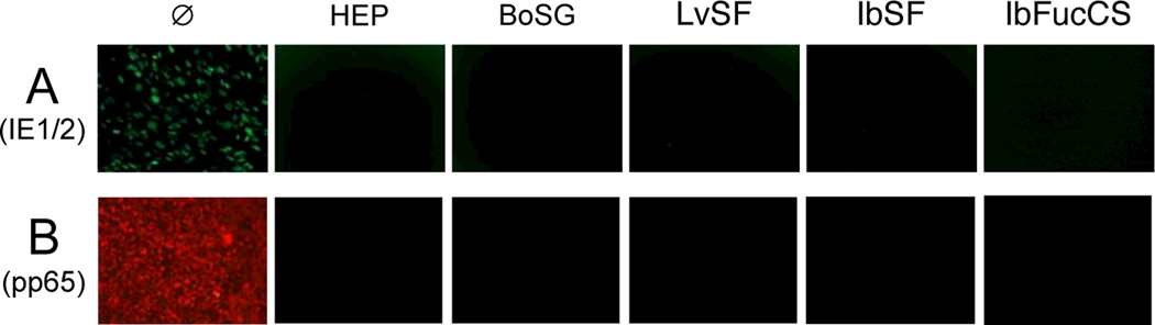
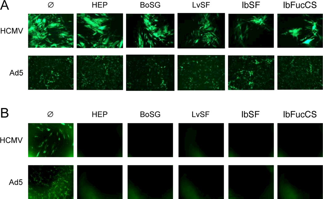
Broad-spectrum antivirals are more needed than ever to provide treatment options for novel emerging viruses and for viruses that lack therapeutic options or have developed resistance. A large number of viruses rely on charge-dependent non-specific interactions with heparan sulfate (HS), a highly sulfated glycosaminoglycan (GAG), for attachment to cell surfaces to initiate cell entry. As such, inhibitors targeting virion-HS interactions have potential to have broad-spectrum antiviral activity. Previous research has explored organic and inorganic small molecules, peptides, and GAG mimetics to disrupt virion-HS interactions. Here we report antiviral activities against both enveloped (the herpesvirus human cytomegalovirus) and non-enveloped (adenovirus) DNA viruses for four defined marine sulfated glycans: a sulfated galactan from the red alga Botryocladia occidentalis; a sulfated fucan from the sea urchin Lytechinus variegatus, and a sulfated fucan and a fucosylated chondroitin sulfate from the sea cucumber Isostichopus badionotus. As evidenced by gene expression, time of addition, and treatment/removal assays, all four novel glycans inhibited viral attachment and entry, most likely through interactions with virions. The sulfated fucans, which both lack anticoagulant activity, had similar antiviral profiles, suggesting that their activities are not only due to sulfation content or negative charge density but also due to other physicochemical factors such as the potential conformational shapes of these carbohydrates in solution and upon interaction with virion proteins. The structural and chemical properties of these marine sulfated glycans provide unique opportunities to explore relationships between glycan structure and their antiviral activities.
Keywords: Adenovirus; Antiviral; Broad-spectrum; Cytomegalovirus; Marine sulfated glycans; Virion attachment.
Copyright © 2021 Elsevier B.V. All rights reserved.
Figures
Similar articles
-
Antiviral activity of marine sulfated glycans against pathogenic human coronaviruses.Sci Rep. 2023 Mar 23;13(1):4804. doi: 10.1038/s41598-023-31722-5. Sci Rep. 2023. PMID: 36959228 Free PMC article.
-
SPR Sensor-Based Analysis of the Inhibition of Marine Sulfated Glycans on Interactions between Monkeypox Virus Proteins and Glycosaminoglycans.Mar Drugs. 2023 Apr 25;21(5):264. doi: 10.3390/md21050264. Mar Drugs. 2023. PMID: 37233458 Free PMC article.
-
Marine sulfated glycans inhibit the interaction of heparin with S-protein of SARS-CoV-2 Omicron XBB variant.Glycoconj J. 2024 Apr;41(2):163-174. doi: 10.1007/s10719-024-10150-1. Epub 2024 Apr 20. Glycoconj J. 2024. PMID: 38642280 Free PMC article.
-
Specific sulfation and glycosylation-a structural combination for the anticoagulation of marine carbohydrates.Front Cell Infect Microbiol. 2014 Mar 6;4:33. doi: 10.3389/fcimb.2014.00033. eCollection 2014. Front Cell Infect Microbiol. 2014. PMID: 24639954 Free PMC article. Review.
-
Sulfated polysaccharides extracted from sea algae as potential antiviral drugs.Gen Pharmacol. 1997 Oct;29(4):497-511. doi: 10.1016/s0306-3623(96)00563-0. Gen Pharmacol. 1997. PMID: 9352294 Review.
Cited by
-
Inhibition of Cytomegalovirus by Pentacta pygmaea Fucosylated Chondroitin Sulfate Depends on Its Molecular Weight.Viruses. 2023 Mar 28;15(4):859. doi: 10.3390/v15040859. Viruses. 2023. PMID: 37112839 Free PMC article.
-
Antiviral activity of marine sulfated glycans against pathogenic human coronaviruses.Sci Rep. 2023 Mar 23;13(1):4804. doi: 10.1038/s41598-023-31722-5. Sci Rep. 2023. PMID: 36959228 Free PMC article.
-
Docking and Molecular Dynamics Simulations Clarify Binding Sites for Interactions of Novel Marine Sulfated Glycans with SARS-CoV-2 Spike Glycoprotein.Molecules. 2023 Sep 3;28(17):6413. doi: 10.3390/molecules28176413. Molecules. 2023. PMID: 37687244 Free PMC article.
-
Structural and kinetic analyses of holothurian sulfated glycans suggest potential treatment for SARS-CoV-2 infection.J Biol Chem. 2021 Oct;297(4):101207. doi: 10.1016/j.jbc.2021.101207. Epub 2021 Sep 17. J Biol Chem. 2021. PMID: 34537241 Free PMC article.
-
Safety and Pharmacokinetics of Intranasally Administered Heparin.medRxiv [Preprint]. 2022 Feb 17:2021.07.05.21259936. doi: 10.1101/2021.07.05.21259936. medRxiv. 2022. Update in: Pharm Res. 2022 Mar;39(3):541-551. doi: 10.1007/s11095-022-03191-4. PMID: 35194614 Free PMC article. Updated. Preprint.
References
-
- Anderson RA, Zaneveld LJD, Usher TC, 2000. Cellulose sulfate for use as antimicrobial and contraceptive agent.
-
- Astrup T, Alkjaersig N, 1950. Polysaccharide polysulphic acids as antihyaluronidases. Nature 166, 568–569. - PubMed
-
- Astrup T, Galsmar I, 1944. On the anticoagulant acitivty of heparin and synthetic polysaccharide sulfuric acids. Acta Physiol. Scand 8, 361–364.
-
- Baba M, Snoeck R, Pauwels R, De Clercq E, 1988. Sulfated polysaccharides are potent and selective inhibitors of various enveloped viruses, including herpes simplex virus, cytomegalovirus, vesicular stomatitis virus, and human immunodeficiency virus. Antimicrob. Agents Chemother 32, 1742–1745. 10.1128/AAC.32.11.1742 - DOI - PMC - PubMed
Publication types
MeSH terms
Substances
Grants and funding
LinkOut - more resources
Full Text Sources
Other Literature Sources
Research Materials
Miscellaneous
