. 2021 May:106:338-347.
doi: 10.1016/j.ijid.2021.04.034. Epub 2021 Apr 14.

Spike is the most recognized antigen in the whole-blood platform in both acute and convalescent COVID-19 patients

Affiliations

Spike is the most recognized antigen in the whole-blood platform in both acute and convalescent COVID-19 patients

Alessandra Aiello et al. Int J Infect Dis. 2021 May.

Abstract

Objectives: To identify the best experimental approach to detect a SARS-CoV-2-specific T cell response using a whole-blood platform.

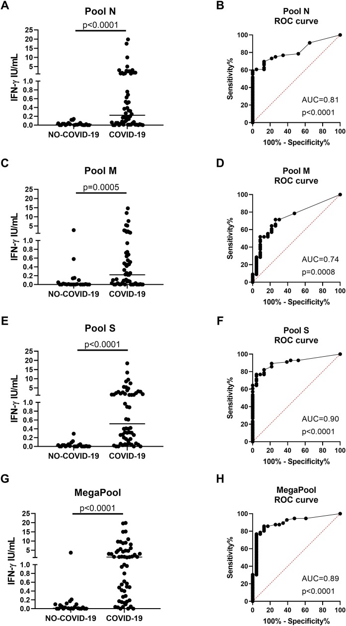
Methods: Whole-blood from 56 COVID-19 and 23 "NO-COVID-19" individuals were stimulated overnight with different concentrations (0.1 or 1 μg/mL) of SARS-CoV-2 PepTivator® Peptide Pools, including spike (pool S), nucleocapsid (pool N), membrane (pool M), and a MegaPool (MP) of these three peptide pools. ELISA was used to analyse interferon (IFN)-γ levels.

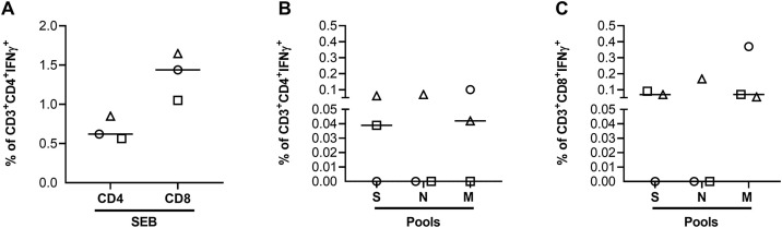
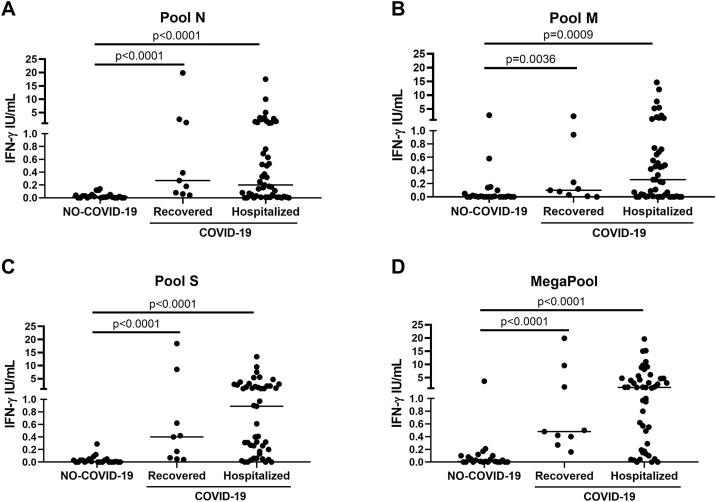
Results: The IFN-γ-response to every SARS-CoV-2 peptide pool was significantly increased in COVID-19 patients compared with NO-COVID-19 individuals. Pool S and MegaPool were the most potent immunogenic stimuli (median: 0.51, IQR: 0.14-2.17; and median: 1.18, IQR: 0.27-4.72, respectively) compared with pools N and M (median: 0.22, IQR: 0.032-1.26; and median: 0.22, IQR: 0.01-0.71, respectively). The whole-blood test based on pool S and MegaPool showed a good sensitivity of 77% and a high specificity of 96%. The IFN-γ-response was mediated by both CD4+ and CD8+ T cells, and independently detected of clinical parameters in both hospitalized and recovered patients.

Conclusions: This easy-to-use assay for detecting SARS-CoV-2-specific T cell responses may be implemented in clinical laboratories as a powerful diagnostic tool.

Keywords: COVID-19; IFN-γ-release assay (IGRA); Membrane protein; Nucleocapsid protein; SARS-CoV-2; Spike protein; T cell response; Whole-blood.


Figures

Figure 1
Figure 1
Spike and MegaPool peptides were the most immunogenic SARS-CoV-2 antigens in the whole-blood platform. The IFN-γ level in response to SARS-CoV-2 peptides was significantly increased in COVID-19 patients compared with NO-COVID-19 individuals after stimulating whole-blood with 1 μg/mL of pool N (A) and 0.1 μg/mL of pool M (C), pool S (E) and MegaPool (G) antigens. The ROC analysis shows significant AUC results for pool N (AUC = 0.81, p < 0.0001, B), pool M (AUC = 0.74, p = 0.0008, D), but mainly for pool S (AUC = 0.90, p < 0.0001, F) and MegaPool (AUC = 0.89, p < 0.0001, H) antigens. For scoring purposes, the cut-off of 0.13 IU/mL and of 0.19 IU/mL were chosen for pools N and M antigens, respectively, that led to 61% sensitivity/96% specificity for pool N and 52% sensitivity/91% specificity for pool M. For scoring purposes, the cut-off of 0.13 IU/mL and of 0.24 IU/mL were chosen for pool S and MegaPool antigens, respectively, that led to 77% sensitivity/96% specificity for both antigens. IFN-γ was measured by ELISA in stimulated plasma. The horizontal lines represent the median; statistical analysis was performed using the Mann–Whitney test and ROC analysis, and p-value was considered significant if ≤0.05. IFN, interferon; COVID-19: coronavirus disease 19; N: nucleocapsid; M: membrane; S: spike; ROC: receiver operating curve.
Figure 2
Figure 2
The Sars-CoV-2-specific response was mediated by both CD4+ and CD8+ T cells. Freshly isolated PBMCs were stimulated overnight with different stimuli to evaluate by flow cytometry the CD4+ or CD8+ T cell-specific IFN-γ response. A) Percentage of CD4+ or CD8+ T cells producing IFN-γ in response to the positive control SEB. B) Percentage of IFN-γ+ CD4+ T cells in response to pools S, N and M. C) Percentage of IFN-γ+ CD8+ T cells in response to pools S, N and M. In all graphs the IFN-γ values were subtracted from the unstimulated control. The horizontal lines represent the median and statistical analysis was performed using the Friedman test and p ≤ 0.05 was considered significant. Footnotes: IFN, interferon; N, nucleocapsid; M, membrane; S, spike.
Figure 3
Figure 3
The IFN-γ response to pools N, M, S and MegaPool was detectable in both recovered and hospitalized COVID-19 patients. Analysis of the IFN-γ response to pools N (A), M (B), S (C) and MegaPool (D) antigens in COVID-19 patients either hospitalized (n = 47) or recovered (n = 9). No significant differences were observed in response to all stimuli between the two groups of COVID-19 patients. IFN-γ was measured by ELISA in stimulated plasma. Statistical analysis was performed using the Mann-Whitney test and p ≤ 0.05 was considered significant. IFN, interferon; COVID-19: coronavirus disease 19; N, nucleocapsid; M, membrane; S, spike.
Figure 4
Figure 4
The IFN-γ response to pool S and MegaPool peptides can be detected in COVID-19 patients with a negative IgG/IgM serology. Evaluation of the IFN-γ response to pools N (A–B), M (C–D), S (E–F) and MegaPool (G–H) antigens in hospitalized COVID-19 patients (n = 41) stratified according to the IgG and IgM serology score and disease severity (A, C, E, G) or symptom onset (B, D, F, H). The IFN-γ response to pool S and MegaPool antigens can be detected in COVID-19 patients with negative IgG and IgM serology. IFN-γ was measured by ELISA in stimulated plasma. The horizontal lines represent the median; statistical analysis was performed using the Mann–Whitney test, and p-value was considered significant if ≤0.008. IFN, interferon; N, nucleocapsid; M, membrane; S, spike.

Similar articles

Cited by

References

    1. Braun J., Loyal L., Frentsch M., Wendisch D., Georg P., Kurth F. SARS-CoV-2-reactive T cells in healthy donors and patients with COVID-19. Nature. 2020;587:270–274. doi: 10.1038/s41586-020-2598-9. - DOI - PubMed
    1. Costela-Ruiz V.J., Illescas-Montes R., Puerta-Puerta J.M., Ruiz C., Melguizo-Rodríguez L. SARS-CoV-2 infection: the role of cytokines in COVID-19 disease. Cytokine Growth Factor Rev. 2020;54:62–75. doi: 10.1016/j.cytogfr.2020.06.001. S1359-6101(20)30109-X. - DOI - PMC - PubMed
    1. Dammermann W., Bentzien F., Stiel E.M., Kühne C., Ullrich S., Schulze Zur Wiesch J. Development of a novel IGRA assay to test T cell responsiveness to HBV antigens in whole blood of chronic Hepatitis B patients. J Transl Med. 2015;13:157. doi: 10.1186/s12967-015-0513-1. - DOI - PMC - PubMed
    1. Dan J.M., Mateus J., Kato Y., Hastie K.M., Yu E.D., Faliti C.E. Immunological memory to SARS-CoV-2 assessed for up to 8 months after infection. Science. 2021;371 doi: 10.1126/science.abf4063. Epub 2021 January 6. eabf4063. - DOI - PMC - PubMed
    1. Echeverría G., Guevara Á, Coloma J., Ruiz A.M., Vasquez M.M., Tejera E. Pre-existing T-cell immunity to SARS-CoV-2 in unexposed healthy controls in Ecuador, as detected with a COVID-19 interferon-gamma release assay. Int J Infect Dis. 2021;105:21–25. S1201-9712(21)00120-X. - PMC - PubMed