Observational Study
. 2021 Apr 17;22(1):363.
doi: 10.1186/s12891-021-04228-2.

Intra- and inter-observer reliability of implant positioning evaluation on a CT-based three-dimensional postoperative matching system for total knee arthroplasty

Affiliations
Observational Study

Intra- and inter-observer reliability of implant positioning evaluation on a CT-based three-dimensional postoperative matching system for total knee arthroplasty

Shotaro Watanabe et al. BMC Musculoskelet Disord. .

Abstract

Background: The evaluation of postoperative total knee arthroplasty (TKA) alignment mainly relies on measurement data obtained from plain radiographs. The aim of this retrospective observational study was to document the intra- and inter-observer reliability in assessment of TKA component positioning after surgery using a three-dimensional (3D) computed tomography (CT) image matching system.

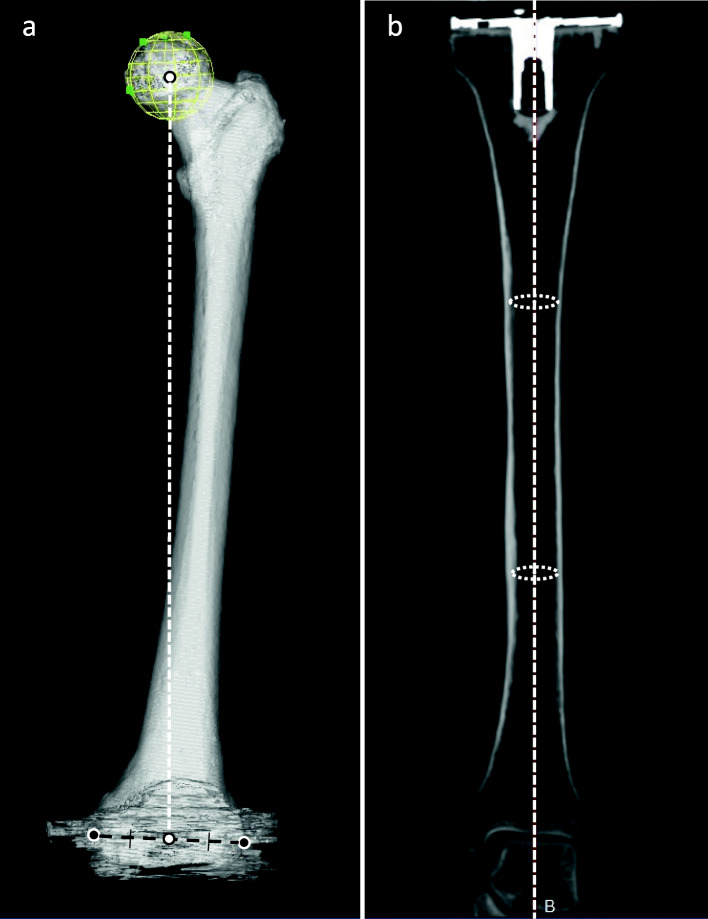
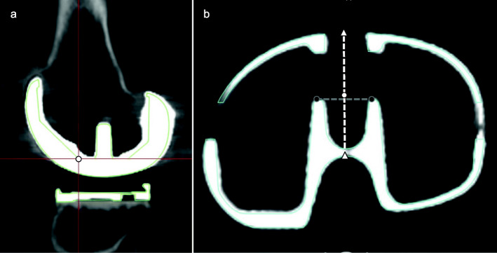
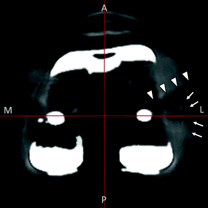
Methods: Fourteen knees from 14 patients who received primary TKA were included, and images were analyzed by blinded readers not associated with the surgeries. The examiner digitized the reference points according to defined landmarks, and the designated size component was superimposed to the 3D reconstructed CT model for measurement. In addition to the evaluation of implant position against the coronal and sagittal lower limb mechanical axes that were defined based on bony landmarks, implant position against axes connecting implant-based reference points that are easier to indicate was evaluated.

Results: The overall intra- and inter-observer reliabilities determined by the intraclass correlation coefficients (ICC) of the implant alignment measurement for both femoral and tibial components were good (ICC > 0.60), except in the direction of femoral flexion and extension, for both mechanical and implant-based axes. The difference between implant alignment measurements according to the traditional mechanical axis and the implant-based axis ranged between means of 0.08o and 1.70o and were statistically significantly different.

Conclusions: The postoperative evaluation of implant position in the coronal and sagittal planes using 3D-CT image matching is reliable and has good reproducibility except for the sagittal alignment assessment of the femoral component. The measured implant position according to the traditional mechanical axis and the implant-based axis were slightly but significantly different.

Keywords: 3D-CT; Implant position; Inter‐observer reliability; Intra‐observer reliability; Postoperative evaluation; Total knee arthroplasty.


Conflict of interest statement

Ryuichiro Akagi and Toshihiro Odera financially supported by Zimmer Biomet Inc. (Warsaw).

Figures

Fig. 1
Fig. 1
Reference points of the traditional mechanical axis . a: Three-dimensional image of the femur from the anterior aspect. The traditional mechanical axis of the femur was defined as the line connecting the white dots, which indicates the center of the femoral head and the center of the medial and lateral epicondyles (black dots). b: Multi-planar reconstruction coronal image of the tibia. Traditional mechanical alignment of the tibia was defined as the axis connecting the center of the circles drawn inside the upper and lower one-third of the medullary canal of the tibia
Fig. 2
Fig. 2
Reference points of the implant-based axis. a: Multi-planar sagittal reconstruction image of the femoral component. The mediolateral center at the distal end of the anterior chamfer was defined as the center of the femoral component. b: Multi-planar reconstruction axial image of the tibial tray. A perpendicular bisector of a line segment connecting the anterior end of the dovetails (black dots) and the center of the anteroposterior edges of the tibial tray (white triangles) was defined as the center of the tibial tray (white dot)
Fig. 3
Fig. 3
Experimental design and analyses. Each examiner independently performed the measurement of sagittal and coronal alignments twice for all images, with at least four weeks between measurements. The intra-observer ICC was calculated for each of the three observers, and the inter-observer ICC was calculated using the average of the two measurements of each observer. Implant alignment measurements were compared between the traditional mechanical axis and the implant-based axis
Fig. 4
Fig. 4
A case in which the anatomical landmarks are difficult to identify because of implant halation The lateral epicondyle (arrow) is obscured due to the overlapping implant halation (arrowheads)

Similar articles

Cited by

References

    1. Lee BS, Cho HI, Bin S, Il, Kim JM, Jo BK. Femoral Component Varus Malposition is Associated with Tibial Aseptic Loosening after TKA. Clin Orthop Relat Res. 2018;476:400–7. doi: 10.1007/s11999.0000000000000012. - DOI - PMC - PubMed
    1. Ritter MA, Faris PM, Keating EM, Meding JB. Postoperative alignment of total knee replacement. Its effect on survival. Clin Orthop Relat Res. 1994;:153–6. - PubMed
    1. Yuan FZ, Wang SJ, Zhou ZX, Yu JK, Jiang D. Malalignment and malposition of quadriceps-sparing approach in primary total knee arthroplasty: A systematic review and meta-analysis. J Orthop Surg Res. 2017;12:1–7. doi: 10.1186/s13018-016-0501-z. - DOI - PMC - PubMed
    1. Meric G, Gracitelli GC, Aram LJ, Swank ML, Bugbee WD. Variability in Distal Femoral Anatomy in Patients Undergoing Total Knee Arthroplasty: Measurements on 13,546 Computed Tomography Scans. J Arthroplasty. 2015;30:1835–8. doi: 10.1016/j.arth.2015.04.024. - DOI - PubMed
    1. Maestro A, Harwin SF, Sandoval MG, Vaquero DH, Murcia A. Influence of intramedullary versus extramedullary alignment guides on final total knee arthroplasty component position: A radiographic analysis. J Arthroplasty. 1998;13:552–8. doi: 10.1016/S0883-5403(98)90055-9. - DOI - PubMed

Publication types