. 2021 Jul;59(4):446-453.
doi: 10.1016/j.resinv.2021.03.002. Epub 2021 Mar 29.

Diagnostic prediction of COVID-19 based on clinical and radiological findings in a relatively low COVID-19 prevalence area

Affiliations

Diagnostic prediction of COVID-19 based on clinical and radiological findings in a relatively low COVID-19 prevalence area

Yosuke Amano et al. Respir Investig. 2021 Jul.

Abstract

Background: Distinguishing coronavirus disease 2019 (COVID-19) pneumonia from other lung diseases is often difficult, especially in a highly comorbid patient population in a low prevalence region. We aimed to distinguish clinical data and computed tomography (CT) images between COVID-19 and other lung diseases in an advanced care hospital.

Methods: We assessed clinical characteristics, laboratory data, and chest CT images of patients with COVID-19 and non-COVID-19 patients who were suspected of having COVID-19 between February 20 and May 21, 2020, at the University of Tokyo Hospital.

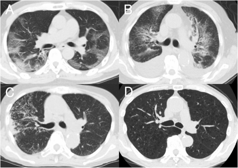
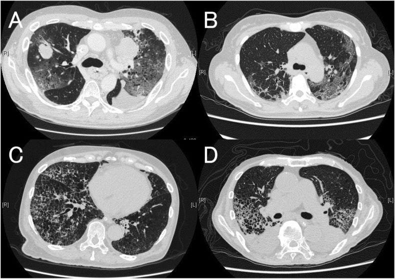
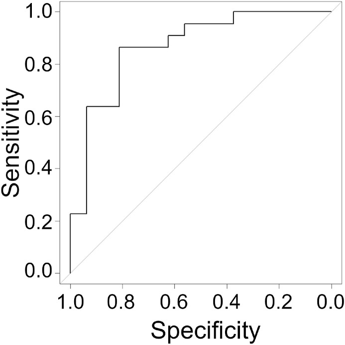
Results: Typical appearance for COVID-19 on CT images were found in 24 of 29 COVID-19 cases and 21 of 168 non-COVID-19 cases, according to the Radiological Society of North America Expert Consensus Statement (for predicting COVID-19, sensitivity 0.828, specificity 0.875, positive predictive value 0.533, negative predictive value 0.967). When we focused on cases with typical CT images, loss of taste or smell, and close contact with COVID-19 patients were exclusive characteristics for the COVID-19 cases. Among laboratory data, high fibrinogen (P < 0.01) and low white blood cell count (P < 0.01) were good predictors for COVID-19 with typical CT images in multivariate analysis.

Conclusions: In a relatively low prevalence region, CT screening has high sensitivity to COVID-19 in patients with suspected symptoms. When chest CT findings are typical for COVID-19, close contact, loss of taste or smell, lower white blood cell count, and higher fibrinogen are good predictors for COVID-19.

Keywords: COVID-19; Fibrinogen; Interstitial lung diseases; Taste disorder; White blood cell count.


Conflict of interest statement

Conflict of Interest The authors declare that they have no conflicts of interest for this study.

Figures

Fig. 1
Fig. 1
Typical images for each CT finding category of RSNA Expert Consensus Statement [6]. A: Cov19Typ is characterized mainly by peripheral, bilateral ground-glass opacity, or findings of organizing pneumonia. B: Cov19Ind is characterized mainly by diffuse, perihilar, or unilateral ground-glass opacity. C: Cov19Aty is characterized mainly by isolated consolidation, discrete small nodules, lung cavitation, or smooth interlobular septal thickening with pleural effusion. D: Cov19Neg is characterized by no CT features to suggest pneumonia. Abbreviations: CT, computed tomography.
Fig. 2
Fig. 2
Examples of non-COVID-19 with typical CT images (A, B) and COVID-19 cases with other than typical CT images (C, D). A: 55-year-old-male with nivolumab induced pneumonia arising during the treatment of esophageal cancer. CT images showed bilateral peripheral-dominant ground-glass opacities on pulmonary emphysema. Lung metastasis in the right upper lobe and preexisting left pleural effusion was also seen. B: 53-year-old-female with idiopathic pulmonary syndrome arising 6 months after an allogeneic hematopoietic stem cell transplant for acute myeloid leukemia. CT images showed bilateral-dominant peripheral ground-glass opacities. C: 68-year-old-male, current smoker, with chronic obstructive pulmonary disease. Two of three radiologists classified the CT images as Cov19Ind, the other Cov19Aty. D: 76-year-old-male, former smoker, with a history of recurrent aspiration pneumonia and chronic heart failure due to atrial fibrillation. All three radiologists classified the CT images as Cov19Ind. CT images of both cases were difficult to interpret because of the superimposition of severe emphysema. Abbreviations: COVID-19, coronavirus disease 2019; CT, computed tomography.
Fig. 3
Fig. 3
The receiver operating characteristics (ROC) curve for COVID-19 in Cov19typ CT images. The predicted probability (P) of COVID-19 among cases with Cov19typ images was estimated according to the results of binomial logistic regression analysis: P = 1/(1+e-x), where X = −0.5426 + 0.0085 ∗ fibrinogen (mg/dL) – 0.0005 ∗ WBC (/μL). The area under the ROC curve was 0.866 (95% CI, 0.745–0.988). When a cut-off point for X was defined as a value ≥ −0.0074, the sensitivity and the specificity were 0.863 and 0.813, respectively.

Similar articles

Cited by

References

    1. World Health organization coronavirus disease dashboard, https://www.covid19.who.int [accessed on January 31, 2021].
    1. Woloshin S., Patel N., Kesselheim A.S. False negative tests for SARS-CoV-2 infection - challenges and implications. N Engl J Med. 2020;383:e38. doi: 10.1056/NEJMp2015897. - DOI - PubMed
    1. McGarry B.E., SteelFisher G.K., Grabowski D.C., Barnett M.L. COVID-19 test result turnaround time for residents and staff in US nursing homes. JAMA Intern Med. 2020 doi: 10.1001/jamainternmed.2020.7330. - DOI - PMC - PubMed
    1. Xu B., Xing Y., Peng J., Zheng Z., Tang W., Sun Y. Chest CT for detecting COVID-19: a systematic review and meta-analysis of diagnostic accuracy. Eur Radiol. 2020;30:5720–5727. - PMC - PubMed
    1. Francone M., Iafrate F., Masci G.M., Coco S., Cilia F., Manganaro L. Chest CT score in COVID-19 patients: correlation with disease severity and short-term prognosis. Eur Radiol. 2020:1–10. doi: 10.1007/s00330-020-07033-y. - DOI - PMC - PubMed