Review
. 2021 Oct;44(5):1696-1712.
doi: 10.1007/s10753-021-01467-2. Epub 2021 Apr 18.

New Insights and Novel Therapeutic Potentials for Macrophages in Myocardial Infarction

Affiliations
Review

New Insights and Novel Therapeutic Potentials for Macrophages in Myocardial Infarction

Zenglei Zhang et al. Inflammation. 2021 Oct.

Abstract

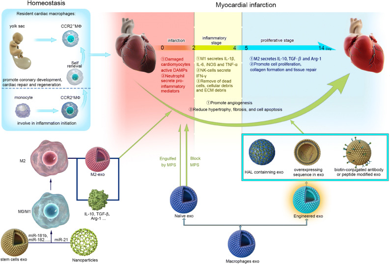
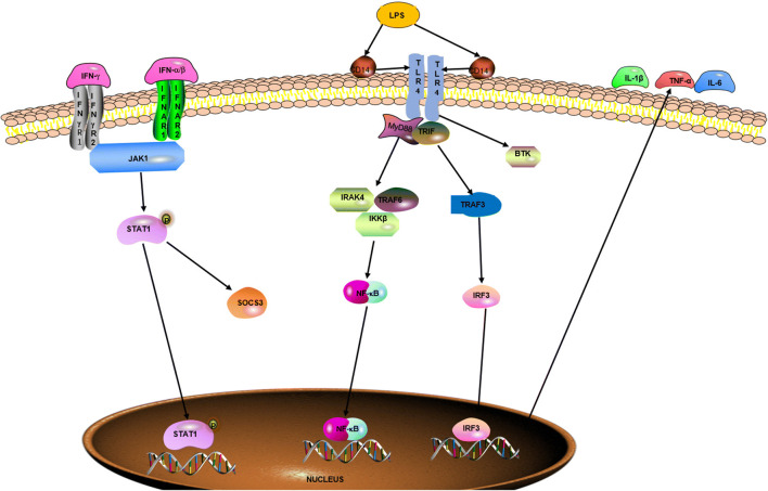
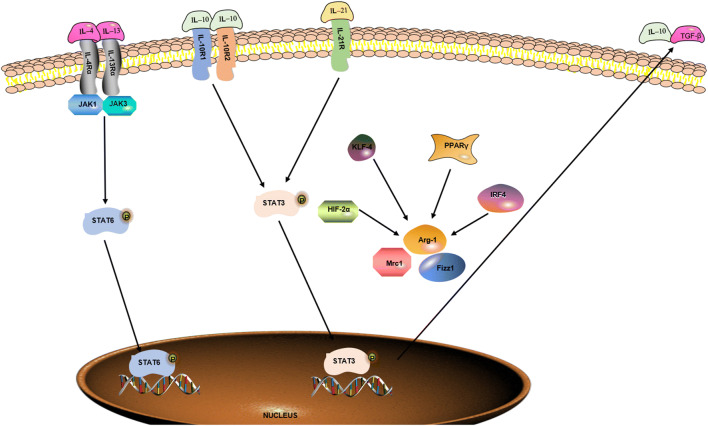
Cardiovascular disease (CVD) has long been the leading cause of death worldwide, and myocardial infarction (MI) accounts for the greatest proportion of CVD. Recent research has revealed that inflammation plays a major role in the pathogenesis of CVD and other manifestations of atherosclerosis. Overwhelming evidence supports the view that macrophages, as the basic cell component of the innate immune system, play a pivotal role in atherosclerosis initiation and progression. Limited but indispensable resident macrophages have been detected in the healthy heart; however, the number of cardiac macrophages significantly increases during cardiac injury. In the early period of initial cardiac damage (e.g., MI), numerous classically activated macrophages (M1) originating from the bone marrow and spleen are rapidly recruited to damaged sites, where they are responsible for cardiac remodeling. After the inflammatory stage, the macrophages shift toward an alternatively activated phenotype (M2) that promotes cardiac repair. In addition, extensive studies have shown the therapeutic potential of macrophages as targets, especially for emerging nanoparticle-mediated drug delivery systems. In the present review, we focused on the role of macrophages in the development and progression of MI, factors regulating macrophage activation and function, and the therapeutic potential of macrophages in MI.

Keywords: cardiac repair; macrophages; myocardial infarction; polarization; therapeutic strategies.


Conflict of interest statement

The authors declare no competing interests.

Figures

Fig. 1
Fig. 1
Macrophages function in homeostasis and in/post-myocardial infarction. The three overlapping stages are involved in the repair response after obstruction of the blood flow: infarction, inflammatory, and proliferative. In the infarction phase, damaged cardiomyocytes, active DAMPs, and neutrophils are recruited to the infarcted site and release many inflammatory mediators, which are indispensable for the subsequent inflammatory stage. In the inflammatory stage, proinflammatory subset M1 macrophages and NK cells secrete inflammatory cytokines, such as IL-1β, IL-6, iNOS, TNF-α, and IFN-γ, which promote clearance of dead cells and cellular and ECM debris. Last, the proliferative stage involves cell proliferation, collagen formation, and tissue repair that mainly contribute to anti-inflammatory subset M2 macrophages that secrete IL-10, TGF-β, and Arg-1. Modulation (i.e., self-assembly/engineered extracellular vesicles including exosomes[exo], nanoparticles, stem cells) of macrophages may repair damaged myocardium by promoting angiogenesis and reducing hypertrophy, fibrosis, and cell apoptosis.
Fig. 2
Fig. 2
Signaling pathways regulating M1 macrophage polarization. Naïve macrophage is induced into M1 macrophage by LPS, IFN-γ, and IFN-α/β through specific receptors, such as TLR4, IFNγR, and IFNAR. And the related signaling pathways such as STAT1, NF-κB, and IRF3 have an important role in the process, which results in the secretion of proinflammatory cytokines, such as IL-1β, TNF-α, and IL-6.
Fig. 3
Fig. 3
Signaling pathways regulating M2 macrophage polarization. Naïve macrophage is induced into M2 macrophage by IL-4, IL-13, IL-10, and IL-21 through interacting with specific receptors, such as IL-4Rα, IL-13Rα, IL-10R, and il-21R. And the related signaling pathways such as STAT6, STAT3, HIF-2α, KLF-4, PPARγ, and IRF4 are activated in the process, which promotes the secretion of anti-inflammatory cytokines, such as TGF-β and IL-10.

Similar articles

Cited by

References

    1. Sadek H, Olson EN. Toward the goal of human heart regeneration. Cell Stem Cell. 2020;26:7–16. doi: 10.1016/j.stem.2019.12.004. - DOI - PMC - PubMed
    1. Kura B, Bacova BS, Kalocayova B, Sykora M, Slezak J. Oxidative stress-responsive microRNAs in heart injury. International Journal of Molecular Sciences. 2020;21(1):358. doi: 10.3390/ijms21010358. - DOI - PMC - PubMed
    1. Roth GA, Johnson C, Abajobir A, Abd-Allah F, Abera SF, Abyu G, Ahmed M, et al. Global, regional, and national burden of cardiovascular diseases for 10 causes, 1990 to 2015. Journals of the American College of Cardiology. 2017;70:1–25. doi: 10.1016/j.jacc.2017.04.052. - DOI - PMC - PubMed
    1. Braunwald E. The war against heart failure: the Lancet lecture. Lancet. 2015;385(9970):812–824. doi: 10.1016/S0140-6736(14)61889-4. - DOI - PubMed
    1. Poss KD, Wilson LG, Keating MT. Heart regeneration in zebrafish. Science. 2002;298:2188–2190. doi: 10.1126/science.1077857. - DOI - PubMed

MeSH terms

LinkOut - more resources