. 2021 Aug;205(2):232-245.
doi: 10.1111/cei.13603. Epub 2021 Jun 1.

16α-Bromoepiandrosterone as a new candidate for experimental diabetes-tuberculosis co-morbidity treatment

Affiliations

16α-Bromoepiandrosterone as a new candidate for experimental diabetes-tuberculosis co-morbidity treatment

Manuel Othoniel López-Torres et al. Clin Exp Immunol. 2021 Aug.

Abstract

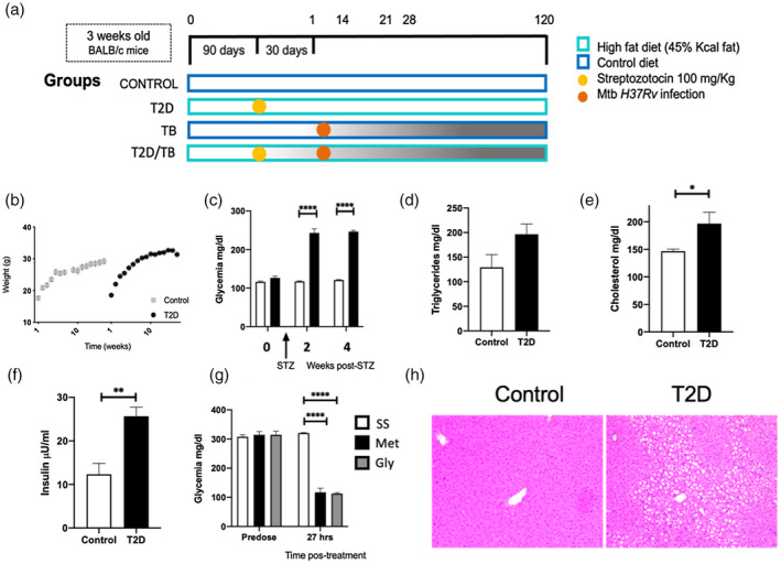
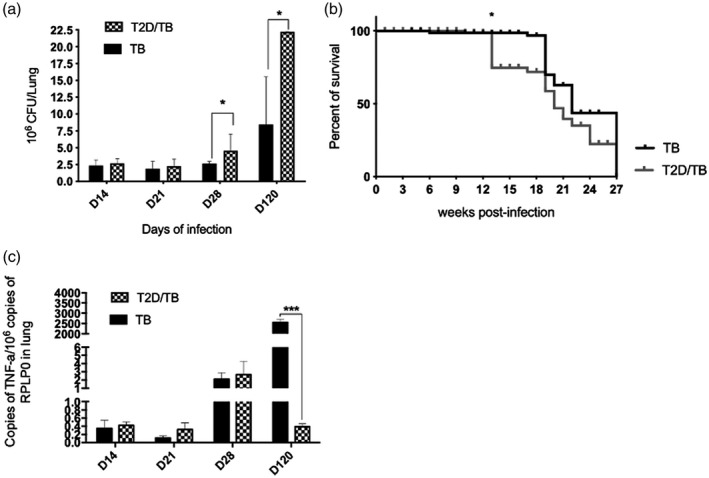
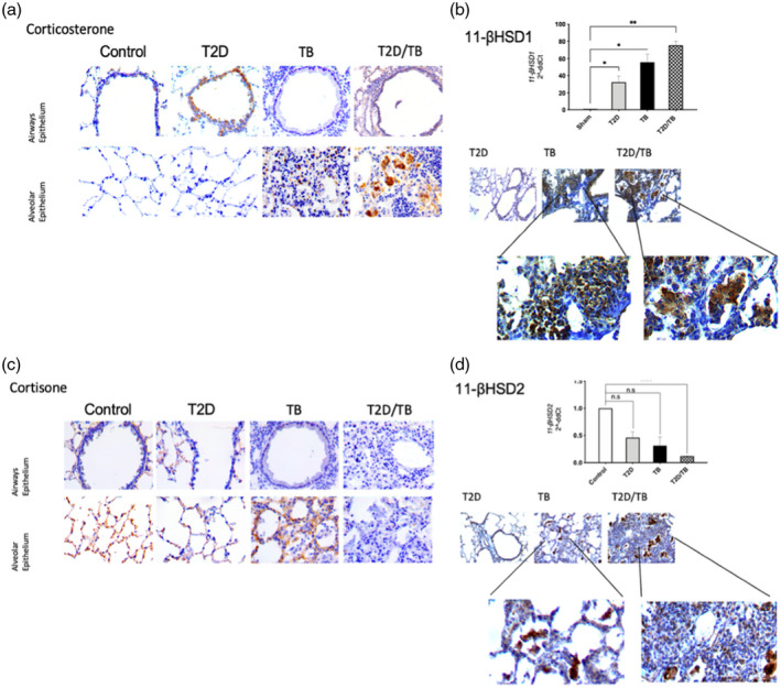
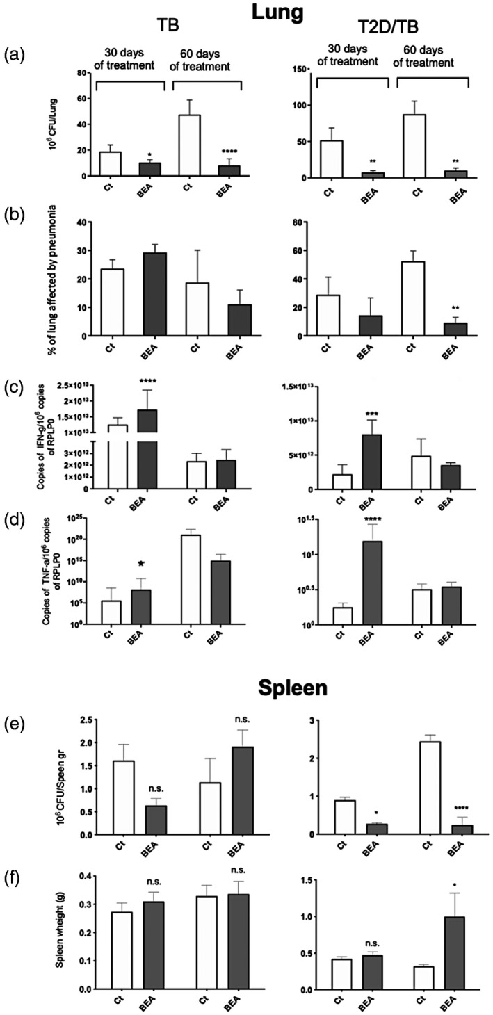
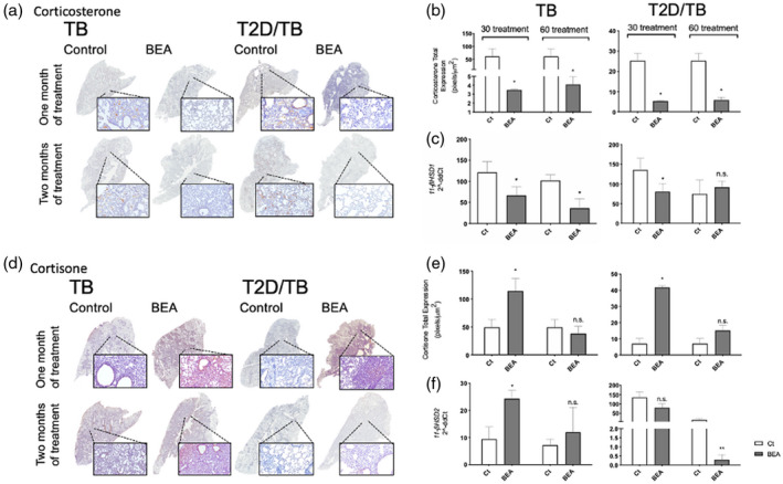
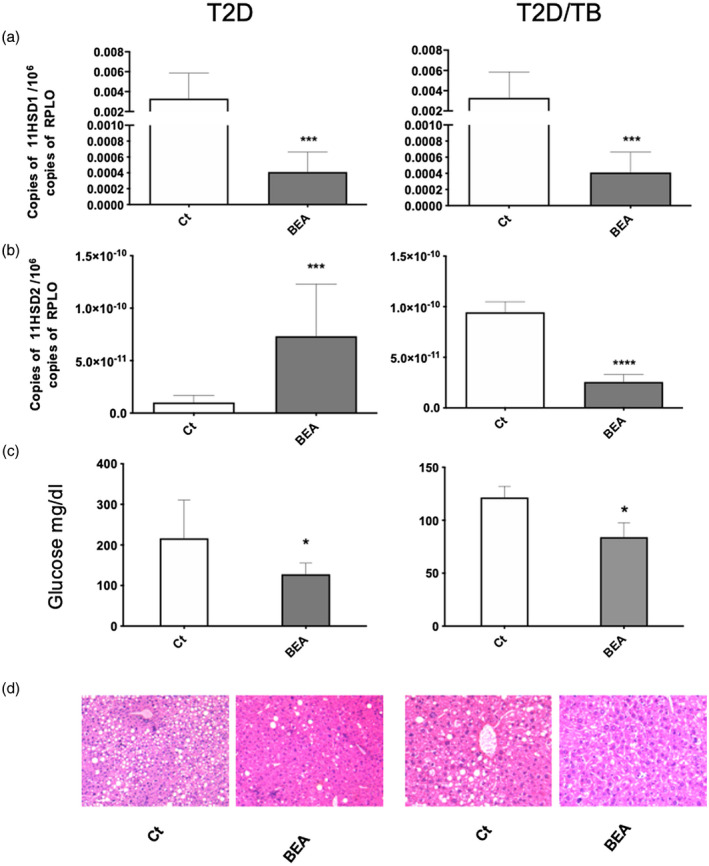
Tuberculosis (TB) is the leading cause of death from a single bacterial infectious agent and is one of the most relevant issues of public health. Another pandemic disease is type II diabetes mellitus (T2D) that is estimated to affect half a billion people in the world. T2D is directly associated with obesity and a sedentary lifestyle and is frequently associated with immunosuppression. Immune dysfunction induced by hyperglycemia increases infection frequency and severity. Thus, in developing countries the T2D/TB co-morbidity is frequent and represents one of the most significant challenges for the health-care systems. Several immunoendocrine abnormalities are occurring during the chronic phase of both diseases, such as high extra-adrenal production of active glucocorticoids (GCs) by the activity of 11-β-hydroxysteroid dehydrogenase type 1 (11-βHSD1). 11-βHSD1 catalyzes the conversion of inactive cortisone to active cortisol or corticosterone in lungs and liver, while 11-β-hydroxysteroid dehydrogenase type 2 (11-βHSD2) has the opposite effect. Active GCs have been related to insulin resistance and suppression of Th1 responses, which are deleterious factors in both T2D and TB. The anabolic adrenal hormone dehydroepiandrosterone (DHEA) exerts antagonistic effects on GC signaling in immune cells and metabolic tissues; however, its anabolic effects prohibit its use to treat immunoendocrine diseases. 16α-bromoepiandrosterone (BEA) is a water miscible synthetic sterol related to DHEA that lacks an anabolic effect while amplifying the immune and metabolic properties with important potential therapeutic uses. In this work, we compared the expression of 11-βHSD1 and the therapeutic efficacy of BEA in diabetic mice infected with tuberculosis (TB) (T2D/TB) with respect to non-diabetic TB-infected mice (TB). T2D was induced by feeding mice with a high-fat diet and administering a single low-dose of streptozotocin. After 4 weeks of T2D establishment, mice were infected intratracheally with a high-dose of Mycobacterium tuberculosis strain H37Rv. Then, mice were treated with BEA three times a week by subcutaneous and intratracheal routes. Infection with TB increased the expression of 11-βHSD1 and corticosterone in the lungs and liver of both T2D/TB and TB mice; however, T2D/TB mice developed a more severe lung disease than TB mice. In comparison with untreated animals, BEA decreased GC and 11-βHSD1 expression while increasing 11-βHSD2 expression. These molecular effects of BEA were associated with a reduction in hyperglycemia and liver steatosis, lower lung bacillary loads and pneumonia. These results uphold BEA as a promising effective therapy for the T2D/TB co-morbidity.

Keywords: 11-βHSD1; BEA; active glucocorticoids; central nervous system; colony-forming units; diabetes-tuberculosis co-morbidity; immunotherapy.


Conflict of interest statement

We declare no conflicts of interest during the course of this work.

Figures

FIGURE 1
FIGURE 1
Metabolic abnormalities and liver pathology of mice with type II diabetes mellitus in comparison with non‐diabetic animals (sham). (a) General description of the experimental groups. (b) Mice were weighed weekly for weight gain monitoring. Data are presented as mean and standard deviation of five mice per group for 16 weeks. (c) Blood glucose levels were assessed at 2 and 4 weeks after administration of streptozotocin (STZ) intraperitoneally in animals fed with a high‐fat diet (HFD) and in control animals not treated with STZ and fed with rodent chow. Serum concentrations of (d) triglycerides, (e) cholesterol and (f) insulin in control (white bars) and T2D (black bars) mice after 4 months of HFD‐STZ treatment. (g) Effect of hypoglycemic drugs on serum glucose levels in T2D animals, before and after 27 h of oral administration of metformin (Met) or glibenclamide (Gly). Asterisk represents statistical significance [one‐way anlysis of variance (anova), p < 0.05 and two‐way anova for graph 1B, 1C and 1G). (h) Representative micrograph of liver tissue from a control mouse (left) and T2D mouse (right). Extensive cytoplasmic vacuolization of hepatocytes corresponding to steatosis is shown in the liver of T2D mouse
FIGURE 2
FIGURE 2
Comparative course of pulmonary tuberculosis in diabetic mice infected with Mycobacterium tuberculosis (Mtb) (T2D/TB) and non‐diabetic mice infected with Mtb (TB). (a) Five mice per group were euthanized at the indicated time‐points post‐infection and the right lungs were used to determine the bacterial loads by counting colony‐forming units (CFU). A significant increase in the bacillary load of the T2D/TB group was observed after 1 and 4 months of infection in comparison with the control group
FIGURE 3
FIGURE 3
Gene expression and cellular localization of glucocorticoids converting enzymes and glucocorticoid content in late tuberculosis (TB) (day 120). (a) Representative micrographs of immunohistochemical staining of corticosterone in the epithelium of the lung airways and the alveolar epithelium. (b) Comparative gene expression of 11‐β‐hydroxysteroid dehydrogenase type 1 (11‐βHSD1) determined by quantitative polymerase chain reaction (qPCR) in the different experimental groups at day 120 (top panel). Immunohistochemical detection of 11‐βHSD1 (bottom panel). (c) Representative micrographs of immunohistochemical detection of cortisone in bronchial epithelial cells and alveoli. (d) Comparative 11‐βHSD2 gene expression and immunohistochemical staining. Asterisks represent statistical significance [****p < 0.0001, two‐ way analysis of variance (anova) and n.s. = not significant]
FIGURE 4
FIGURE 4
Therapeutic effect of 16α‐bromoepiandrosterone (BEA) in tuberculosis (TB) and type 2 diabetes mellitus (T2D)/TB mice. Groups of TB and T2D/TB mice after 60 days of intratracheal infection with high‐dose Mycobacterium tuberculosis (Mtb) strain H37Rv were treated during 1 or 2 months with BEA, while the control TB or T2D/TB groups received only the vehicle. Lungs were processed for the indicated determination. In comparison with the control TB or T2D/TB mice, BEA induced a significant decrease of pulmonary bacillary loads and tissue damage (pneumonia) in both TB and T2D/TB groups, particularly after 60 days of treatment. After 1 month of treatment, BEA induced higher expression of the protective cytokines interferon (IFN)‐γ and tumor necrosis factor (TNF)‐α. In the spleen, treatment with BEA did not produced significant changes in the weight and colony‐forming units (CFUs) in TB mice, while it induced significant decrease of bacillary burdens and higher weight in T2D/TB mice after 2 months of treatment. Asterisks represent statistical significance [****p < 0.0001, two‐way analysis of variance (anova); n.s. = not significant]
FIGURE 5
FIGURE 5
Effect of 16α‐bromoepiandrosterone (BEA) on lung glucocorticoids (GCs) production and gene expression of their converting enzymes in tuberculosis (TB) and type 2 diabetes mellitus (T2D)/TB mice. (a) Representative low‐power and mild‐power micrographs of immunohistochemistry detection of corticosterone in both groups after 1 and 2 months of BEA treatment. (b) In comparison with the TB or T2D/TB groups, the digital pathology analysis showed significant lower immunostaining of corticosterone induced by BEA treatment after 1 and 2 months in TB mice and T2D/TB mice. (c) BEA treatment induced lower 11‐β‐hydroxysteroid dehydrogenase type 1 (11β‐HSD1) gene expression in TB and T2D/TB in comparison with TB or T2D/TB mice treated with vehicle. (d) Representative micrographs of cortisone detection by immunohistochemistry in the lungs of TB and T2D mice treated with BEA at 1 and 2 months of infection. (e) The digital pathology study shows higher expression of cortisone after 1 month of treatment in the TB and T2D/TB groups. (f) Gene expression of 11b‐HSD2 corticosterone converting enzyme after BEA treatment in TB and T2D/TB animals. Asterisk represents statistical significance (p < 0.05)
FIGURE 6
FIGURE 6
Effect of 16α‐bromoepiandrosterone (BEA) in the liver of tuberculosis (TB) and type 2 diabetes mellitus (T2D)/TB mice. Liver samples from groups of five T2D and T2D/TB mice treated with BEA for 1 month and their respective control mice that received only the vehicle were used to isolate total RNA and 11‐β‐hydroxysteroid dehydrogenase type 1 (1‐βHSD1) (a) and 11‐βHSD2) (b) mRNA determination by reverse transcription–polymerase chain reaction (RT–PCR). (c) Treatment with BEA corrected hyperglycemia in T2D and T2D/TB mice. Asterisks represent statistical significance [***p < 0.0001, ****p < 0.0001, two‐way analysis of variance (anova), n.s. = without significance]. D) Liver tissue sections stained with hematoxylin and eosin (H&E) revealed steatosis in T2D and T2D/TB mice, which was reverted by treatment with BEA

References

    1. Ayelign B, Negash M, Genetu M, Wondmagegn T, Shibabaw T. Immunological impacts of diabetes on the susceptibility of Mycobacterium tuberculosis . J Immunol Res. 2019;2019:1–8. - PMC - PubMed
    1. Ehlers S, Schaible UE. The granuloma in tuberculosis: dynamics of a host–pathogen collusion. Front Immunol. 2012;3:1–9. - PMC - PubMed
    1. Meenakshi P, Ramya S, Lavanya J, Vijayalakshmi V, Sumanlatha G. Effect of IFN‐c, IL‐12 and IL‐10 cytokine production and mRNA expression in tuberculosis patients with diabetes mellitus and their household contacts. Cytokine 2016;81:127–36. https://doi.org/10.1016/j.cyto.2016.03.009. - DOI - PubMed
    1. Cooper AM, Mayer‐Barber KD, Sher A. Role of innate cytokines in mycobacterial infection. Mucosal Immunol. 2011;4:252–60. - PMC - PubMed
    1. Rook WG. Th2 cytokines in susceptibility to tuberculosis. Curr Mol Med. 2007;7:327–37. - PubMed

Publication types

MeSH terms