Heterogeneity of Lipid and Protein Cartilage Profiles Associated with Human Osteoarthritis with or without Type 2 Diabetes Mellitus
- PMID: 33866785
- PMCID: PMC8155553
- DOI: 10.1021/acs.jproteome.1c00186
Heterogeneity of Lipid and Protein Cartilage Profiles Associated with Human Osteoarthritis with or without Type 2 Diabetes Mellitus
Abstract
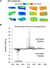
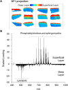
Osteoarthritis (OA) is a multifactorial pathology and comprises a wide range of distinct phenotypes. In this context, the characterization of the different molecular profiles associated with each phenotype can improve the classification of OA. In particular, OA can coexist with type 2 diabetes mellitus (T2DM). This study investigates lipidomic and proteomic differences between human OA/T2DM- and OA/T2DM+ cartilage through a multimodal mass spectrometry approach. Human cartilage samples were obtained after total knee replacement from OA/T2DM- and OA/T2DM+ patients. Label-free proteomics was employed to study differences in protein abundance and matrix-assisted laser desorption/ionization (MALDI) mass spectrometry imaging (MSI) for spatially resolved-lipid analysis. Label-free proteomic analysis showed differences between OA/T2DM- and OA/T2DM+ phenotypes in several metabolic pathways such as lipid regulation. Interestingly, phospholipase A2 protein was found increased within the OA/T2DM+ cohort. In addition, MALDI-MSI experiments revealed that phosphatidylcholine and sphingomyelin species were characteristic of the OA/T2DM- group, whereas lysolipids were more characteristic of the OA/T2DM+ phenotype. The data also pointed out differences in phospholipid content between superficial and deep layers of the cartilage. Our study shows distinctively different lipid and protein profiles between OA/T2DM- and OA/T2DM+ human cartilage, demonstrating the importance of subclassification of the OA disease for better personalized treatments.
Keywords: MALDI-MSI; cartilage; diabetes; label-free proteomics; osteoarthritis; spatially resolved-lipid analysis.
Conflict of interest statement
The authors declare no competing financial interest.
Figures


Similar articles
-
Spatially resolved endogenous improved metabolite detection in human osteoarthritis cartilage by matrix assisted laser desorption ionization mass spectrometry imaging.Analyst. 2019 Oct 21;144(20):5953-5958. doi: 10.1039/c9an00944b. Epub 2019 Aug 16. Analyst. 2019. PMID: 31418440
-
Matrix-assisted laser desorption/ionization mass spectrometry imaging (MALDI-MSI) reveals potential lipid markers between infrapatellar fat pad biopsies of osteoarthritis and cartilage defect patients.Anal Bioanal Chem. 2023 Oct;415(24):5997-6007. doi: 10.1007/s00216-023-04871-9. Epub 2023 Jul 28. Anal Bioanal Chem. 2023. PMID: 37505238 Free PMC article.
-
Matrix assisted laser desorption ionization mass spectrometry imaging identifies markers of ageing and osteoarthritic cartilage.Arthritis Res Ther. 2014 May 9;16(3):R110. doi: 10.1186/ar4560. Arthritis Res Ther. 2014. PMID: 24886698 Free PMC article.
-
Mass Spectrometry Imaging as a Potential Tool to Investigate Human Osteoarthritis at the Tissue Level.Int J Mol Sci. 2020 Sep 3;21(17):6414. doi: 10.3390/ijms21176414. Int J Mol Sci. 2020. PMID: 32899238 Free PMC article. Review.
-
Biomarkers and proteomic analysis of osteoarthritis.Matrix Biol. 2014 Oct;39:56-66. doi: 10.1016/j.matbio.2014.08.012. Epub 2014 Aug 30. Matrix Biol. 2014. PMID: 25179675 Review.
Cited by
-
Expression and clinical significance of LncRNA Kcnq1ot1 in type 2 diabetes mellitus patients with osteoarthritis.BMC Endocr Disord. 2025 May 16;25(1):131. doi: 10.1186/s12902-025-01915-2. BMC Endocr Disord. 2025. PMID: 40380166 Free PMC article.
-
Functional mass spectrometry imaging maps phospholipase-A2 enzyme activity during osteoarthritis progression.Theranostics. 2023 Aug 21;13(13):4636-4649. doi: 10.7150/thno.86623. eCollection 2023. Theranostics. 2023. PMID: 37649605 Free PMC article.
-
Evaluation of the Anti-Inflammatory and Chondroprotective Effect of Celecoxib on Cartilage Ex Vivo and in a Rat Osteoarthritis Model.Cartilage. 2022 Jul-Sep;13(3):19476035221115541. doi: 10.1177/19476035221115541. Cartilage. 2022. PMID: 35932105 Free PMC article.
-
Tissue-specific and spatially dependent metabolic signatures perturbed by injury in skeletally mature male and female mice.bioRxiv [Preprint]. 2025 Feb 7:2024.09.30.615873. doi: 10.1101/2024.09.30.615873. bioRxiv. 2025. PMID: 39975211 Free PMC article. Preprint.
-
Three decades of advancements in osteoarthritis research: insights from transcriptomic, proteomic, and metabolomic studies.Osteoarthritis Cartilage. 2024 Apr;32(4):385-397. doi: 10.1016/j.joca.2023.11.019. Epub 2023 Dec 2. Osteoarthritis Cartilage. 2024. PMID: 38049029 Free PMC article. Review.
References
-
- Helmick C. G.; Felson D. T.; Lawrence R. C.; Gabriel S.; Hirsch R.; Kwoh C. K.; Liang M. H.; Kremers H. M.; Mayes M. D.; Merkel P. A.; Pillemer S. R.; Reveille J. D.; Stone J. H. Estimates of the prevalence of arthritis and other rheumatic conditions in the United States. Arthritis Rheum. 2008, 58, 15–25. 10.1002/art.23177. - DOI - PubMed
Publication types
MeSH terms
Substances
LinkOut - more resources
Full Text Sources
Other Literature Sources
Medical

